Search results (71 results)
-
 Central Retinal Artery Occlusion
Aug 23 2012 by Gerardo Garcia-Aguirre, MD
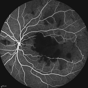
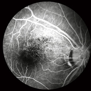
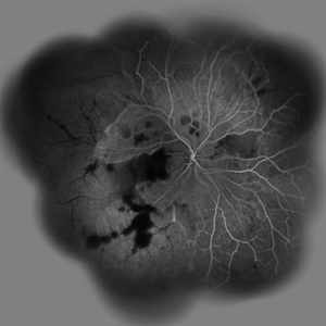
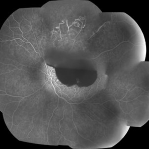
Fluorescein angiogram, late phase, of a central retinal artery occlusion, showing very delayed filling and wide areas of capillary nonperfusion.
Photographer: Noemí Hernández, Asociación para Evitar la Ceguera en México
Condition/keywords: capillary nonperfusion, central retinal artery occlusion (CRAO), vessel sheathing
-
 Acute Idiopathic Occlusive Retinal Vasculitis
May 31 2014 by Hamid Ahmadieh, MD
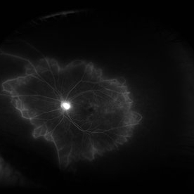
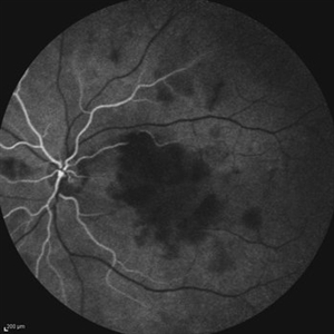
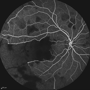
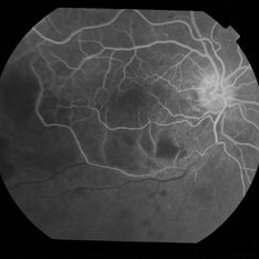
Mid- phase fluorescein angiogram of the left eye of a 28-year-old woman with acute drop of vision due to occlusive retinal vasculitis leading to extensive capillary nonperfusion and macular infarction.
Photographer: Naghmeh Nozhat, Negah Eye Center, Tehran
Imaging device: Heidelberg Spectralis
Condition/keywords: capillary nonperfusion, retinal vasculitis
-
 Coats Disease Fluorescein Angiography
Sep 2 2022 by FLOR ANGELICA JACOME GUTIERREZ
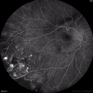
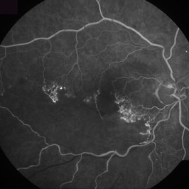
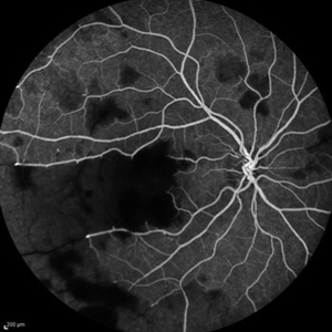
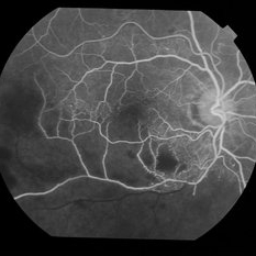
Fluorescein angiography of a patient with Coats disease where we found telangiectatic vessels, aneurysms, peripheral capillary nonperfusion and perivascular leak.
Photographer: Dr.Guillermo Salcedo Villanueva
Imaging device: Zeiss CLARUS 700 (FA)
Condition/keywords: Coats' disease, epiretinal membrane (ERM)
-
 Laser Induced BRAO in IRVAN Syndrome
May 3 2019 by Deependra Vikram Singh, MD FASRS
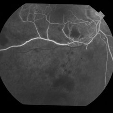
Fundus photograph of a 26-year-old man with IRVAN syndrome referred for vitreous surgery in OS for secondary rhegmatogenous retinal detachment. OD has received laser photocoagulation for capillary nonperfusion areas and retinal artery macroaneurysm associated with retinal vasculitis. Fundus photograph of OD shows laser induced nasal BRAO. Case re-emphasizes why laser for macroaneurysm should be avoided in cases with IRVAN.
Photographer: Deependra V Singh, Eye-Q Superspecialty Eye Hospitals. Gurugram, India
Imaging device: Zeiss Visucam 500
Condition/keywords: arteriolar macroaneurysm, branch retinal artery occlusion (BRAO), laser photocoagulation
-
 Severe Capillary Nonperfusion
Jul 8 2012 by Jeffrey S. Heier, MD
Severe NPDR capillary nonperfusion diabetic retinopathy wide-angle angiography. 40 year history of diabetic retinopathy.
Imaging device: OPTOS
Condition/keywords: nonperfusion diabetic retinopathy
-
 Capillary Nonperfusion
Apr 12 2018 by SUSHIL BHATT
OPTOS ultra wide field angiogram of an 45 years old diabetic male patient shows capillary nonperfusion areas with inadequate laser.
Photographer: Bhatt Sushil PGIMER chandigarh INDIA
Imaging device: OPTOS Ultra wide Field
Condition/keywords: capillary nonperfusion
-
 Proliferative Diabetic Retinopathy - Neovascularization on the Disc
Aug 23 2012 by Gerardo Garcia-Aguirre, MD
Fluorescein angiogram, early phase, showing microaneurysms, wide areas of capillary nonperfusion, and leakage secondary to neovascularization on the disc.
Photographer: Noemí Hernández, Asociación para Evitar la Ceguera en México
Condition/keywords: microaneurysms, neovascularization of the disc (NVD)
-
 Wyburn-Mason Fluorescein Angiography
Apr 29 2018 by Sarina M Amin, MD
Wide-field fluorescein angiography of a 32-year-old woman with Wyburn-Mason syndrome showing temporal periphery capillary nonperfusion.
Photographer: Sarah Ellano, Retinal Consultants of Arizona, Phoenix, Arizona
Imaging device: Optos
Condition/keywords: Wyburn-Mason
-
 Ischemic BRVO
Aug 23 2012 by Gerardo Garcia-Aguirre, MD
Fluorescein angiogram inferior to the macular area, showing wide areas of capillary nonperfusion.
Photographer: Noemí Hernández, Asociación para Evitar la Ceguera en México
Condition/keywords: branch retinal vein occlusion (BRVO), capillary nonperfusion
-
 BRVO FA, Early Phase
Oct 1 2012 by Jeffrey G. Gross, MD, FASRS
BRVO-FA early phase.
Condition/keywords: branch retinal vein occlusion (BRVO), capillary nonperfusion, early phase, microaneurysms
-
 Ischemic BRVO with Neovascularization
Aug 23 2012 by Gerardo Garcia-Aguirre, MD
Fluorescein angiogram of the macula showing wide areas of capillary nonperfusion and leakage in the superotemporal quadrant.
Photographer: Noemí Hernández, Asociación para Evitar la Ceguera en México
Condition/keywords: branch retinal vein occlusion (BRVO), capillary nonperfusion, neovascularization (NV)
-
 Ischemic BRVO with neovascularization
Aug 23 2012 by Gerardo Garcia-Aguirre, MD
Fluorescein angiogram of the temporal periphery showing wide areas of capillary nonperfusion and leakage secondary to neovascularization.
Photographer: Noemí Hernández, Asociación para Evitar la Ceguera en México
Condition/keywords: branch retinal vein occlusion (BRVO), capillary nonperfusion, neovascularization (NV)
-
 Paracentral Acute Middle Maculopathy
Oct 25 2019 by Gayathri Mohan
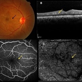
Multimodal images of a case of a 29-year-old female with paracentral acute middle maculopathy. A-color fundus photograph showing multiple confluent white retinal patches. B- On OCT the acute lesions of PAMM characteristically appear as placoid, hyperreflective bands at the level of the INL C-Fundus fluorescein angiography showing a capillary nonperfusion area D-flow void areas in deep capillary plexus
Photographer: Akshar Soni
Imaging device: Heidelberg, Nidek
Condition/keywords: fundus albipunctatus, optical coherence tomography (OCT), paracentral acute middle maculopathy
-
 Proliferative diabetic retinopathy
Dec 15 2012 by Sharon Fekrat, MD FACS FASRS
65-year old-man with proliferative diabetic retinopathy, retinal capillary nonperfusion, and neovascularization elsewhere in the right eye.
Photographer: John Reaves, Ophthalmic Photographer, Durham VA Medical Center Eye Clinic Imaging Suite, Durham, NC
Imaging device: fluorescein angiography
Condition/keywords: retinal neovascularization
-
 Branches Starved of Flow, Yet Nature Strives to Grow
Apr 1 2025 by rohan jain
Tufts of NVE's in a case of Branch Retinal Vein Occlusion
Photographer: Dr. ROHAN JAIN
Condition/keywords: branch retinal vein occlusion (BRVO), capillary nonperfusion, non-perfused branch retinal vein occlusion (BRVO), non-perfusion, NVE, OCT Angiography, ST BRVO
-
 Ischemic BRVO
Aug 23 2012 by Gerardo Garcia-Aguirre, MD
Fluorescein angiogram of the posterior pole showing wide areas of capillary nonperfusion involving the fovea.
Photographer: Noemí Hernández, Asociación para Evitar la Ceguera en México
Condition/keywords: branch retinal vein occlusion (BRVO), capillary nonperfusion
-
 Acute Idiopathic Occlusive Retinal Vasculitis
May 31 2014 by Hamid Ahmadieh, MD
Early phase fluorescein angiogram of the left eye of a 28-year-old woman with acute drop of vision due to occlusive retinal vasculitis leading to extensive capillary nonperfusion and macular infarction.
Photographer: Naghmeh Nozhat, Negah Eye Center, Tehran
Imaging device: Heidelberg Spectralis
Condition/keywords: retinal vasculitis
-
 Acute Idiopathic Occlusive Retinal Vasculitis
May 31 2014 by Hamid Ahmadieh, MD
Wide- field fluorescein angiogram of the right eye of a 28-year-old woman with acute drop of vision due to occlusive retinal vasculitis leading to extensive capillary nonperfusion and macular infarction.
Photographer: Naghmeh Nozhat, Negah Eye Center, Tehran
Imaging device: Heidelberg Spectralis
Condition/keywords: capillary nonperfusion, retinal infarction, retinal vasculitis
-
 Acute Idiopathic Occlusive Retinal Vasculitis
May 31 2014 by Hamid Ahmadieh, MD
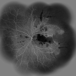
Mid phase fluorescein angiogram of the right eye of a 28-year-old woman with acute drop of vision due to occlusive retinal vasculitis leading to extensive capillary nonperfusion and macular infarction.
Photographer: Naghmeh Nozhat, Negah Eye Center, Tehran
Imaging device: Heidelberg Spectralis
Condition/keywords: capillary nonperfusion, retinal vasculitis
-
 Acute Idiopathic Occlusive Retinal Vasculitis
May 31 2014 by Hamid Ahmadieh, MD
Early phase fluorescein angiogram of the right eye of a 28-year-old woman with acute drop of vision due to occlusive retinal vasculitis leading to extensive capillary nonperfusion and macular infarction.
Photographer: Naghmeh Nozhat, Negah Eye Center, Tehran
Imaging device: Heidelberg Spectralis
Condition/keywords: retinal vasculitis
-
 Acute Idiopathic Occlusive Retinal Vasculitis
May 31 2014 by Hamid Ahmadieh, MD
Wide- field fluorescein angiogram of the left eye of a 28-year-old woman with acute drop of vision due to occlusive retinal vasculitis leading to extensive capillary nonperfusion and macular infarction.
Photographer: Naghmeh Nozhat, Negah Eye Center, Tehran
Imaging device: Heidelberg Spectralis
Condition/keywords: capillary nonperfusion, retinal infarction, retinal vasculitis
-
 Branch Retinal Vein Occlusion with Acute on Chronic Subhyaloid Hemorrhage
Oct 24 2019 by Nichole Lewis
60-year-old male with a branch retinal vein occlusion and subhyaloid hemorrhage and retinal neovascularization. VA HM.
Photographer: Nichole
Condition/keywords: branch retinal vein occlusion (BRVO), capillary nonperfusion, retinal neovascularization, subhyaloid hemorrhage
-
 Branch Retinal Vein Occlusion With Nonperfusion
May 4 2014 by Mallika Goyal, MD
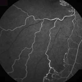
Right eye fluorescein angiogram of a 59-year-old male patient with inferotemporal branch retinal vein occlusion shows delayed venous filling and capillary nonperfusion in the affected quadrant.
Photographer: Mallika Goyal, MD, Apollo Health City, Jubilee Hills, Hyderabad, India
Condition/keywords: branch retinal vein occlusion (BRVO)
-
 Branch Retinal Vein Occlusion With Nonperfusion
May 4 2014 by Mallika Goyal, MD
Right eye fluorescein angiogram of a 59-year-old male patient with inferotemporal branch retinal vein occlusion shows delayed venous filling and capillary nonperfusion in the affected quadrant.
Photographer: Mallika Goyal, MD, Apollo Health City, Jubilee Hills, Hyderabad, India
Condition/keywords: branch retinal vein occlusion (BRVO)
 Loading…