Search results (181 results)
-
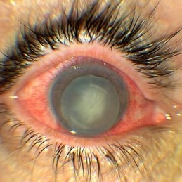
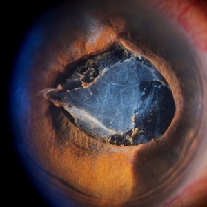 Inflammatory pupillary membrane in patient with endophthalmitis
Jan 28 2023 by Kingston Rodolfo Ureña-Wong, MD, Opht, MSc
Anterior segment photography of a 54-year-old woman with post phacoemulsification endophthalmitis. She did not improve after first intravitreal antibiotics injection and develop an inflammatory pupillary membrane. After two vitrectomies, and a complete three intravitreal injections scheme, we decided to remove the intraocular lens and capsules.
Photographer: Marco Antonio Rubio-Atonal,UNAM, Asociación para evitar la ceguera en México
Imaging device: Zeiss Clarus 700
Condition/keywords: endophthalmitis, pupillary membranes
-
 Hourglass in an Eye
Apr 22 2025 by KRISHNENDU NANDI, MS
A twenty-five-year-young male presented with a decrease in vision in the right eye following a blunt trauma with a football. On examination the BCVA in the right eye was CFCF and the left eye was 6/6, N6. The anterior segment was within normal limits. AT was 12 and 10 mm of Hg in the right and left eyes, respectively. Fundus examination reveals subhyaloid haemorrhage in the right eye with an attached retina. The fundus of the left eye was within normal limits. YAG laser hyaloidotomy was done with an energy of 2 mJ in the right eye. After 3 weeks the BCVA in the right eye improved to 6/9, N6.
Photographer: Dr. Krishnendu Nandi
Imaging device: Topcon
Condition/keywords: Trauma, YAG HYALOIDOTOMY, Young Male
-
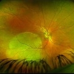
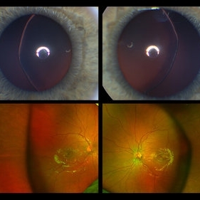 Ectopia Lentis
Jan 21 2021 by Jamin S. Brown, MD
This image serial demonstrates a patient with simple ectopia lentis. Anterior segment photographs in the upper panel demonstrate nasally subluxated crystalline lenses. Widefield fundus photography shows a "pseudo-buckle" which is the result of an optical effect due to the lens subluxation (artifactual image enlargement). Also note the juvenile macular reflex in this young patient. Ectopia lentis can present isolated ("simple") or in combination with various systemic defects (Marfan's syndrome, Weil-Marchesani syndrome or Ehlers-Danlos syndrome to name a few). Isolated ectopia lentis can be hereditary and causative genes have been identified as ADAMTSL4 located on chromosome 4 and FBN1 gene located on chromosome 15. Defects in the genes cause weakness in the zonular fibers which can lead to lens dislocation. Lastly, various ocular disorders such as Aniridia, Axenfeld-Rieger, Pseudoexfoliation or Trauma may also result in lens dislocation or subluxation.
Photographer: Stefanie Palmer CRA, Retina Vitreous Surgeons of CNY
Condition/keywords: dislocated lens, ectopia lentis
-
 Waardenberg Syndrome
Jan 21 2021 by Jamin S. Brown, MD
9-year-old African American female, fellow eye color blue.
Photographer: Stefanie Palmer CRA, Retina Vitreous Surgeons of CNY
Condition/keywords: anterior segment, iris, waardenburg syndrome
-
 4 Point Scleral Fixation Akreos AO60 With Gore Tex Suture
May 21 2021 by Jesus Lozano, MD
Anterior segment photo of a 54-year-old man after 4 point scleral fixation Akreos AO60 with Gore Tex suture plus PPV who had a severe traumatic iris defect and was aphakic after ocular trauma.
Photographer: Luigi Zinn, Hadassah Medical Center, Jerusalem.
Condition/keywords: aphakia, cornea rupture, lens, penetrating trauma
-
 Anterior Segment Gas Bubble and PFC Interface
Jun 21 2018 by Maria Stephanie R. Jardeleza, MD
Anterior segment photographs of 30-year-old male who underwent superior rhegmatogenous retinal detachment repair with intraocular gas tamponade. Perfluorocarbon was used to flatten the macula to prevent a macular fold and was removed during PFC/air exchange. Post operative week two visit shows gas migration into the anterior chamber with retained PFC on the posterior aspect of the gas bubble/anterior surface of the lens. Patient had been maintaining face down positioning.
Photographer: Andy Zepeda, COA, Retina Clinic, San Antonio Eye Center, San Antonio, TX
Condition/keywords: retained perfluorocarbon, vitreous substitutes
-
000---thumb.jpg/image-square;max$300,300.ImageHandler) Anterior Segment Photo of Emulsified Silicone Oil
Anterior Segment Photo of Emulsified Silicone Oil
Dec 25 2013 by Dong Yoon Kim, MD
47-year-old woman underwent vitrectomy and silicone oil tampoande for tractional retinal detachment due to proliferative diabetic retinopathy. 8 months after silicone oil tamponade, silicone oil was emulsified. And emulsified silicone oil was observed at anterior chamber.
Condition/keywords: silicone oil, tractional retinal detachment
-
 BDUMP
BDUMP
Dec 11 2018 by John S. King, MD
67-year-old white female with normal vision four months ago, consulted for dry AMD. She reported that vision in the left eye had worsened over the last two months and had progressively gotten worse. Denied history of cancer, or her primary eye doctor ever mentioning choroidal nevi. Va cc was 20/30 OD and 20/100 OS. No RAPD. IOP 9-10 OU. Anterior segment had some stellate like pigmented dusting of the endothlium, a/c was quiet, 2+NSC OU. Vitreous quiet; multiple, flat, pigmented choroidal lesions varying in size was seen the in fundus. Area in the temporal macula extending up to the superior arcade in the left eye that was suspicious for a mass; it did have a "giraffe like" pattern on one of the early FA pics; the OCT in this area showed thickening of the choroid without a definite mass lesion, and overlying thickening of the RPE, or exudative like scar, with SRF directly above. Consulted with Dr. Matt Wilson, who confirmed diagnosis, and had patient evaluated by oncology, who diagnosed non-small cell lung cancer.
Photographer: Stacey Coleman
Imaging device: Topcon
Condition/keywords: bilateral diffuse uveal melanocytic proliferation (BDUMP)
-
 Giant Retinal Tear
Apr 1 2016 by Nichole Lewis
Giant retinal tear montaged on Anterior Segment due to the Detachment being very bullous.
Photographer: Nichole Lewis - Pennsylvania Retina Specialists, Camp Hill, PA
Condition/keywords: giant retinal tear, retinal tear
-
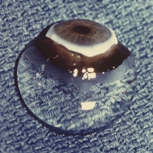 Human Vitreous Body
Sep 1 2020 by J. Sebag, MD, FACS, FRCOphth, FARVO
The sclera, choroid and retina were peeled off the vitreous body which remains attached to the anterior segment in this 9 month-old child. Due to this young age, the vitreous body maintains its solid gel structure in spite of being situated on a surgical towel (blue) in room air. [Cover photo – Sebag J: The Vitreous- Structure, Function, and Pathobiology, Springer-Verlag, New York, 1989. Specimen courtesy of the New England Eye Bank; image © Springer Nature, reprinted with permission]
Condition/keywords: choroid, retina, sclera, vitreous
-
 Hyaloid Artery, Anterior Segment
May 3 2018 by Alexandr Stepanov
Hyaloid artery, anterior segment.
Photographer: Alexandr Stepanov MD, PhD, FEBO, Faculty Hospital Hradec Kralove, Czech Republic
Condition/keywords: hyaloid artery
-
 Luxated lens to anterior segment
Sep 7 2022 by JEFFERSON R SOUSA, Tecg.º (Biomedical Systems Technology)
Patient 61 years old, Female, subta low vision after blunt trauma. In the anterior segment photograph, the presence of a lens in the anterior chamber is observed. In the previous follow-up OCT, the disorganization of this follow-up is clear. Above all, the documentation of these cases is essential for future decisions. This patient was urgently referred for a surgical procedure, mainly to control the intraocular pressure, which was at 60 IOP.
Photographer: JEFFERSON ROCHA DE SOUSA - Retinal Department at Instituto Dr. Suel Abujamra Sao Paulo-Brazil.
Imaging device: Clarus 700 - Zeiss,
Condition/keywords: lens luxation, Luxated lens to anterior segment, subluxation of lens
-
 Metastatic Breast Carcinoma
Jan 21 2021 by Jamin S. Brown, MD
This anterior segment photograph was taken with a smartphone camera attached to a regular Haag Streit slit lamp ocular demonstrates unusual clustering of white cells on the posterior surface of the intraocular lens. The clinical diagnosis is metastatic breast carcinoma to the vitreous, which is very rare.
Photographer: Stefanie Palmer CRA, Retina Vitreous Surgeons of CNY
Imaging device: Cell phone camera
Condition/keywords: anterior segment, breast cancer, cell phone camera, slit lamp photo
-
 MIDD (Maternally Inherited Diabetes and Deafness) - Left AF
Nov 30 2024 by John S. King, MD
Both right and left eyes have symmetrical ring of mottled hypo/hyper AF around the fovea and disc. The HyperAF areas correspond to RPE deposits on OCT as well as areas of blockage on FA, and drusenoid deposits seen on fundus photos 57 yo WF referred for AMD vs Pattern Dystrophy that was diagnosed 10 years ago. Reported some slow progressive vision loss in both eyes for distance and near. Denies nyctalopia or hemeralopia. Background medical history includes HTN, CVD, and DM. No family history of eye problems. Denied pentosan use. Anterior segment showed moderate cataracts (OD>OS). Posterior segment exam showed macular changes and mild NPDR. The macular appearance showed a symmetrical, paramacular ring of fleck-like drusenoid material with some faint focal areas of RPE hyperplasia. Fundus Photos, AF, OCT were performed as well as a gene test. Further questioning showed revealed that her mother and maternal grandmother had both diabetes mellitus and sensorineural hearing loss. The patient developed diabetes in her teens, and some high frequency hearing loss in her early twenties. She had not had a previous genetic test or diagnosis of MIDD. Gene testing is pending for the mitochondrial component. Invitae's retinal panel, which does not include mitochondrial disorders, only showed a variant of uncertain significance, HMCN1. I discussed this case with Dr. Freund, and it is similar to a the case report : Inoue M, Kiss S, Freund KB. MACULAR PIGMENT RINGS AS THE PRESENTING FINDING OF MITOCHONDRIAL MYOPATHY, ENCEPHALOPATHY, LACTIC ACIDOSIS, AND STROKELIKE EPISODES. Retin Cases Brief Rep. 2015 Fall;9(4):260-4. doi: 10.1097/ICB.0000000000000182. PMID: 26200388.
Photographer: Grace Melton and Carley Gunn
Imaging device: Clarus
Condition/keywords: Macular Dystrophy, Maternally Inherited Diabetes and Deafness, MIDD, Mitochondrial Disorder
-
 Mild Patton's Lines in IIH - Initial Photos
Jan 16 2019 by John S. King, MD
18-year-old African American female with increased BMI with a history of headaches, nausea, transient diplopia and vision loss that she notices when getting up from her bed (and goes away after standing upright) for the last two weeks. Went to PCP and was treated for the flu, and after no improvement and visual symptoms known, was sent to ED. MRI did not show any masses and showed empty sella turcia. Vision 20/30 OD and 20/20 OS; no RAPD; IOP 15OU; no anterior segment or vitreous inflammation; discs are elevated with obscuration of the disc margins and some of the smaller vessels; there are no SVPs; there are mild Patton's lines temporally (see Initial Photos). The optic disc cube shows 360 degrees of RNFL thickening (see OCT). Was referred to near-ophthalmologist, Dr. Doyle. She obtained additional work-up, and LP opening pressure was high, and MRV showed bilateral transverse sinus stenosis. Patient showed steady improvement with medical therapy, that included weight loss and oral diamox. On her last visit with Dr. Doyle, vision has remained stable at 20/20-20/25 without an enlarged blindspot; there are SVPs and optic disc edema has resolved (see Post Treatment Photos); she is currently on 1000 mg of diamox and has lost 15 pounds, and no stinting procedure needed.
Photographer: Gretchen Harper
Imaging device: Topcon 50
Condition/keywords: idiopathic intracranial hypertension, optic disc edema, papilledema, Patton's Lines
-
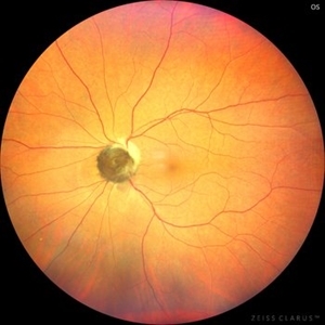 Optic Nerve Melanocytoma
May 4 2025 by KANWALJEET HARJOT MADAN, M.S. (Ophthalmology); FAICO (Vitreous - Retina)
This is a fundus picture of a young 42-year male who visited for a routine eye exam. His BCVA was 20/20 in both eyes. Anterior segment examination was normal. His left eye showed grey-black pigmentation at the infero-nasal margin of the optic disc. Fundus of the right eye was normal. The patient was diagnosed to have optic disc melanocytoma on multimodal imaging and was advised regular follow-up. Optic nerve melanocytoma is typically a benign tumor made up of melanocytes and melanin. It can grow, but rarely transforms into a malignancy. Patients with Optic Nerve Melanocytoma should be periodically examined for evidence of growth, loss of visual field and optic nerve compression.
Photographer: Dr. Kanwaljeet Harjot Madan, Thind Eye Hospital, Jalandhar City (Punjab) INDIA.
Imaging device: Zeiss Fundus Camera
Condition/keywords: melanocytoma, melanoma, optic nerve
-
 Ozurdex
Mar 20 2025 by T. P . VIGNESH, MBBS,MS
Photo of the anterior segment of left eye of a 50 year old woman with intravitreal Ozurdex implant seen attached to the posterior capsule.
Photographer: T.P. VIGNESH
Condition/keywords: ozurdex, Ozurdex implant
-
 Posterior Placoid Chorioretinopathy
Dec 19 2020 by John S. King, MD
44-year-old white female seen over the weekend complaining of a "spot" in her vision centrally OD for three days. She was referred over by another eye doctor who was concerned about a possible retinal detachment vs ARN in that eye. Her past medical history includes adrenal insufficiency for which she takes a low dose of hydrocortisone, thyroxine (post thyroidectomy), Plaquenil (inflammatory arthritis). She is divorced with one partner and denies any IVDU. Va 20/200 OD and 20/20 OS, IOP 12 OU, pupils mydriatic post gtts (light desaturation OD). There was 1+ A/C cell OD, O/W unremarkable anterior segment OU; in the posterior segment OD there was 1+ vitritis with a diffusely swollen optic disc and a large yellowish placoid lesion in the macula with yellowish border and extended out past the arcades inferiorly, as well as another lesion smaller in the IN periphery, and two possible smaller spots SN (See Photo above). There was a trace vitreous cell OS with a large, granular placoid lesion nasally. The OCT showed mild subfoveal fluid with nodular areas in the RPE and some overlying irregular architecture of the outer retina. Syphilis was a concern at this point. She denied any hand or foot rash, and said that she was recently working on the house, and her hands were dried out. There did appear to be a rash on the hand, and later learned that she had a rash on the soles of her feet. She was sent to ED for a work-up and her syphilis IgG was positive and VDRL 1:128, and negative for HIV. She was started on a course IV Penicillin (40mg PO steroid two days after tx started). She has responded well. A few days after treatment her visual acuity has improved to 20/60 OD; there was no anterior segment inflammation OU, and decreased vitreous cell OU. Disc edema was improved. The large placoid lesion in the macula of the right eye was slightly enlarged, but more granular in appearance without a distinct yellowish border, and the smaller lesions SN had dissipated. OCT showed resolution of the subfoveal fluid and an improved appearance of the outer retina and RPE layer.
Imaging device: Optos CA
Condition/keywords: acute syphilitic posterior placoid chorioretinitis, syphilis
-
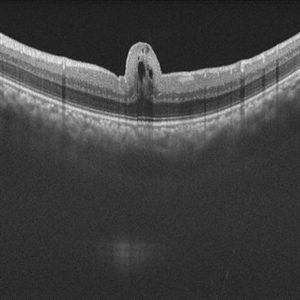 Retinal Fold in Posterior Microphthalmos
Mar 1 2025 by Hemanth Murthy, MBBS, MD, FASRS
Swept source OCT image of left eye of 34 year male patient with high hypermetropia(+14). BCVA 20/20 in right eye and 20/60 in left eye. Anterior segment was normal. There is loss of foveal pit with omega shaped elevation of inner retinal layers.
Photographer: Mr Veda Vyas
Condition/keywords: posterior microphthalmos
-
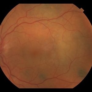
 Serpigenous Choroidopathy in a 68-Year-Old Male
Feb 15 2013 by Roy Schwartz, MD
A 68-year-old healthy male presented with a few years of decreased vision bilaterally. Visual acuity in OD was 1/36 and in OS 20/40. Anterior segments were normal except for bilateral mild nuclear sclerosis and pseudoexfoliation in OS. In the fundus of OD a large atrophy with pigmentary scars were seen in the macula and nasally to the optic disc while OS presented with the same clinical picture but an island of normal appearing retina was seen in the fovea. On fluorscein angiography no leakage was shown. A diagnosis of Serpigenous choroidopathy was made.
Photographer: Galit Yair-Pur
Condition/keywords: macula serpiginous choroidopathy, serpiginous choroiditis
-
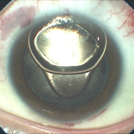
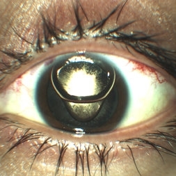
 Silicone oil in traumatic aniridia
Apr 19 2022 by Thais Bastos
A 27-year-old patient who developed aniridia, aphakia and retinal detachment after ocular trauma in the left eye. She underwent vitrectomy with silicone oil. Photo of the anterior segment 3 months after surgery showing a double meniscus made of silicone oil. Note red reflex, the retina is totally attached.
Photographer: Thaís Azeredo Bastos, CBCO Hospital de Olhos, Goiânia - Brazil
Imaging device: Zeiss Clarus 700
Condition/keywords: aniridia, ocular trauma, silicone oil
-
 Choroidal Granuloma
Apr 23 2019 by Purva Patwari
22-year-old male patient presented with blurring of vision in the right eye noticed since last one week. He was asymptomatic a week ago when he noticed the blurring in his right eye. On examination his vision was 6/6 in both eyes. Anterior segment was normal. Posterior segment was normal for the left eye. Right eye examination revealed a clear vitreous cavity with choroidal granulomas scattered throughout the fundus. The present picture shows choroidal granulomas with OCT segment passing through the parafoveal lesion showing subretinal fluid accumulation and corresponding thickening of the retinal layers. CT scan reveals heterogeneously enhancing lymph nodes showing conglomerationin the hilar region-possibility of tubercular etiology.
Photographer: Dr Purva Patwari, Patwari Retina Center
Imaging device: Zeiss Visu 500
Condition/keywords: choroidal granuloma, choroiditis, granulomatous choroiditis, tubercular choroidal granuloma, tuberculosis
-
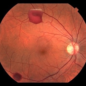 A Motor Vehicle Accident Causing Valsalva Retinopathy OD, While Racing A Side By Side 4 Wheel Off-Road Vehicle
Apr 29 2020 by John S. King, MD
43-year-old white male who was injured while racing a side by side 4-wheel off-road vehicle (see Video: https://imagebank.asrs.org/file/53854/sxs-crash-during-a-race-causing-valsalva-retinopathy-od). He presented about three weeks after the injury. He was being seen by his local eye doctor who wanted an evaluation for the retinal heme and scotoma. His main complaint was a central/parcentral scotoma described as a greyish area in vision. Va 20/50 OD, nomotensive, no APD (by technician), anterior segment u/r; see picture for the fundus exam - of note there are superficial/preretinal heme, with layering of the heme superiorly, and small superficial heme at nasal edge of the optic disc; in the parafoveal region nasally there is some mottling of the RPE that may indicate an area of prior commotio retinae (also possible to have TON), which may account for his scotoma. Really bad accident (video), and amazingly, he had no LOC or injuries other than the right retina. Helmet and racing harness seat belt were used.
Photographer: Asli Ahmed
Imaging device: Topcon 50
Condition/keywords: valsalva retinopathy
-
 Serpigenous Choroidopathy in a 68-Year-Old Male
Feb 15 2013 by Roy Schwartz, MD
A 68-year-old healthy male presented with a few years of decreased vision bilaterally. Visual acuity in OD was 1/36 and in OS 20/40. Anterior segments were normal except for bilateral mild nuclear sclerosis and pseudoexfoliation in OS. In the fundus of OD a large atrophy with pigmentary scars were seen in the macula and nasally to the optic disc while OS presented with the same clinical picture but an island of normal appearing retina was seen in the fovea. On fluorscein angiography no leakage was shown. A diagnosis of Serpigenous choroidopathy was made.
Photographer: Galit Yair-Pur
Condition/keywords: macula serpiginous choroidopathy, serpiginous choroiditis
-
 Anterior Chamber Gas and PFC Migration
Jun 21 2018 by Maria Stephanie R. Jardeleza, MD
Anterior segment photographs of 30-year-old male who underwent superior rhegmatogenous retinal detachment repair with intraocular gas tamponade. Perfluorocarbon was used to flatten the macula to prevent a macular fold and was removed during PFC/air exchange. Post operative week two visit shows gas migration into the anterior chamber with retained PFC layered in a tear drop shape posterior to the gas bubble and anterior to the lens. Patient had been maintaining face down positioning.
Photographer: Andy Zepeda, COA, Retina Clinic, San Antonio Eye Center, San Antonio, TX
Condition/keywords: retained perfluorocarbon, retina surgery complications, vitreous substitutes
 Loading…