Search results (17 results)
-
 Ectopia Lentis
Jan 21 2021 by Jamin S. Brown, MD
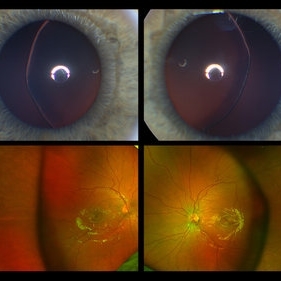
This image serial demonstrates a patient with simple ectopia lentis. Anterior segment photographs in the upper panel demonstrate nasally subluxated crystalline lenses. Widefield fundus photography shows a "pseudo-buckle" which is the result of an optical effect due to the lens subluxation (artifactual image enlargement). Also note the juvenile macular reflex in this young patient. Ectopia lentis can present isolated ("simple") or in combination with various systemic defects (Marfan's syndrome, Weil-Marchesani syndrome or Ehlers-Danlos syndrome to name a few). Isolated ectopia lentis can be hereditary and causative genes have been identified as ADAMTSL4 located on chromosome 4 and FBN1 gene located on chromosome 15. Defects in the genes cause weakness in the zonular fibers which can lead to lens dislocation. Lastly, various ocular disorders such as Aniridia, Axenfeld-Rieger, Pseudoexfoliation or Trauma may also result in lens dislocation or subluxation.
Photographer: Stefanie Palmer CRA, Retina Vitreous Surgeons of CNY
Condition/keywords: dislocated lens, ectopia lentis
-
 Silicone oil in traumatic aniridia
Apr 19 2022 by Thais Bastos
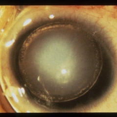
A 27-year-old patient who developed aniridia, aphakia and retinal detachment after ocular trauma in the left eye. She underwent vitrectomy with silicone oil. Photo of the anterior segment 3 months after surgery showing a double meniscus made of silicone oil. Note red reflex, the retina is totally attached.
Photographer: Thaís Azeredo Bastos, CBCO Hospital de Olhos, Goiânia - Brazil
Imaging device: Zeiss Clarus 700
Condition/keywords: aniridia, ocular trauma, silicone oil
-
 Aniridic Fibrosis Syndrome - #3 of 7
Jan 24 2013 by Christopher D. Riemann, MD
6-year-old pseudophakic girl with aniridic fibrosis syndrome. Nasal view with HD endoscope. Note: increasing fibrosis fibrosis clearly extending onto the ciliary body and enveloping ciliary processes.
Photographer: Christopher Riemann MD, Cincinnati Eye Institute, University of Cincinnati
Imaging device: Endoscope
Condition/keywords: aniridia, epiciliary membrane
-
 Aniridia and Dislocated Lens
Oct 18 2012 by Larry Halperin, MD
Aniridia and dislocated lens
Condition/keywords: aniridia, dislocated lens
-
 Aniridia Cloudy Cornea
Jul 29 2013 by H. Michael Lambert, MD
Aniridia cloudy cornea.
Condition/keywords: aniridia cloudy cornea
-
---thumb.jpg/image-square;max$300,300.ImageHandler) Aniridia With Glaucoma
Aniridia With Glaucoma
Jan 2 2014 by David Callanan, MD
32-year-old male, aniridia with glaucoma, 20/200 OU.
Condition/keywords: aniridia, glaucoma
-
---thumb.jpg/image-square;max$300,300.ImageHandler) Aniridia With Glaucoma
Aniridia With Glaucoma
Jan 2 2014 by David Callanan, MD
32-year-old male, aniridia with glaucoma, 20/200 OU.
Condition/keywords: aniridia, glaucoma
-
---thumb.jpg/image-square;max$300,300.ImageHandler) Aniridia With Glaucoma
Aniridia With Glaucoma
Jan 2 2014 by David Callanan, MD
32-year-old male, aniridia with glaucoma, 20/200 OU.
Condition/keywords: aniridia, glaucoma
-
 Aniridic Fibrosis Syndrome #2 of 7
Jan 24 2013 by Christopher D. Riemann, MD
6-year-old pseudophakic girl with aniridic fibrosis syndrome. Superonasal view with HD endoscope. Note: 20 gauge sclerotomy and very mild fibrosis barely extending onto the ciliary body.
Photographer: Christopher Riemann MD, Cincinnati Eye Institute, University of Cincinnati
Imaging device: Endoscope
Condition/keywords: aniridia, epiciliary membrane
-
 Aniridic Fibrosis Syndrome #4 of 7
Jan 24 2013 by Christopher D. Riemann, MD
6-year-old pseudophakic girl with aniridic fibrosis syndrome. Close up nasal view with HD endoscope. Note: increasing fibrosis fibrosis clearly extending onto the ciliary body and enveloping ciliary processes.
Photographer: Christopher Riemann MD, Cincinnati Eye Institute, University of Cincinnati
Imaging device: Endoscope
Condition/keywords: aniridia, epiciliary membrane
-
 Aniridic Fibrosis Syndrome #6 of 7
Jan 24 2013 by Christopher D. Riemann, MD
6-year-old girl with Aniridic Fibrosis Syndrome. Note: endoscopic view of 20 gauge vitreous cutter engaging epiciliary membrane.
Photographer: Christopher Riemann MD, Cincinnati Eye Institute, University of Cincinnati
Imaging device: Endoscope
Condition/keywords: aniridia, epiciliary membrane
-
 Aniridic Fibrosis Syndrome - #1 of 7
Jan 24 2013 by Christopher D. Riemann, MD
6-year-old pseudophakic girl with aniridic fibrosis syndrome. Superior view with HD endoscope. Note: complete absence of fibrosis, a normal ciliary body, normal pars plana and normal anterior retina.
Photographer: Christopher Riemann MD, Cincinnati Eye Institute, University of Cincinnati
Imaging device: Endoscope
Condition/keywords: aniridia, epiciliary membrane
-
 Aniridic Fibrosis Syndrome - #5 of 7
Jan 24 2013 by Christopher D. Riemann, MD
6-year-old pseudophakic girl with aniridic fibrosis syndrome. Inferior view with HD endoscope. Note: severe inferior fibrosis fibrosis clearly extending across the ciliary body and obliterating the inferior ciliary processes and migrating onto the anterior pars plana .
Photographer: Christopher Riemann MD, Cincinnati Eye Institute, University of Cincinnati
Imaging device: Endoscope
Condition/keywords: aniridia, epiciliary membrane
-
 Aniridic Fibrosis Syndrome - #7 of 7
Jan 24 2013 by Christopher D. Riemann, MD
6-year-old pseudophakic girl with aniridic fibrosis syndrome. Superonasal view with HD endoscope. Note: 20 gauge probe applying endocautery to ciliary body bleed resulting from stripping of epiciliary membranes
Photographer: Christopher Riemann MD, Cincinnati Eye Institute, University of Cincinnati
Imaging device: Endoscope
Condition/keywords: aniridia, epiciliary membrane
-
 Blunt Trauma
May 18 2016 by Andrea Arriola-Lopez, MD MSc
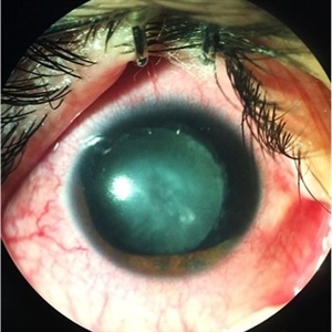
26-year-old man, old ocular blunt trauma. VA HM OD. IOP 14mmHg. Traumatic partial aniridia, cataract and phacodonesys. Ophthalmoscopy showed diffuse hemovitreous, Retina remained attached.
Photographer: Andrea E. Arriola-López MD MSc
Condition/keywords: aniridia, cataract, trauma, traumatic cataract
-
 Slide 4-26
Feb 20 2019 by Lancaster Course in Ophthalmology
Congenital aniridia. Clinical appearance showing the equator of the lens and some of the zonules.
Condition/keywords: aniridia, lens, zonules
-
 Slide 4-27
Feb 20 2019 by Lancaster Course in Ophthalmology
Congenital aniridia. Histologic section indicating the presence of small, rudimentary iris tags ( x16). (Scheie Eye Institute, No. 478.)
Condition/keywords: aniridia, iris tags
 Loading…