Search results (473 results)
-
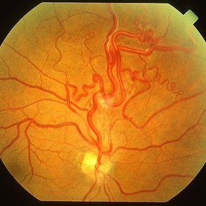
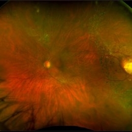
 Wyburn Mason
Jun 23 2018 by Caesar K. Luo, MD, FASRS
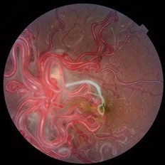
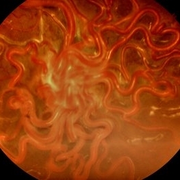
Fundus photograph of a 16-year-old female patient with poor vision in one eye demonstrates racemose angiomatosis as seen in Wyburn-Mason Syndrome.
Photographer: Joseph Trabucco, Progressive Vision Institute, Allentown, PA
Condition/keywords: Wyburn-Mason
-
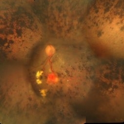 Retinitis Pigmentosa With Hemangioma CF
Dec 15 2016 by Manish Nagpal, MD, FRCS (UK), FASRS
Fluorescein angiography OS of a patient having retinitis pigmentosa with a hemangioma inferiorly.
Condition/keywords: hemangioma, retinitis pigmentosa
-
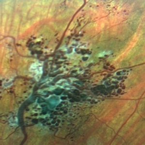
 Retinal Arteriovenous Malformations (Racemose Hemangiomatosis)
Mar 30 2018 by Rameez N Hussain, MD
A 7-years-old Portuguese girl with unilateral retinal arteriovenous malformations composed of dilated, tortuous vessels with normal vision.
Photographer: Thambi Durai. Consultant Optometrist, Orbit Health Care - Dr Agarwal's Eye Hospital, Maputo, Mozambique
Imaging device: TOPCON
Condition/keywords: racemose hemangioma, retinal arteriovenous malformations, Wyburn-Mason
-
 Wyburn-Mason Syndrome (Racemose Hemangiomatosis)
Mar 30 2018 by Rameez N Hussain, MD
A 7-year-old Portuguese girl with unilateral retinal arteriovenous malformations composed of dilated, tortuous vessels with normal vision.
Photographer: Thambi Durai
Imaging device: TOPCON
Condition/keywords: arteriovenous malformation, racemose hemangioma, Wyburn-Mason
-
 Hemangioma Capilar Retina
Apr 9 2023 by Gustavo Aguirre-Suarez
Fundus photograph composition of a Retinal Capilar Hemangioma
Photographer: Dr. Gustavo Aguirre-Suarez
Imaging device: Visucam 500
Condition/keywords: hemangioma, Von Hippel-Lindau
-
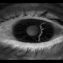 Iris Racemose Hemangioma
Jan 1 2023 by Maxwell J Wingelaar, MD
Fluorescein Angiogram of 66 year old female presented with an iris racemose hemangioma
Photographer: Ken Huff
Condition/keywords: Racemose hemangioma
-
 VHL With Capillary Hemangioma Pre-Rx
Dec 29 2016 by Manish Nagpal, MD, FRCS (UK), FASRS
VHL with hemangioma with feeder vessels.
Photographer: rakesh juneja
Condition/keywords: cryotherapy, hemangioma, laser, Von Hippel-Lindau
-
 Wyburn Mason Syndrome
May 2 2013 by Henry J. Kaplan, MD
Racemose angioma of the retina in Wyburn Mason syndrome.
Condition/keywords: racemose hemangioma
-
 Capillary Hemangioma
Dec 14 2016 by Young Hee Yoon, MD, PhD
Wide fundus photo of a 35-year-old man with huge capillary hemagioma in the right eye. He is diagnosed with Von Hippel-Lindau disease. His best-corrected visual acuity was 20/50.
Photographer: Yu Jin Jang and Hun Eui Hong, Asan Medical Center
Imaging device: Wide fundus camera
Condition/keywords: Von Hippel-Lindau
-
 Coats' Disease - Stage 3A
Aug 21 2019 by Victor M Villegas, MD
Coats' Disease - stage 3A.
Condition/keywords: abnormal retina, Coats' disease, diffuse lipid exudate, edema, foveal hard exudates, pediatic retina, retcam, retinal angioma
-
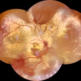 Coats' Disease Montage
Feb 5 2021 by Akansha Sharma
Fundus photograph of a 5-year-old male child who presented with unilateral diminution of vision since one month.
Photographer: Dr. Nivesh Gupta, M.S., Retina Foundation, Ahmedabad
Condition/keywords: angiomatosis retinae, Coats' disease, exudative detachment, subretinal exudates
-
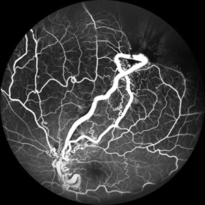
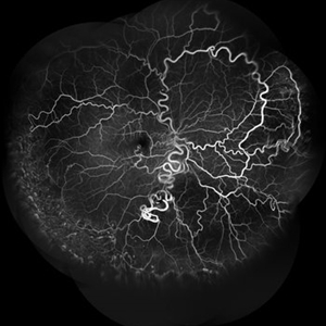 Racemose Angioma
Jan 23 2025 by SHILPI H NARNAWARE, ICO ( Retina) , FAICO ( Vitreo-Retina)
42 year old male, presented with blurred vision . Examination revealed Racemose angioma. FFA was done which revealed tortuosity of blood vessels.
Photographer: Shilpi Narnaware, Sarakshi Netralaya , Nagpur, Maharashtra , India
Imaging device: Mirante ( by Nidek)
Condition/keywords: FFA in a case of Racemose angioma
-
 Von Hippel-Lindau Syndrome
Mar 12 2016 by Sjakon G Tahija, MD
Fundus photograph of a patient with Von Hipple Lindau Disease and retinal angiomas.
Photographer: Avris Siahaan, Klinik Mata Nusantara
Condition/keywords: Von Hippel-Lindau
-
 Wyburn-Mason Syndrome (Racemose Angioma)
Mar 23 2024 by Pushkar Mahale
Fundus photograph of a 10 year old child presenting with no perception of light in right eye. Fundus examination revealed dilated and tortuous retinal vessels suggestive of Racemose Hemangioma.
Photographer: Dr Pushkar Mahale
Condition/keywords: racemose hemangioma, Wyburn -Mason Syndrome
-
---thumb.jpg/image-square;max$300,300.ImageHandler) Cavernous Hemangioma of the Retina
Cavernous Hemangioma of the Retina
Sep 20 2013 by Hector E. Ibanez, MD, FACS, FASRS, FABO, FAAO
Fundus photograph of an 8-year-old boy with a cavernous hemangioma of the retina.
Photographer: Hector E. Ibanez, MD, FACS
Imaging device: Topcon TRC 50
Condition/keywords: cavernous hemangioma of the retina
-
 Circumscribed Choroidal Hemangioma with Serous Macular Retinal Detachment
Oct 2 2023 by Aditya S Kelkar, MS, FRCS, FASRS,FRCOphth
Fundus photograph of a 43-year-old male with a circumscribed choroidal hemangioma with serous macular retinal detachment associated with diminision of vision.
Photographer: Dr. Harsh Jain, National Institute of Ophthalmology
Imaging device: Clarus 500
Condition/keywords: choroidal hemangioma
-
 Coats' Disease
Mar 30 2022 by Aditya S Kelkar, MS, FRCS, FASRS,FRCOphth
Colour Fundus photograph of a 3-year-old boy presenting with complaints of slowly progressive divergent squint of his left eye which on funduscopy revealed presence of Coats' Disease in the same eye.
Photographer: Dr. Pranali Surawase. National Institute of Ophthalmology, Pune, Maharashtra, India
Imaging device: Zeiss Clarus 500
Condition/keywords: angioma, cholesterol crystals, Coats' disease
-
 Focal Chroidal Hemangioma
Sep 18 2018 by Somnath Chakraborty, MD
Right eye fundus photo montage of a 17-year-old boy showing a focal choridal hemangioma temporally.
Photographer: Saptarshi Mehta, Retina Institute of Bengal
Condition/keywords: choroidal hemangioma
-
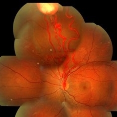
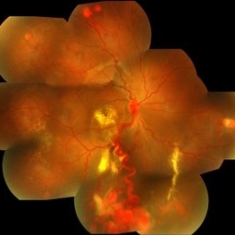
 From Artery to Vein, No Detour: Meet the AV Maverick: Racemose Hemangioma
Jul 1 2025 by rohan jain
A case of 10 year old girl with defective vision in LE (6/60) who presented us with this condition.
Photographer: Dr. ROHAN JAIN
Imaging device: mirante
Condition/keywords: arteriovenous malformation, FFA in a case of Racemose angioma, racemose hemangioma
-
 Hemangioma
Feb 9 2021 by Kim Barrett
66-year-old female with a history of thyroid and uterine cancer in her 30's. She has a family history of cancers also. Current VA 20/40-2 PH OS. Patient and doctor chose observation at this time with possible surgical intervention in the future. She also has a small Hemangioma temporally in the right eye. Von Hippel-Lindau is also suspected and genetic testing was suggested.
Photographer: Kim Barrett C.O.A. Retina Specialists of Michigan, Grand Rapids, MI
Imaging device: Optos California
Condition/keywords: cancer, genetic testing, Optos, retinal hemangioblastoma, Von Hippel-Lindau
-
 Hemangioma of Retina
Sep 11 2018 by Carolyn Daley
50 degree OCT imaging of a 20-year-old with multiple bilateral hemangiomas. Patient was diagnosed with Von Hippel-Lindau Syndrome.
Photographer: Carolyn Daley, Retina Specialists of Michigan
Imaging device: Heidelberg Spectralis
Condition/keywords: 50 degrees, edema, hemangioma, optical coherence tomography (OCT), Von Hippel-Lindau
-
 Hemangioma of Retina
Mar 5 2025 by Virginia Gebhart
64 year old male with choroidal hemangioma in the macula and STA. Persistent IRF and new cuff of SRF compared to previous photos. BCVA CF@face. Pt has had PDT in the past with no significant improvement. Will observe closely
Photographer: Virginia Gebhart, Retina Consultants of Carolina
Imaging device: Optos California
Condition/keywords: hemangioma, inferior subretinal fluid
-
 Hemangioma of Retina (FAF)
Mar 5 2025 by Virginia Gebhart
Fundus autofluorescence of 64 year old male with choroidal hemangioma in the macula and STA. Persistent IRF and new cuff of SRF compared to previous photos. BCVA CF@face. Pt has had PDT in the past with no significant improvement. Will observe closely
Photographer: Virginia Gebhart, Retina Consultants of Carolina
Imaging device: Optos California
Condition/keywords: autofluorescence imaging, hemangioma, inferior subretinal fluid
-
 Indocyanine Green (ICG) of Circumscribed Choroidal Hemangioma (CCH)
Feb 6 2025 by Jack B Margines, MD, MHCI
Peripheral patchy hyperfluorescence is seen on this early image of ICG-A on a 53-year-old asymptomatic with an extramacular circumscribed choroidal hemangioma.
Photographer: W Ryan Miliam, CRA, OCT-C, University of California, Irvine Gavin Herbert Eye Institute
Imaging device: Optos
Condition/keywords: choroidal hemangioma, indocyanine green (ICG) angiography
-
 Multiple Cavernous Hemangioma
Jan 20 2020 by Sarah Oelrich
Multiple Cavernous Hemangioma
Photographer: Sarah Oelrich CRA COT OCT-C Southeastern Retina Associates
Condition/keywords: cavernous hemangioma of the retina
 Loading…