Search results (473 results)
-
 Capillary Hemangioma
Dec 20 2025 by DR Rohit Gupta
A 1 year old male child , presented with his parents , with a red color swelling below eyelid.
Photographer: Dr Rohit gupta
Imaging device: Samsung S21
Condition/keywords: hemangioendothelioma, Hemangioma
-
 Racemose Hemangioma
Dec 16 2025 by Seif Allah Anwar
18 year-old female with dilated, tortuous arteriovenous communication without an intervening capillary bed. Vessels may appear coiled or spaghetti-like extending into the foveal region with no associated retinal hemorrhages, exudates, or edema On OCT : the anomalous vessels appear hyper reflective spanning the whole retinal thickness with ILM draping, No associated subretinal or intraretinal fluid.
Photographer: Seif Anwar , KING SALMAN INTERNATIONAL UNIVERSITY
Imaging device: TOPCON
Condition/keywords: racemose hemangioma
-
 The Eye Siren - A Case of VHL
Dec 3 2025 by Surabhi Gupta, MS, DNB, FVRS
A 25 year old man presented with chief complaints of dimuntion of vision in right eye for past 2 weeks. Best corrected visual acuity in right eye was 6/12, N8 and left eye was 6/6, N6. Montage color fundus photograph shows bilateral multiple capillary hemangioma with epiretinal membrane causing traction over macula in right eye. On PET-CT multiple metabolically inactive cystic lesion were noted in the pancreas and seminal vesicle with low grade metabolically active cystic lesion with enhancing septations in kidneys which were suspicious of malignant etiology. MRI brain showed presence of a small cystic lesion anterior to celebellar vermis suggestive of CNS hemangioblastoma. A diagnosis of Von Hippel- Lindau syndrome was made the patient is under oncologist care for suspected RCC.
Photographer: Mr Brajesh Kumar
Imaging device: zeiss visucam 500
-
 Branch Retinal Vein Occlusion with Retinal Neovascularization
Oct 20 2025 by Jason Gayoski
Prev. getting injections, last one in 2024 at external provider. Hx of liver hemangiomas on CT/MRI in 2018: no known follow-up for this. Hx of migraine headaches longstanding without hemangiomas noted on brain MRI in 2018. CTA head/neck neg in 2021. No hx or Fhx of VHL. NVE/shunt vessels inferiorly on FA today. Discussed option of restarting antiVEGF or laser. Pt would like to observe for now given stability. Will consider antiVEGF/Laser/belzutifan in future. Referral to medical genetics to eval for VHL. Recommend obtain MRI brain, spine w/wo con, CTAbd w/wo con to eval for systemic hemangiomas. Will likely refer to Ocular Onc pending these results. Also consider GI referral given liver hemangiomas since 2018. Pt already established with Neurology for migraines.
Photographer: Jason Gayoski COA
Imaging device: Optos
Condition/keywords: branch retinal vein occlusion (BRVO), retinal neovascularization
-
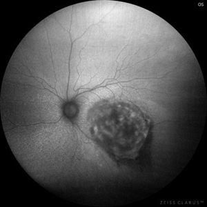
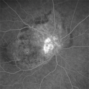 Juxtapapillary Retinal Capillary Hemangioblastoma
Oct 16 2025 by Sara Mayoral Sánchez
A hyperfluorescent lesion located superotemporal to the optic disc, consistent with a juxtapapillary retinal capillary hemangioma.
Photographer: Sara Mayoral Sánchez, H.U.Puerta del Mar, Cádiz
Condition/keywords: angiography with fluorescein, hemangioma, optic disc, retina capillary hemangioblastoma
-
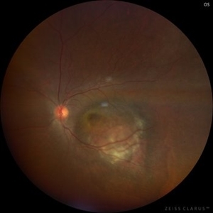
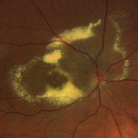 Juxtapapillary Retinal Capillary Hemangioblastoma
Oct 16 2025 by Sara Mayoral Sánchez
Peripapillary circinate lipid exudation secondary to retinal capillary hemangioblastoma.
Photographer: Sara Mayoral Sánchez, H.U.Puerta del Mar, Cádiz
Condition/keywords: circinate lipid ring, hemangioma, massive lipid exudation, retinal capillary hemangioblastoma
-
 Amelanotic Melanoma
Aug 12 2025 by César Adrián Gómez Valdivia, MD
This case highlights an amelanotic melanoma, an atypical presentation of a choroidal melanoma lacking the characteristic pigmentation. These lesions can easily be mistaken for choroidal hemangiomas, metastases, or inflammatory masses. Clinically, the lesion appears as a dome-shaped, yellowish subretinal mass, often associated with subretinal fluid, lipofuscin deposition, or retinal detachment. The absence of pigment can delay diagnosis, making multimodal imaging essential. Diagnostic tools: • B-scan ultrasound: low to medium internal reflectivity • OCT: overlying subretinal fluid and RPE elevation • FAF: orange pigment and RPE disruption • ICG/FA: variable, often hypofluorescent core Important: Prompt referral to ocular oncology is critical for management and prognosis.
Photographer: @eyemissu2
Imaging device: TOPCON TRC-50DX
Condition/keywords: amelanotic melanoma
-
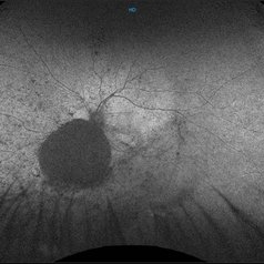 Choroidal Hemangioma (AF)
Jul 5 2025 by Gustavo Uriel Fonseca Aguirre
This wide-field fundus autofluorescence image demonstrates a mushroom-shaped choroidal melanoma adjacent to the optic nerve head, exhibiting hypo-autofluorescence (melanin). Vitreous pigment dispersion (tobacco dust sign) is evident, indicating tumor activity.
Photographer: Gustavo U. Fonseca Aguirre, Hospital Conde de Valenciana, Ciudad de México
Condition/keywords: choroidal melanoma
-
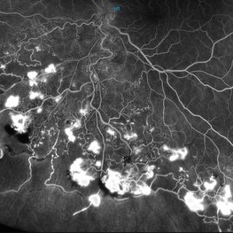
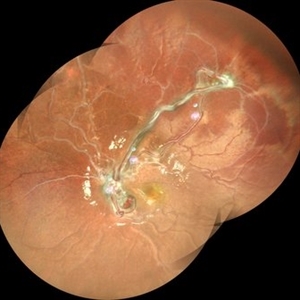
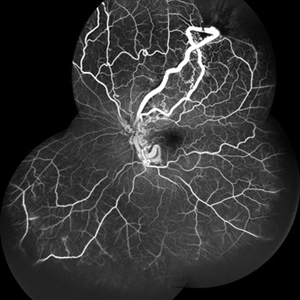
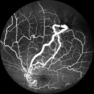
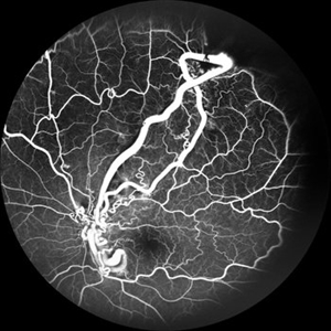
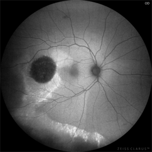
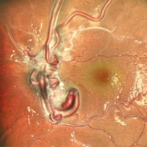 From Artery to Vein, No Detour: Meet the AV Maverick: Racemose Hemangioma
Jul 1 2025 by rohan jain
A case of 10 year old girl with defective vision in LE (6/60) who presented us with this condition.
Photographer: Dr. ROHAN JAIN
Imaging device: mirante
Condition/keywords: arteriovenous malformation, FFA in a case of Racemose angioma, racemose hemangioma
-
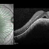 Choroidal Hemangioma
Jun 18 2025 by Moazzam Parvez
An OCT image of a 42 year old man presenting with a vision of 20/80 and complaining of distortion. OCT reveals serous retinal detachment with RPE alteration and disruption of outer retinal layers.
Photographer: Moazzam Parvez , Netralayam , Kolkata
Imaging device: Heidelberg Spectralis
Condition/keywords: Choroidal Hemangioma, Sub retinal fluid, tumor
-
 Choroidal Hemangioma
Jun 18 2025 by Moazzam Parvez
Multicolor and infrared reflectance image of a 42 year old gentleman with a Choroidal hemangioma lesion temporal to the fovea complaining of distortion in his right eye . Fundus imaging revealed a well-circumscribed ,elevated, reddish orange lesion within the choroid involving the posterior pole temporally .
Photographer: Moazzam Parvez, Netralayam , Kolkata
Imaging device: Heidelberg Spectralis
Condition/keywords: Choroidal Hemangioma, tumor
-
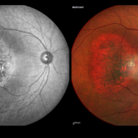
 Choroidal Hemangioma 4 Ways
Mar 13 2025 by Virginia Gebhart
Color fundus, FAF, late FA, late ICG of 64 year old male with choroidal hemangioma. Early hyperfluorescence with late leakage on FA, early hypercyanescence with late washout (25 min) on ICG.
Photographer: Virginia Gebhart, Retina Consultants of Carolina
Imaging device: Optos California
Condition/keywords: autofluorescence imaging, choroidal hemangioma, FA late phase, Fluorescein angiography, hemangioma, indocyanine green (ICG) angiography
-
 Choroidal Hemangioma
Mar 13 2025 by Virginia Gebhart
64 year old male referred for lesion in the STA with worsening SRF. Pt had been receiving injections for wetAMD q4weeks for 7 months. Reddish, elevated choroidal lesion, chronic SRF and pigment clumping consistent with hemangioma. FA/ICG/Bscan ultrasound also performed to confirm. Pt scheduled for PDT
Photographer: Virginia Gebhart, Retina Consultants of Carolina
Imaging device: Optos California
Condition/keywords: choroidal hemangioma, hemangioma, subretinal fluid
-
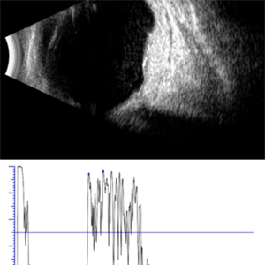 Choroidal Hemangioma, Ecography
Mar 11 2025 by Gustavo Uriel Fonseca Aguirre
45-year-old female with choroidal hemangioma in the macular area. A B-mode ultrasound study is presented showing a well-circumscribed, elevated lesion at the level of the choroid, involving the posterior pole, with intralesional fluid. The standardized A-mode ultrasound shows a lesion with medium-high reflectivity characteristic of choroidal hemangioma.
Photographer: Gustavo U. Fonseca Aguirre, Hospital Conde de Valenciana, Ciudad de México
Condition/keywords: Choroidal Hemangioma, ecography
-
 Choroidal Hemangioma, Autofluorescence
Mar 11 2025 by Gustavo Uriel Fonseca Aguirre
45-year-old female with choroidal hemangioma in the macular area. An autofluorescence image of the lesion is presented, with heterogeneous hypoautofluorescent characteristics.
Photographer: Gustavo U. Fonseca Aguirre, Hospital Conde de Valenciana, Ciudad de México
Condition/keywords: Choroidal Hemangioma
-
 Choroidal Hemangioma
Mar 11 2025 by Gustavo Uriel Fonseca Aguirre
45-year-old female with choroidal hemangioma in the macular area.
Photographer: Gustavo U. Fonseca Aguirre, Hospital Conde de Valenciana, Ciudad de México
Condition/keywords: Choroidal Hemangioma
-
 Hemangioma of Retina (FAF)
Mar 5 2025 by Virginia Gebhart
Fundus autofluorescence of 64 year old male with choroidal hemangioma in the macula and STA. Persistent IRF and new cuff of SRF compared to previous photos. BCVA CF@face. Pt has had PDT in the past with no significant improvement. Will observe closely
Photographer: Virginia Gebhart, Retina Consultants of Carolina
Imaging device: Optos California
Condition/keywords: autofluorescence imaging, hemangioma, inferior subretinal fluid
-
 Hemangioma of Retina
Mar 5 2025 by Virginia Gebhart
64 year old male with choroidal hemangioma in the macula and STA. Persistent IRF and new cuff of SRF compared to previous photos. BCVA CF@face. Pt has had PDT in the past with no significant improvement. Will observe closely
Photographer: Virginia Gebhart, Retina Consultants of Carolina
Imaging device: Optos California
Condition/keywords: hemangioma, inferior subretinal fluid
-
 FAF-G Circumscribed Choroidal Hemangioma
Mar 1 2025 by Vishal Agrawal, MD, FRCS,FACS,FASRS
A 37-year-old male presented with decreased vision in the right eye. This is the fundus autofluorescence (FAF-G) of the right eye showing hypo auto fluorescent lesion with surrounding hyper auto fluorescence extending inferiorly corresponding to the fluid tract.
Photographer: Dr Ayushi Gupta
Imaging device: Clarus 700
Condition/keywords: Circumscribed Choroidal Hemangioma, fundus autofluorescence (FAF)
-
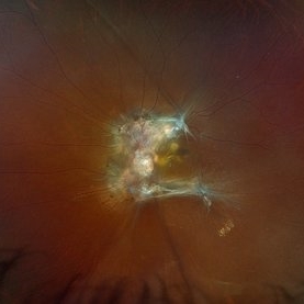 VHL Syndrome with Capillary Hemangioblastomas
Feb 26 2025 by Virginia Gebhart
39 year old female with choroidal hemangioma with capillary hemangioblastomas. Positive genetic testing for Von Hippel-Lindau Syndrome. Hemangioblastomas are stable compared to initial imaging in 2021. Pt started Welireg in Dec 2024, CNS tumors have started shrinking. No lesions in OD
Photographer: Virginia Gebhart, Retina Consultants of Carolina
Imaging device: Optos California
Condition/keywords: retinal capillary hemangioblastoma, Von Hippel-Lindau
 Loading…