Search results (9 results)
-
 Massive Submacular Hemorrhage
Sep 28 2012 by Joseph M. Civantos, MD
82-year-old gentleman who developed this massive submacular hemorrhage 3 days after his 16th Lucentis injection. Visual acuity dropped from 20/80 to LP.
Condition/keywords: subretinal hemorrhage
-
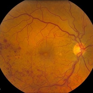 Branch Retinal Vein Occlusion
Jul 14 2013 by Jason S. Calhoun
Branch retinal vein occlusion in the right eye. Proceeded with Lucentis and will be followed up in 1-month
Photographer: Jason S. Calhoun, Department of Ophthalmology, Mayo Clinic Jacksonville, Florida
Imaging device: TOPCON TRC 50-EX
Condition/keywords: branch retinal vein occlusion (BRVO)
-
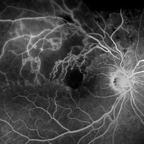 Branch Retinal Vein Occlusion- Fluorescein Angiogram, Montage
Apr 15 2016 by James B. Soque, CRA, OCT-C, COA, FOPS
A fluorescein angiogram of an 80-year-old white female with a superotemporal branch retinal vein occlusion, and retinal edema of the right eye. Currently receiving Lucentis 0.5 injection therapy.
Photographer: James Soque, CRA OCT-C COA, Island Retina, Shirley, NY
Imaging device: Topcon TRC, MERGE Imaging Software V. 11.2.0
Condition/keywords: branch retinal vein occlusion (BRVO), montage, non-perfused branch retinal vein occlusion (BRVO)
-
 Lucentis Endophthalmitis
Oct 8 2012 by David R. Chow, MD, FRCS(C)
Condition/keywords: endophthalmitis, Lucentis
-
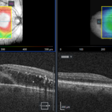
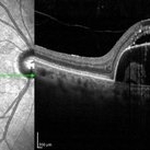 Pigment Epithelium Detachment, Secondary to AMD
Mar 17 2023 by Ceara Donovan
Optical coherence tomography of a 76 year old woman with a Pigment Epithelium Detachment, Secondary to AMD affecting her right eye. Patient had no significant response to Avastin, Eylea, Lucentis 0.5, or Vabysmo and was switched to Beovu. Following Beovu intravitreal injection her edema improved on OCT. Patient's vision was sc20/200+1 at the time the image was taken.
Photographer: Ceara Donovan
Imaging device: Heidelberg Spectralis
Condition/keywords: exudative age-related macular degeneration, heidelberg spectralis, macular degeneration, optical coherence tomography (OCT), pigment epithelial detachment (PED), Sub-retinal fluid
-
 Astrocytic Hamartoma
Apr 30 2015 by Mariam A Al-Feky, MD
A 15-year-old boy with history of seizures controlled on treatment. C/O: OD painless DV 10/7 ago (accidental discovery) O/E: BCVA OD: 6/60 ,, OS 6/6. AS: NAD OU. Pupil: RRR no RAPD OU. Fundus examination OD showed a retinitis like lesion with an overlying corkscrew vessel well evident on FFA with late leakage and CSR and OCT through the retinitis like lesion shows diffuse hypereflective thickeninig in the superficial NFL. Thorough history taking revealed that patient has seizures and MRI lesions suggestive of tuberous sclerosis. So this is exudative hamartoma secondary to tuberous sclerosis with marked resolution after single IVI of Lucentis. Retinitis like lesion with corkscrew vessels in FFA is typical together with the homogenous hypereflective thickening in the NFL.
Photographer: Mariam AL-Feky
Imaging device: Optical coherence tomography
Condition/keywords: astrocytic hamartoma
-
 DME Treated with Lucentis
Sep 26 2017 by Theodore Leng, MD, MS, FASRS
DME treated with Lucentis.
Condition/keywords: diabetic macular edema, Lucentis
-
 Hemi Vein Occlusion, Fluorescein Angiogram, Montage
Dec 17 2015 by James B. Soque, CRA, OCT-C, COA, FOPS
74-year-old woman, with recurrent superior hemi vein occlusion, montage image of fluorescein angiogram left eye. Currently receiving Lucentis injections OS.
Photographer: James Soque, CRA COA
Imaging device: Topcon RC 50 DX Fundus Camera with MERGE Winstation Software for Fluorescen Angiography
Condition/keywords: montage, occlusion of retinal vein, superior arcade
-
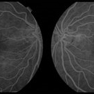 JXT with PDR
Jan 26 2022 by ASRS Staff
Fluorescein angiography pictures of same patient in 2009. At that time she had leakage in left eye and 3 intravitreal Lucentis injections were given. Patient was stabilized.
Photographer: Dr. Manish Nagpal, Retina Foundation, Ahmedabad
Imaging device: Nidek Mirante
Condition/keywords: fluorescein angiogram (FA), fluorescein leakage, juxtafoveal telangiectasis
 Loading…