Search results (40 results)
-
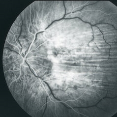
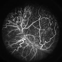 Coats' Disease FA
Apr 27 2018 by Brenda Fallas
3-year-old boy with unilateral Coats' Disease FA photo.
Photographer: Brenda Fallas, Bascom Palmer Eye Institute, Miami, FL
Imaging device: Retcam III 130 degree lens
Condition/keywords: Coats' disease, FA early phase, fluorescein angiogram (FA), retinal telangiectasia
-
 Acute Posterior Multifocal Placoid Pigment Epitheliopathy
Feb 20 2024 by Soobien Lee
Fluorescein angiogram of a 20-year-old caucasian female with viral prodrome and vision loss OS>OD secondary to Acute Posterior Multifocal Placoid Pigment Epitheliopathy (APPME). Early blockage with late hyperfluorescent leakage can be seen on fluorescein angiography of the left eye.
Photographer: Ashley Metzger, Elman Retina Group
Imaging device: Optos Ultra-Widefield Fluorescein Angiography
Condition/keywords: acute posterior multifocal placoid pigment epitheliopathy (APMPPE), bacilliary layer detachment, FA, FA early phase, fluorescein angiogram (FA), Optos, uveitis, white dot syndrome
-
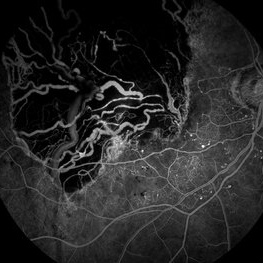
 Retinal Arterio-Venous Malformations
Apr 7 2017 by Deepak Bhojwani, MS
Multimodal imaging of a 16-year-old boy with retinal arterio-venous malformations(AVM). He also had cerebral AVM's on MRI-contrast studies suggesting Wyburn-Mason syndrome.
Photographer: DEEPAK BHOJWANI, RAGHUDEEP EYE HOSPITAL, AHMEDABAD.
Imaging device: Zeiss VISUCAM
Condition/keywords: color fundus photograph, FA early phase, optical coherence tomography (OCT), Wyburn-Mason
-
 Acute Zonal Occult Outer Retinopathy (AZOOR) FA, Fluorescein Angiography, Peripheral Vasculitis
Jan 19 2022 by James B. Soque, CRA, OCT-C, COA, FOPS
Acute Zonal Occult Outer Retinopathy (AZOOR). Peripheral Vasculitis OD. Fluorescein angiography showing vasculitis in the far right periphery 8-10 o'clock. 46-year-old white male, VA CC 20/16, 20/12.5, has had recurrent vasculitis for 11 years. No treatment.
Photographer: James Soque, CRA, OCT-C, COA, FOPS, Island Retina, Shirley, NY
Imaging device: Optos California
Condition/keywords: acute zonal occult outer retinopathy (AZOOR), FA early phase, fluorescein angiogram (FA), Peripheral Vasculitis, ultra-wide field imaging
-
 Central Retinal Vein Occlusion with Retinal Neovascularization
Jan 19 2022 by Olivia Rainey
Ultra-widefield fluorescein angiogram of a 56-year-old male with a Central Retinal Vein Occlusion with Retinal Neovascularization affecting his left eye. The patient presented on 1/19/2022 with scNLP vision in the left eye. The patient has good PRP, however areas of ischemia still remain untreated by laser. He also has severe neovascular glaucoma contributing to his poor vision.
Photographer: Olivia Rainey, OCT-C, COA
Imaging device: Optos California
Condition/keywords: central retinal vein occlusion (CRVO), FA early phase, fluorescein angiogram (FA), hemorrhage, ischemic CRVO, left eye, neovascular glaucoma, Optos, pan-retinal photocoagulation (PRP), retinal ischemia, retinal neovascularization, ultra-wide field imaging
-
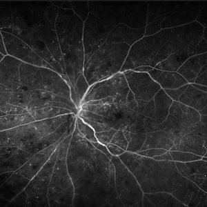
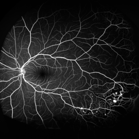
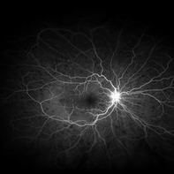
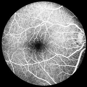 Normal FA Early Phase
-
 Sickle SC Sea Fan
Oct 8 2012 by Jeffrey G. Gross, MD, FASRS
Sickle SC sea fan, partial regression, FA early phase.
Condition/keywords: FA early phase, partial regression, sickle cell
-
 Sickle SC Sea Fan FA Early Phase
Oct 8 2012 by Jeffrey G. Gross, MD, FASRS
Sickle SC sea fan, FA, early phase.
Condition/keywords: early phase, sea fan, sickle cell
-
 BRVO FA, Early Phase
Oct 1 2012 by Jeffrey G. Gross, MD, FASRS
BRVO-FA early phase.
Condition/keywords: branch retinal vein occlusion (BRVO), capillary nonperfusion, early phase, microaneurysms
-
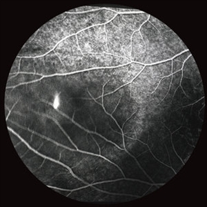
 Circumscribed Choroidal Hemangioma
Oct 20 2012 by Hyung-Woo Kwak, MD
Fundus and OCT examination showed an oval mass at the posterior pole with indistinct margins that blend with surrounding choroid. FA early phase showed hyperfluorescence.
-
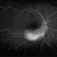
 Acute Multifocal Placoid Pigment Epitheliopathy
Sep 15 2014 by Thomas A. Ciulla, MD, MBA, FASRS
AMPPE in a 42-year-old woman. Early phase angiography show blockage of multiple focal lesions in the superior macula and peripapillary region of the right eye.
Photographer: Thomas Steele
Condition/keywords: acute multifocal placoid pigment epitheliopathy (AMPPE), FA early phase
-
 BRVO and VMT Vitreo Macular Traction, FA Early Phase
Apr 18 2013 by James B. Soque, CRA, OCT-C, COA, FOPS
Early FA photo, 50 degrees, mag 2X of 79-year old white female, VA sc 20/40, with BRVO OS, and VMT OS, diagnosed on exam and SD OCT. See accompanying FC and RF photos reveal BRVO IT OS, and SD OCT reveal VMT OS.
Photographer: James B. Soque, CRA, COA, Island Retina. Shirley, NY
Imaging device: Topcon TRC-50DX with MERGE software
Condition/keywords: branch retinal vein occlusion (BRVO), vitreomacular traction (VMT)
-
 BRVO, FA, Hemorrhage, Diabetic
Mar 13 2014 by James B. Soque, CRA, OCT-C, COA, FOPS
51-year-old white male, diabetes, and with BRVO left eye, early phase 36 seconds. Flame heme from ON, showing microaneurysims, and fine capillary detail of this FA.
Photographer: James B Soque, CRA COA
Imaging device: Topcon TRC 50DX with MERGE software
Condition/keywords: branch retinal vein occlusion (BRVO), diabetes, FA early phase, microaneurysms
-
 C-R Folds
Mar 26 2019 by Gary R. Cook, MD, FACS
Early phase FA frame of the left eye of a WM with bilateral C-R folds showing alternating hyper- and hypofluorescent bands.
Imaging device: Topcon VT-50
Condition/keywords: bilateral chorioretinal folds, chorioretinal fold, FA early phase, fluorescein angiogram (FA)
-
 Central Retinal Vein Occlusion With Waldenstroms macroglobulinemia
Jun 18 2025 by Korey Starkey
64-year-old patient presents with CRVO with secondary macular edema in both eyes. Venous beading present in 2/4 quadrants OU. Patient diagnosed with Waldenstroms macroglobulinemia, found on SPEP and bone marrow biopsy. Treatment recommended of anti-vegF intravitreal injections OU.
Photographer: Korey Starkey
Imaging device: Optos
Condition/keywords: attenuated vessels, central retinal vein occlusion (CRVO), CRVO, FA early phase, FLUORESCEIN ANGIOGRAPHY, macular edema, Optomap, OPTOS CALIFORNIA, severe NPDR, venous beading, Waldenstroms macroglobulinemia
-
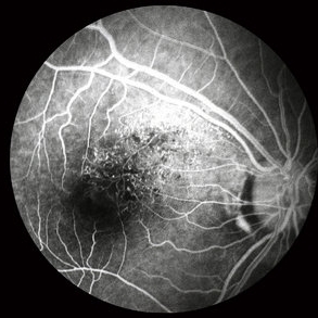
 Central Serous Chorioretinopathy with Smokestack FA Early Phase
Oct 8 2012 by Jeffrey G. Gross, MD, FASRS
CSCR with smokestack, FA, early phase.
Condition/keywords: central serous chorioretinopathy (CSCR), FA early phase, smokestack
-
 Choroidal Melanoma
Jan 4 2024 by Virginia Gebhart
57 year old female with new choroidal melanoma. Early hyperfluorescence with vascularity and minimal late leakage on FA.
Photographer: Virginia Gebhart
Imaging device: Optos California
Condition/keywords: FA, FA early phase, fluorescein angiogram (FA), Fluorescein angiography
-
 Choroidal Melanoma - Stable, Fluorescein Angiogram, Early Phase
Mar 13 2019 by James B. Soque, CRA, OCT-C, COA, FOPS
Early FA, right eye, with choroidal melanoma-stable, and a few tiny microaneurysms showing leakage in re-circulation phase.
Photographer: James Soque, CRA, OCT-C, FOPS
Imaging device: Topcon TRC-50DX with MERGE Eye Station software
Condition/keywords: FA early phase, fluorescein angiogram (FA), MERGE, microaneurysms
-
 Circumscribed Choroidal Hemangioma
Oct 20 2012 by Hyung-Woo Kwak, MD
Fundus and OCT examination showed an oval mass at the posterior pole with indistinct margins that blend with surrounding choroid. FA early phase showed hyperfluorescence.
-
 Circumscribed Choroidal Hemangioma
Oct 20 2012 by Hyung-Woo Kwak, MD
Fundus and OCT examination showed an oval mass at the posterior pole with indistinct margins that blend with surrounding choroid. FA early phase showed hyperfluorescence.
-
 Circumscribed Choroidal Hemangioma
Oct 20 2012 by Hyung-Woo Kwak, MD
Fundus and OCT examination showed an oval mass at the posterior pole with indistinct margins that blend with surrounding choroid. FA early phase showed hyperfluorescence.
-
 Coats' Disease
Mar 2 2020 by Stephanie Burke
15-year-old female with Coats' disease.
Photographer: Stephanie Burke, CRA, OCT-C
Condition/keywords: Coats' disease, FA early phase, ultra-wide field imaging
-
 Coats' disease early fluorescein angiogram of telangiectasia
Apr 3 2018 by Victor M Villegas, MD
6-year-old male with unilateral exudative retinopathy.
Photographer: Brenda Fallas
Imaging device: RetCam3
Condition/keywords: Coats' disease, FA early phase, fluorescein angiogram (FA), fluorescein leakage
-
 Coccidioides Choroiditis - Early FA
Mar 2 2020 by Scott C. Oliver, MD
Coccidioides choroiditis - early FA.
Photographer: UCLA
Imaging device: Optos
Condition/keywords: coccidiomycosis, FA early phase
-
 FA Early Phase Optic Disc Edema
Oct 1 2012 by Jeffrey G. Gross, MD, FASRS
FA early phase optic disc edema with blockage of hemorrhages in patient with subdural hematoma.
Condition/keywords: subdural hematoma
 Loading…