Search results (120 results)
-
 New Iris Melanoma
Oct 10 2024 by Virginia Gebhart
56 year old male with new amelanotic melanoma emanating from the ciliary body through the posterior iris epithelium. CT scan showed no evidence of metastatic disease. Pt scheduled for radioactive plaque and tumor biopsy
Photographer: Virginia Gebhart, Retina Consultants of Carolina
Imaging device: Samsung Galaxy
Condition/keywords: amelanotic melanoma, iris melanoma
-
 Plaquenil Toxicity
Apr 30 2013 by Theodore Leng, MD, MS, FASRS
SD-OCT scan from a 44-year-old woman with bilateral plaquenil toxicity. There is damage visible in the outer retina in a perifoveal distribution.
Condition/keywords: hydroxychloroquine toxicity, plaquenil toxicity
-
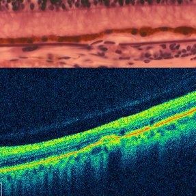
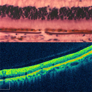 Angioid streaks - Light microscopy and OCT
Jan 11 2013 by Gerardo Garcia-Aguirre, MD
Top: Light microscopy of an angioid streak. Note: Bruch's membrane below the RPE, which is thickened and fractured. Bottom: OCT scan of an angioid streak (not of the same patient) showing fracture of the RPE.
Photographer: Alfredo Gomez-Leal, MD (top) and Gerardo Garcia-Aguirre, MD (bottom)
Condition/keywords: angioid streaks
-
 Candida Endophthalmitis
Jan 26 2020 by Marlon García Roa, MD
Female, 30-years-old with <<< V Pregnancy Currently with 18 weeks gestation. Pathological personal history 1 month prior hospitalization for complicated acute appendicitis + pyelonephritis + severe thrombocytopenia (autoimmune treated with corticosteroids) with septic shock, appendectomy was performed, due to torpid evolution, intensive care unit with placement of central venous catheter treated with intravenous antibiotics is performed, CT scan is performed of thorax, abdomen and pelvis in search of aggregate pathology; finding multiple renal lithiasis that conditions hydronephrosis and reactivation of pyelonephritis, so he continued with antibiotic therapy and underwent endoscopic lithotomy, due to febrile persistence and with a positive blood culture result for candida Albicans, intravenous antifungals (anidulafungin) were started for 1 week, with improvement satisfactory for what was decided his discharge. During hospitalization it was required to transfuse 2 globular packages and platelet plasmapheresis as well as replacement of calcium, phosphorus and potassium. It refers to approximately 3 weeks of visual loss of the left eye associated with myodisopsia. visual acuity 20/100 Vitritis +, with floating vitreous abscess on the posterior pole, round papilla, slightly erased edges, excavation 0.3, macula without foveolar luster, conserved vein artery relationship, with vessels with multiple mineralization areas and superior peripheral lesion of ¼ diameter of papilla as ball of snow applied to retina.
Photographer: MARLON GARCIA ROA, INSTITUTO DE RETINA DEL BAJIO (INDEREB), QUERETARO, MEXICO
Condition/keywords: candida endophthalmitis
-
 Ciliary Body Melanoma
Nov 2 2024 by Virginia Gebhart
53 year old male with a large mass behind the lens as well as prominent scleral vessels. Clinical exam and ultrasound findings consistent with melanoma. Pt will be scheduled for enucleation pending CT scan results. Edit: Sadly patient has canceled all appointments and has requested no further contact
Photographer: Virginia Gebhart, Retina Consultants of Carolina
Imaging device: Optos California
Condition/keywords: ciliary body mass, ciliary body melanoma, ciliary body tumor
-
 Ciliary Body Metastasis
Mar 26 2025 by Virginia Gebhart
54 year old female referred for iris mass. UBM shows large solid mass originating in the ciliary body and eroding into the anterior chamber under the iris epithelium. Recent CT scans revealed multiple bilateral pulmonary and hepatic nodules. Pt has been scheduled for PET scan and liver biopsy by radiation oncologist.
Photographer: Virginia Gebhart, Retina Consultants of Carolina
Imaging device: Samsung Galaxy
Condition/keywords: choroidal metastasis, ciliary body mass, metastatic cancer
-
 Evolution of VMT - OCT 2 at 6 months
Dec 23 2012 by Alex P. Hunyor, MD
OCT scan 6 months later, showing further separation of posterior cortical vitreous with early VMT.
Condition/keywords: vitreomacular traction (VMT)
-
 Fibrovascular Membrane
Apr 5 2018 by Mohamed Tawfik, MD
16 mm wide field OCT scan of a case of fiber-vascular membrane demonstrate the point of attachment of membrane.
Photographer: Mohamed A,Tawfik MD,FRCSed
Condition/keywords: fibrotic neovascularization, fibrous proliferation, fibrovascular change
-
 Horizontal OCT Scan of Sub ILM Hemorrhage
Mar 8 2017 by Manish Nagpal, MD, FRCS (UK), FASRS
Patient with a macroaneurysm leading to a sub ILM hemorrhage near fovea showing an interesting horizontal scan passing through the central area.
Photographer: pranita chaudhary
Condition/keywords: hemorrhage, macroaneurysm
-
 Optic Disc Coloboma
Apr 25 2017 by Nimrod Dar
9 year-old patient, noticed a gradual deterioration in her visual acuity at her LE (6/15). On her examination, a double optic disc can be seen. OCT scan revealed an intra retinal fluid and macular schisis.
Photographer: Nimrod Dr, MD
Condition/keywords: coloboma of the optic nerve
-
 Plaquenil Toxicity
Apr 30 2013 by Theodore Leng, MD, MS, FASRS
SD-OCT scan from a 44-year-old woman with bilateral plaquenil toxicity. There is damage visible in the outer retina in a perifoveal distribution.
Condition/keywords: hydroxychloroquine toxicity, plaquenil toxicity
-
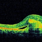
 Severe vitreomacular traction
Dec 23 2012 by Alex P. Hunyor, MD
OCT scan of the right eye of an 82-year-old male with 20/40 vision despite severe vitreomacular traction (VMT).
Condition/keywords: vitreomacular traction (VMT)
-
 Choroidal Granuloma Secondary to Tuberculosis
Mar 14 2013 by Eduardo Torres-Porras, MD
OCT scan through the granuloma shows attachment of the retinal pigment epithelial-choriocapillaris layer and the neurosensory retina over the granuloma (“contact” sign), inflammatory retinal infiltrate in the deeper retinal layers and subretinal fluid.
Photographer: Eduardo Torres Porras
Imaging device: Cirrus
Condition/keywords: optical coherence tomography (OCT), tubercular choroidal granuloma
-
 Solar Retinopathy
Mar 1 2013 by Theodore Leng, MD, MS, FASRS
OCT scan of a 15-year-old male with solar retinopathy after staring directly at a solar eclipse. Note the hyperreflectivity in the foveola.
Photographer: Erich Hagan
Imaging device: Heidelberg
Condition/keywords: solar retinopathy
-
 Choroidal Granuloma
Apr 23 2019 by Purva Patwari
22-year-old male patient presented with blurring of vision in the right eye noticed since last one week. He was asymptomatic a week ago when he noticed the blurring in his right eye. On examination his vision was 6/6 in both eyes. Anterior segment was normal. Posterior segment was normal for the left eye. Right eye examination revealed a clear vitreous cavity with choroidal granulomas scattered throughout the fundus. The present picture shows choroidal granulomas with OCT segment passing through the parafoveal lesion showing subretinal fluid accumulation and corresponding thickening of the retinal layers. CT scan reveals heterogeneously enhancing lymph nodes showing conglomerationin the hilar region-possibility of tubercular etiology.
Photographer: Dr Purva Patwari, Patwari Retina Center
Imaging device: Zeiss Visu 500
Condition/keywords: choroidal granuloma, choroiditis, granulomatous choroiditis, tubercular choroidal granuloma, tuberculosis
-
 Choroidal Granuloma Secondary to Tuberculosis
Mar 14 2013 by Eduardo Torres-Porras, MD
OCT scan through the granuloma shows attachment of the retinal pigment epithelial-choriocapillaris layer and the neurosensory retina over the granuloma (“contact” sign), inflammatory retinal infiltrate in the deeper retinal layers and subretinal fluid.
Photographer: Eduardo Torres Porras, Laser y ultrasonido ocular de Puebla
Imaging device: Cirrus
Condition/keywords: optical coherence tomography (OCT), tubercular choroidal granuloma
-
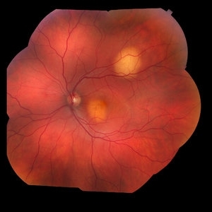 Choroidal Metastasis
Jan 24 2018 by Olivia Rainey
Color fundus montage of a 35-year-old male with choroidal metastasis from the lung. Before the diagnosis was confirmed, the patient had multiple CT scans revealing only pneumonia, with no signs of cancer.
Photographer: Olivia Rainey
Imaging device: Topcon 50DX
Condition/keywords: choroidal metastasis, color fundus photograph, lung cancer metastasis, montage
-
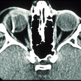 Intraocular Foreign Body, Metallic, CT Scan Orbits
Oct 1 2012 by Jeffrey G. Gross, MD, FASRS
IOFB, metallic, CT scan orbits.
Condition/keywords: CT scan, intraocular foreign body, orbits
-
---thumb.JPG/image-square;max$300,300.ImageHandler) Central Retinal Vein Occlusion
Central Retinal Vein Occlusion
Feb 28 2013 by Theodore Leng, MD, MS, FASRS
OCT scan at presentation showing a large amount of cystoid macular edema.
Imaging device: Zeiss Cirrus HD-OCT
Condition/keywords: central retinal vein occlusion (CRVO), cystoid macular edema (CME)
-
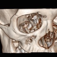 3D Image Of Intraocular Foreign Body
Sep 14 2014 by Mehul A Shah
14-year-old girl presented with penetrating injury to LE.
Photographer: Drashti Netralaya,Dahod
Imaging device: CT scan
Condition/keywords: intraocular foreign body
-
 Angioid streaks - Light microscopy and OCT
Jan 11 2013 by Gerardo Garcia-Aguirre, MD
Top: Light microscopy of an angioid streak. Note: Bruch's membrane below the RPE, which is thickened, elevated and fractured. Bottom: OCT scan of an angioid streak (not of the same patient) showing elevation and fracture of the RPE.
Photographer: Alfredo Gomez-Leal, MD (top) and Gerardo Garcia-Aguirre, MD (bottom)
Condition/keywords: angioid streaks
-
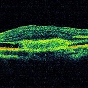 Angioid Streaks With CNVM OCT LE
Jun 17 2014 by Neha Goel, MS DNB FRCS (Glasg)
Horizontal OCT scan through the left macula.
Photographer: Neha Goel
Imaging device: RTVue
Condition/keywords: angioid streaks, choroidal neovascularization (CNV)
-
 Angioid Streaks With CNVM OCT RE
Jun 17 2014 by Neha Goel, MS DNB FRCS (Glasg)
Horizontal OCT scan through the right macula.
Photographer: Neha Goel
Imaging device: RTVue
Condition/keywords: angioid streaks, choroidal neovascularization (CNV)
-
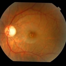 Bilateral Myopic Foveoschisis
Feb 10 2016 by Mallika Goyal, MD
Left eye fundus of a 22-year-old lady with bilateral myopic foveoschisis who presented with complaint of left eye vision drop 4 months prior to presentation. BCVA was 20/400. OCT scan through foveal centre revealed an outer lamellar macular hole accounting for vision loss.
Photographer: Mallika Goyal, MD, Apollo Health City, Hyderabad, India
Condition/keywords: myopic foveoschisis
-
 Bilateral Myopic Foveoschisis
Feb 10 2016 by Mallika Goyal, MD
Left eye OCT of a 22-year-old lady with bilateral myopic foveoschisis who presented with complaint of left eye vision drop 4 months prior to presentation. BCVA was 20/400. This OCT scan is taken superior to foveal centre. The scan through foveal centre reveals an outer lamellar macular hole accounting for vision loss.
Photographer: Mallika Goyal, MD, Apollo Health City, Hyderabad, India
Condition/keywords: myopic foveoschisis
 Loading…