Search results (35 results)
-
 Traumatic Macular Hole pre and post repair
Nov 25 2024 by Shobhit Chawla, M.S.
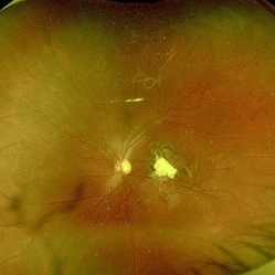
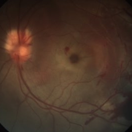
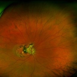
31 year-old male reported with h/o of blunt trauma over right eye ,from cricket ball. On examination DVA RE 6/60,LE 6/18,ant segment BE :WNL,FUNDUS RE:Sub retinal hemorrhage at macula with chroidal tear,LE :WNL. Undwer went 25G vitrectomy+sub retinal TPA+C3F8(RE).Post op 1 month DVA RE:6/24 ,ANT SEGMENT:WNL,FUNDUS:resolved sub retinal haem with traumatic macular hole. Under went repeat vit+autologous retinal transplant +SOI RE.POST SOR AFTER4monthsV/A :6/18 RE
Photographer: Ranjit Ray
Imaging device: Clarus 500
Condition/keywords: Macular hole, retinal graft, subretinal hemorrhage
-
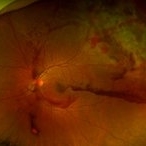
 Traumatic Macular Hole Closed With Amniotic Membrane Graft
Aug 18 2024 by Hemanth Murthy, MBBS, MD, FASRS
A 24 year old male patient presented with history of injury at workplace followed by loss of vision. He had a intraocular foreign body with a large traumatic macular hole. Patient was operated and the intraocular foreign body was removed. The hole was too large to close by ILM so an AMG graft was used. Patient regained 20/120 vision.
Photographer: Mr Veda Vyas
Imaging device: Optos Daytona
Condition/keywords: human amniotic graft, traumatic macular hole
-
 Traumatic Macular Hole
Aug 18 2024 by Hemanth Murthy, MBBS, MD, FASRS
A 24 year old male patient presented with history of injury at workplace followed by loss of vision. He had a intraocular foreign body with a large traumatic macular hole. Patient was operated and the intraocular foreign body was removed. The hole was too large to close by ILM so an AMG graft was used. Patient regained 20/120 vision.
Photographer: Mr Veda Vyas
Imaging device: Topcon Triton
Condition/keywords: human amniotic graft, traumatic macular hole
-
 Traumatic Macular Hole
Aug 18 2024 by Hemanth Murthy, MBBS, MD, FASRS
A 24 year old male patient presented with history of injury at workplace followed by loss of vision. He had a intraocular foreign body with a large traumatic macular hole. Patient was operated and the intraocular foreign body was removed. The hole was too large to close by ILM so an AMG graft was used. Patient regained 20/120 vision.
Photographer: Mr Veda Vyas
Imaging device: Optos Daytona
Condition/keywords: human amniotic graft, traumatic macular hole
-
 Traumatic macular hole
Nov 4 2022 by rodrigo torres
Traumatic macular hole
Photographer: Rodrigo Torres
Condition/keywords: choroidal rupture, macular hole, Trauma
-
 Fundus Photograph (Follow-up)
May 8 2021 by Jazli Tan
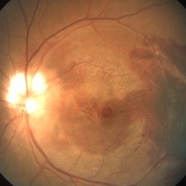
Follow-up fundoscopic examination revealed decreased subretinal blood and oedema at one week post injury, with persistent macular hole.
Condition/keywords: traumatic macular hole
-
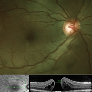
 Optical Coherence Tomography
May 8 2021 by Jazli Tan
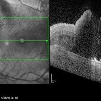
Optical Coherence Tomography (OCT) of a 31-year-old man with a traumatic macular hole from a badminton racket injury. OCT with enhanced depth imaging showed full thickness macular hole with subretinal fluid.
Condition/keywords: traumatic macular hole
-
 Traumatic Macular Hole
May 8 2021 by Jazli Tan
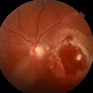
Fundus photograph of a 31-year-old man with a traumatic macular hole from a badminton racket injury. Fundoscopic examination revealed macular hole with associated sub-retinal, sub-retinal pigment epithelium, and sub-hyaloid bleed temporal to macula. Myelinated nerve fibre layer of the optic disc was noted superiorly with splinter hemorrhages.
Condition/keywords: traumatic macular hole
-
 Traumatic Macular Hole And Bruch Membrane Rupture
Jan 22 2021 by Renata Garcia Franco, Md
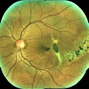
Male with history of ocular blunt injury, full-thickness macular hole, RPE changes and Bruch membrane rupture.
Photographer: Fatima Hernandez, Instituto de la Retina del Bajio SC
Imaging device: Zeiss
Condition/keywords: traumatic macular hole
-
 Traumatic Macular Hole And Bruch Membrane Rupture
Jan 22 2021 by Renata Garcia Franco, Md
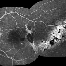
FA shows hypofluorescence in early frames due to a break in choriocapillaris and choroidal vessels at the rupture site with staning at late phases.
Photographer: Fatima Hernandez, Instituto de la Retina del Bajio SC
Imaging device: Zeiss
Condition/keywords: traumatic macular hole
-
 TRD with Macular Hole
Dec 8 2020 by Priya Rasipuram Chandrasekaran, MBBS, DO, DNB, FRCS
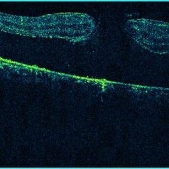
Horizontal 5 line raster scan through the macula shows full thickness macular hole along with separation of neuro sensory retina from the retinal pigment epithelium.
Condition/keywords: acute retinal detachment, traumatic macular hole
-
 Very Large Choroidal Melanoma in Monocular Patient - Widefield Color (Non-Tumor Eye)
Feb 13 2020 by Michael Seider, MD
Very large choroidal melanoma in the left eye of a 51-year-old man with long-standing poor vision in the right eye from a childhood injury (with traumatic macular hole and chorioretinal scarring). Note the large superior choroidal tumor with overlying subretinal hemorrhage and extensive inferior exudative retinal detachment. B-Scan ultrasound shows the collar-stud shape of the lesion and the overlying subretinal hemorrhage which is hyper-reflective compared to the vitreous and slightly hypo-reflective compared to the tumor. The patient has optic disk drusen in both eyes. He elected enucleation.
-
 Very Large Choroidal Melanoma in Monocular Patient - BScan
Feb 13 2020 by Michael Seider, MD
Very large choroidal melanoma in the left eye of a 51-year-old man with long-standing poor vision in the right eye from a childhood injury (with traumatic macular hole and chorioretinal scarring). Note the large superior choroidal tumor with overlying subretinal hemorrhage and extensive inferior exudative retinal detachment. B-Scan ultrasound shows the collar-stud shape of the lesion and the overlying subretinal hemorrhage which is hyper-reflective compared to the vitreous and slightly hypo-reflective compared to the tumor. The patient has optic disk drusen in both eyes. He elected enucleation.
-
 Very Large Choroidal Melanoma in Monocular Patient - Widefield Color (Eye with Tumor)
Feb 13 2020 by Michael Seider, MD
Very large choroidal melanoma in the left eye of a 51-year-old man with long-standing poor vision in the right eye from a childhood injury (with traumatic macular hole and chorioretinal scarring). Note the large superior choroidal tumor with overlying subretinal hemorrhage and extensive inferior exudative retinal detachment. B-Scan ultrasound shows the collar-stud shape of the lesion and the overlying subretinal hemorrhage which is hyper-reflective compared to the vitreous and slightly hypo-reflective compared to the tumor. The patient has optic disk drusen in both eyes. He elected enucleation.
-
 Blunt Ocular Trauma with Commotio Retinae
Nov 5 2019 by Nichole Lewis
11-year-old male with blunt ocular trauma from a soccer ball. Commotio Retinae, retinal hemorrhages, vitreous hemorrhage, multiple retinal tears and a traumatic macular hole. VA 20/70.
Photographer: Nichole Lewis
Imaging device: Optos
Condition/keywords: blunt trauma, commotio retinae, retinal hemorrhage, retinal tear, traumatic macular hole, vitreous hemorrhage
-
 Traumatic Macular Hole
Mar 27 2019 by Gary R. Cook, MD, FACS
7-year-old white male with a traumatic macular hole and secondary epiretinal membrane formation OS; hit in the eye with a rock; V.A.= counting fingers.
Imaging device: Topcon VT-50
Condition/keywords: epiretinal membrane formation, full thickness macular hole, macular hole, traumatic macular hole
-
 Traumatic Macular Hole
Mar 27 2019 by Gary R. Cook, MD, FACS
14-year-old white male with a traumatic macular hole OS 23 days post hockey puck injury; V.A.= 20/200.
Imaging device: Topcon VT-50
Condition/keywords: full thickness macular hole, macular hole, trauma, traumatic macular hole
-
 OCT - Traumatic Full Thickness Macular Hole
Feb 6 2019 by awaneesh m upadhyay, MBBS, DNB
Right eye OCT image of an 8-year-old boy shows full thickness macular hole following blunt trauma of 1 week duration.
Photographer: Awaneesh Upadhyay
Imaging device: HEILDERBERG - HRA
Condition/keywords: traumatic macular hole
-
 Traumatic Macular Hole
Feb 6 2019 by awaneesh m upadhyay, MBBS, DNB
Fundus photograph of an 8-year-old boy with vision of <20/200 in OD following blunt trauma of 10 days duration show macular hole with Berlin's edema.
Photographer: Dr. Awaneesh Upadhyay
Condition/keywords: Berlin's edema
-
 Traumatic Macular Hole with OCT
Jun 29 2018 by Gareth Lema, MD, PhD
Traumatic macular hole caused by the screw cap of a baby formula bottle. Pressure had built up in the container and propelled the cap into the patient's eye after she partially unscrewed it.
Photographer: Sandra Boglione, Ross Eye Institute, University at Buffalo Jacobs School of Medicine, Buffalo, NY
Imaging device: Optos
Condition/keywords: blunt trauma, traumatic macular hole
-
 Traumatic Macular Hole
Oct 2 2017 by Mehul A Shah
A 43-year-old male presented with history of blunt trauma before 6 months. Clinical picture was presented to us.
Photographer: Mehul Shah
Condition/keywords: traumatic macular hole
-
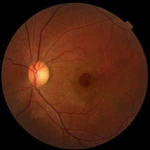 Traumatic Macular Hole and Choroidal Ruptures
Mar 15 2017 by Hamid Ahmadieh, MD
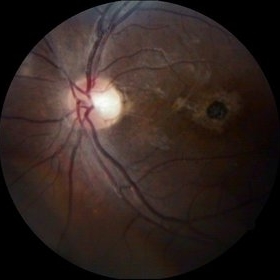
Color fundus photograph of the left eye of a 30 -year-old woman with a history of closed eye injury leading to a large traumatic macular hole and two concentric choroidal ruptures.
Photographer: Solmaz Shahmohammad, Negah Eye Center, Tehran,Iran
Condition/keywords: choroidal rupture, color fundus photograph, traumatic macular hole
-
 Traumatic Macular Hole and Choroidal Ruptures
Mar 15 2017 by Hamid Ahmadieh, MD
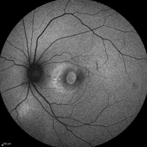
Fundus autofluorescence ( FAF) image of the left eye of a 30 -year-old woman with a recent history of closed eye injury leading to a large traumatic macular hole and two concentric choroidal ruptures.
Photographer: Solmaz Shahmohammad, Negah Eye Center, Tehran,Iran
Condition/keywords: choroidal rupture, fundus autofluorescence (FAF), traumatic macular hole
-
 Traumatice Macular Hole
Oct 4 2014 by Mehul A Shah
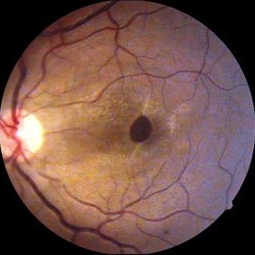
A 27-year-old patient presented with history of blunt trauma and patient was examined few months later.
Photographer: Drashti Netralaya,Dahod
Imaging device: Zeiss ff450
Condition/keywords: traumatic macular hole
-
 Traumatic Macular Hole
Sep 14 2014 by Mehul A Shah
20-year-old presented with cricket ball injury.
Photographer: Drashti Netralaya,Dahod
Imaging device: Zeiss ff450
Condition/keywords: traumatic macular hole
 Loading…