Search results (43 results)
-
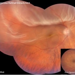
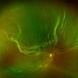 Rhegmatogenous Retinal Detachment
Sep 20 2025 by Aditya S Kelkar, MS, FRCS, FASRS,FRCOphth
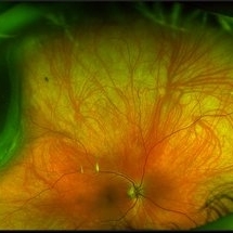
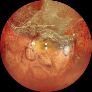
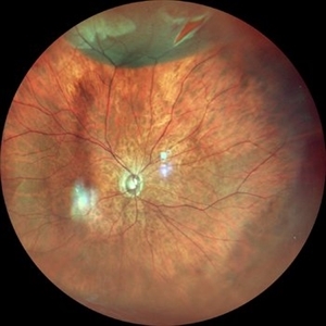
A 56-year-old patient diagnosed with rhegmatogenous retinal detachment presents with two distinct retinal breaks observed on fundus imaging.
Photographer: Dr. muskan
Imaging device: optos daytona
Condition/keywords: rhegmatogenous retinal detachment, superior retina
-
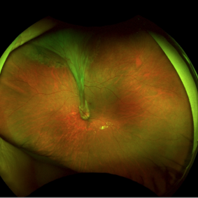
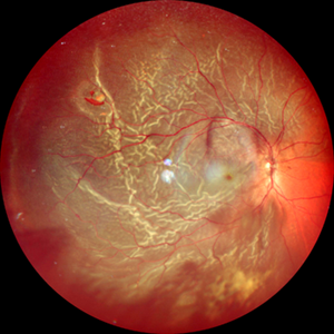 Retinal Detachment Secondary to Anomalous PVD
Mar 13 2025 by Fabricio Dolores
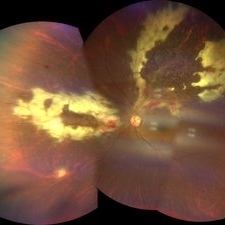
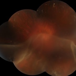
This color wide-field clinical image depicts the right eye of a female patient who experienced a sudden loss of vision one month earlier. She was initially diagnosed with a vitreous hemorrhage and managed with conservative treatment. Upon presentation to our institute one month later, a superior rhegmatogenous retinal detachment was identified, extending across the 12 o’clock meridian. This was accompanied by an inferior vitreous hemorrhage and a solitary superior retinal lesion located at M11 in the superior triangle of the ora serrata, in alignment with Lincoff's second law.
Photographer: Fabricio Dolores-Villanueva, MD
Imaging device: Nidek Mirante
Condition/keywords: Retinal Detachment
-
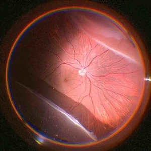 Giant Retinal Tear
Sep 28 2024 by Anjana Mirajkar, MS Ophthalmology
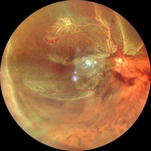
An intra operative image of the right eye showing a giant retinal tear with a superior retinal detachment.
Photographer: Dr. Anjana Mirajkar -Retina Foundation, Ahmedabad
Condition/keywords: GIANT RETINAL TEAR
-
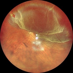 Superior Retinal Detachment With Horse Shoe Tear
Jul 18 2024 by Anjana Mirajkar, MS Ophthalmology
A widefield photo of LE showing superior retinal detachment involving the macula with a horse shoe tear at 11 'o clock
Photographer: Dr. Anjana Mirajkar -Retina Foundation, Ahmedabad
Imaging device: Mirante-Nidek
-
 Retinal Detachment with Tear
Jun 19 2024 by Sanauddin Samejo , Diploma (Ophthalmic Technician Training Course)
Fundus photo of a 47 year-old male who came to Dr. Rao's clinic and was diagnosed with retinal detachment with superior retinal tear.
Photographer: Sanauddin Samejo
Imaging device: Optos Silver Stone
Condition/keywords: Retinal tear with detachment
-
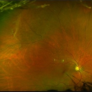
 Choroidal Melanoma
Mar 26 2024 by Xitlali Caterina
Ultra-widefield fundus photograph of a 40-year-old woman with Choroidal Melanoma in right eye. Patient present with 20/50+2 vision in the right eye. Patient reported having frequent headaches located frontal area of their head and sometimes radiated to the right side as well. Patient also noted eye pain in both eyes that has remained constant for many years, as well as light sensitivity. The physician stated that since this is a medium-sized tumor, the treatment options include I-125 brachytherapy or enucleation. He recommended I-125 brachytherapy.
Photographer: Xitlali Caterina
Imaging device: Optos California RGB
Condition/keywords: fundus photography, Optos, OPTOS CALIFORNIA, superior retina, ultra-wide field imaging, ultra-widefield image
-
 CMV Retinitis
Feb 17 2024 by Eloy Mata-Cortes, MD
Fundus photograph of left eye showing Cytomegalovirus retinitis of a 40-year-old male with positive HIV history. He presented with CD4 cell count of 50 cells/mm3 and decreased vision of left eye. In the photograph we can see the three typical patterns in this retinitis: a hemorrhagic appearance in superior temporal arcade and between nasal arcades, granular pattern in superior temporal retina, and a “frosted branch” angiitis surrounding the retinal vessels in nasal and superior retina.
Photographer: Eloy Mata-Cortes, Instituto Mexicano de Oftalmologia, Queretaro, Mexico
Imaging device: Clarus 700
Condition/keywords: CMV retinitis, cytomegalovirus (CMV), frosted branch angiitis, Frosted Branch Angitis
-
 Superior Retinal Detachment
Jan 30 2024 by Akansha Sharma
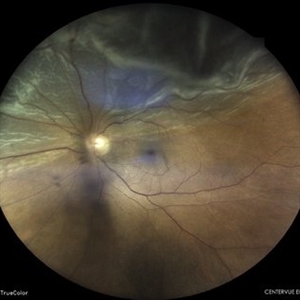
Color fundus photograph of a 62 year old male patient with superior retinal detachment.
Photographer: Dr. Akansha Sharma, Bharati Eye Hospital
Condition/keywords: RD
-
 Superior Retinal Detachment
Jan 30 2024 by Akansha Sharma
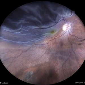
Color fundus photograph of a 72 year old female with superior retinal detachment with macula off.
Photographer: Dr. Akansha Sharma, Bharati Eye Hospital
Condition/keywords: RD, Retinal Detachment
-
 Superior retinal detachment with break
Nov 20 2023 by ANKIT JAIN
Widefield fundus image of LE showing superior retinal detachment with break
Photographer: Dr Ankit Jain
Imaging device: MIRANTE
-
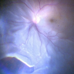
 SUNSET THROUGH A VEIL
Oct 12 2023 by Deepti A Kulkarni, M.B.B.S., D.N.B., F.V.R.
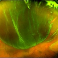
HIGH MYOPE WITH A CORRECTION OF -24D. SUDDEN VISION LOSS FOLLOWING TRAUMA. SUPERIOR RETINA FOLDED ON TO THE INFERIOR RETINA MASKING THE DISC IN A TRANSLUCENT VEIL.
Photographer: DEEPTI KULKARNI, DR ANIL KULKARNI EYE HOSPITAL, MIRAJ, INDIA
Imaging device: TOPCON
Condition/keywords: GIANT RETINAL TEAR
-
 Superior Retinal Detachment
Aug 13 2023 by Anjana Mirajkar, MS Ophthalmology
A color photo of RE of a 55 year old male in a case of Superior Retinal Detachment with macula off with a superior horse shoe tear with vitreous Haze
Photographer: Dr. Anjana Mirajkar -Retina Foundation, Ahmedabad
Condition/keywords: chronic retinal detachment
-
 Retinal Detachment
Jul 30 2023 by Maneesh M Bapaye, MD, MBA
Montage fundus photo of superior retinal detachment in right eye of a 45 year old male patient
Photographer: Dr.Maneesh Bapaye
Imaging device: Zeiss Fundus camera
Condition/keywords: montage fundus photo
-
 Total retinal Detachment multiple holes
Sep 26 2022 by Denica Rodriguez
60 year old Male presented with two week old Macula off Retinal detachment with multiple tears.
Photographer: Denica Rodriguez
Imaging device: Optos California
Condition/keywords: color fundus photograph, color photo, macula-off, optos, pseudocolor, Retinal detachment, retinal holes, retinal tear, Retinal tear with detachment, superior arcade, superior field, superior retina, total retinal detachment
-
 Retinal Detachment with a Large Superior Tear
Feb 2 2022 by Manish Nagpal, MD, FRCS (UK), FASRS
Intraoperative photo of a retinal detachment with a large superior retinal tear.
Photographer: Manish Nagpal, Director, Retina Foundation, Ahmedabad
Imaging device: Sony PMW -10 MD surgical camera
Condition/keywords: retina, tear
-
 Choroidal Detachment
Jan 17 2022 by Logan ryzenga
Left ultra-wide field photograph of an 81-year old female with a choroidal detachment affecting her left eye. Patient had a stent placed November, 2021 and following the procedure she complains of variable blurred vision and severe constricted visual fields. She presented at our office with flashes a month prior but without pain or floaters.
Photographer: Logan Ryzenga
Imaging device: Optos California
Condition/keywords: choroidal detachment, fundus photograph, left eye, Optos, pseudocolor, superior retina, ultra-wide field imaging
-
 Ocular Toxocariasis with Peripheral Granuloma
Apr 24 2021 by Alexandre Grandinetti, MD, PhD
8-year-old boy with a retinal fold secondary to peripheral toxocara canis granuloma localized on the superior retina.
Photographer: Corina Skrzek
Imaging device: Optos California
Condition/keywords: toxocariasis
-
 Valslava Retinopathy
Jan 15 2021 by Priya Rasipuram Chandrasekaran, MBBS, DO, DNB, FRCS
This is the fundus photo and red free montage showing preretinal hemorrhage of the left eye along the superior retina, abutting the disc margin and extending as far as the macula. There are few scattered flame shaped hemorrhages superiorly, nasally and inferiorly with a central white spot mimicking Roth spots.
Condition/keywords: valsalva retinopathy
-
 Bullous Retinoschisis with Outer Retinal Holes
Jun 15 2020 by Olivia Rainey
Ultra-widefield pseudocolor fundus photograph of a 56-year-old female with bullous retinoschisis with outer retinal holes affecting her right eye. The physician noted superotemporal retinoschisis in her monoculcar functioning eye. There was no demarcation line and no inner or outer layer breaks at her first appointment in February of 2020. On 6/15/20 she had a new onset outer holes and SRF tracking inferiorly. The physician recommended observation, however if this continues to progress we have discussed indications for barrier laser.
Photographer: Olivia Rainey, OCT-C, COA
Imaging device: Optos California
Condition/keywords: bullous retinoschisis, Optos, outer layer breaks, outer layer hole, pseudocolor, subretinal fluid, superior retina, ultra-wide field imaging
-
 Retinal Detachment with Horseshoe Tear
Jun 10 2020 by Manish Nagpal, MD, FRCS (UK), FASRS
Localized superior retinal detachment with horseshoe tear and minimal fluid.
Photographer: gayathri mohan
Imaging device: nidek slo mirante
-
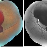
 Traumatic Giant Retinal Tear Associated Retinal Detachment
Nov 9 2019 by Luis J Haddock, MD
This wide field fundus photograph of the left eye shows a traumatic giant retinal tear associated with total retinal detachment. The image shows the torn superior retina folded over the macula with the underside of the retina visible. There is associated peripheral choroidal detachment due to hypotony from giant retinal tear. This patient has history of spondyloepithelial dysplasia with dwarfism and presented with vision loss after a recent blunt trauma with elbow to the eye.
Imaging device: Optos
Condition/keywords: giant retinal tear, traumatic optic neuropathy
-
 Hemi-CRAO
Mar 26 2019 by Gary R. Cook, MD, FACS
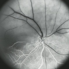
Mid-phase (laminar venous return) fluorescein angiogram image of an embolic superior hemi-CRAO showing marked delay in filling of the superior retinal arteriolar and venous vasculature and total loss of the retinal capillary bed in the superior hemisphere OD.
Condition/keywords: capillary closure, capillary nonperfusion, central retinal artery occlusion (CRAO), FA mid phase, fluorescein angiogram (FA)
-
 Retinal Detachment with Retinal Tears
Dec 11 2018 by Olivia Rainey
Ultra-wide field pseudocolor image of a 56-year-old male with a large superior retinal detachment with retinal tears affecting his right eye.
Photographer: Olivia Rainey
Imaging device: Optos
Condition/keywords: Optos, pseudocolor, retinal tear with detachment
-
 Fresh Superior Macula-On Rhegmatogenous Retinal Detachment
Feb 10 2018 by Deepak Bhojwani, MS
A 60 year old gentlemen came rushing to the retinal clinic with history of sudden onset of loss of inferior visual field since last 3 hours. Fundus photograph indeed corelates with his complaints documenting fresh superior macula-on rhegmatogenous retinal detachment.
Photographer: Dr Deepak Bhojwani, Raghudeep Eye Hospital , Ahmedabad
Condition/keywords: macula-on fresh superior retinal detachment
-
 Choroidal Melanoma
Feb 2 2018 by Olivia Rainey
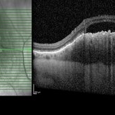
Optical coherence tomography with enhanced depth imaging of a 78-year-old female with choroidal melanoma with subretinal fluid affecting her right eye.
Photographer: Olivia Rainey
Imaging device: Heidelberg Spectralis
Condition/keywords: enhanced depth imaging, infrared image, optical coherence tomography (OCT), subretinal fluid, superior retina
 Loading…