Search results (282 results)
-
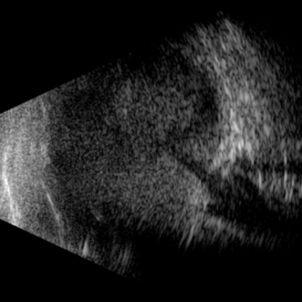
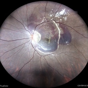
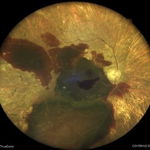
 Advanced Proliferative Diabetic Retinopathy
Apr 9 2025 by Gustavo Uriel Fonseca Aguirre
B-mode ultrasound of a patient with long-standing poorly controlled diabetes demonstrates characteristic findings of advanced proliferative diabetic retinopathy. The examination reveals moderate vitreous hemorrhage appearing as diffuse hyperechoic opacities throughout the vitreous cavity, along with a posterior hyaloid membrane densely infiltrated by hemorrhagic material, showing irregular thickening and increased reflectivity. A mild subhyaloid hemorrhage is visible as a subtle hyphema-like space anterior to the retinal surface. The study documents a total tractional retinal detachment, evidenced by rigid retinal folds with clear insertion points of vitreous strands, accompanied by a significant subretinal hemorrhage seen as a prominent hyperechoic collection beneath the elevated retina. These findings collectively illustrate the severe vitreoretinal interface pathology characteristic of end-stage diabetic eye disease, with predominant tractional components and distinct echographic stratification of hemorrhagic layers - from anterior vitreous involvement to deeper subretinal blood accumulation.
Photographer: Gustavo U. Fonseca Aguirre, Hospital Conde de Valenciana, Ciudad de México
Condition/keywords: diabetic retinopathy, tractional retinal detachment, Vitreous hemorrhage
-
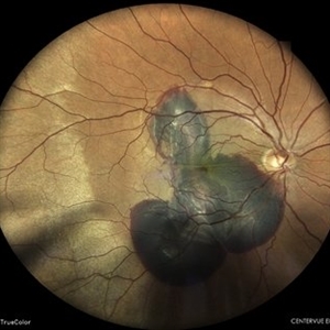
 Negative Retinal Detachment
Apr 7 2025 by Gustavo Uriel Fonseca Aguirre
B-mode ultrasound of a 7-month-old premature infant with a history of perinatal supplemental oxygen therapy reveals a total funnel-shaped retinal detachment appearing as a hypoechoic structure, accompanied by significant hyperechoic subretinal hemorrhage. This distinctive echographic pattern creates the characteristic appearance of a "negative retinal detachment."
Photographer: Gustavo U. Fonseca Aguirre, Hospital Conde de Valenciana, Ciudad de México
Condition/keywords: retinopathy of prematurity
-
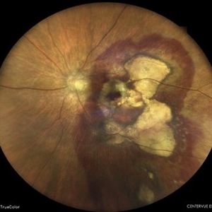
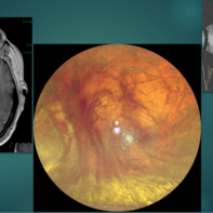 Choroidal Melanoma Masquerading as PEHCR
Mar 3 2025 by Tejaswita Verma
A 65 year old diabetic male presented with large nasal retinal mass giving the appearance of organized dehaemoglobinized subretinal hemorrhage with breakthrough vitreous hemorrhage , with 6/6P vision. Enucleation specimen showed histopathology confirmed choroidal melanoma.
Photographer: DR. TEJASWITA VERMA
Imaging device: MIRANTE
Condition/keywords: vitreous hemorrhage
-
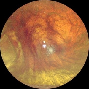 Choroidal Melanoma Masquerading as Subretinal Hemorrhage With Breakthrough VH
Jan 23 2025 by Tejaswita Verma
A 65 year old diabetic male presented with large nasal retinal mass giving the appearance of organized dehaemoglobinized subretinal hemorrhage with breakthrough vitreous hemorrhage , with 6/6P vision. Enucleation specimen showed histopathology confirmed choroidal melanoma.
Photographer: DR. TEJASWITA VERMA
Imaging device: MIRANTE
-
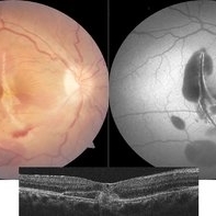
 New Subretinal Hemorrhage in AMD
Jan 8 2025 by Drew Mitchell
HD 1 line 100x OCT scan of a New Subretinal Hemorrhage in a established patient with AMD.
Photographer: Drew Mitchell, OCT-C
Imaging device: Zeiss Cirrus 6000
Condition/keywords: age-related macular degeneration (AMD), OCT, subretinal hemorrhage
-
 Large Subretinal Bleed in Case of Wet ARMD
Sep 28 2024 by Anjana Mirajkar, MS Ophthalmology
An intra operative image showing large sub retinal hemorrhage involving the macular area and along the superior arcade with exudation at the macular area in case of wet ARMD.
Photographer: Dr. Anjana Mirajkar -Retina Foundation, Ahmedabad
Condition/keywords: polypoidal choroidal vasculopathy (PCV), subretinal hemorrhage, wet age-related macular degeneration (wet AMD)
-
 Subretinal Hemorrhage Status Post Blunt Trauma
May 27 2024 by Akansha Sharma
Color fundus photograph of a 41 year old male with subretinal bleed status post blunt trauma.
Photographer: Dr. Akansha Sharma, Bharati Eye Hospital
Condition/keywords: subretinal hemorrhage, trauma
-
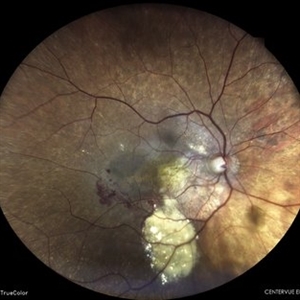
 Sea Fan Neovascular Frond in Rare Case of Fanconi Anemia
May 18 2024 by Amol yuvraj ganvir
The left eye fundus shows a typical sea fan-shaped hyperfluorescent area, confirming neovascularization along the major temporal arcade and blocked fluorescence due to intraretinal and subretinal hemorrhage.
Photographer: Dr. Amol Ganvir
Condition/keywords: Fanconi Anemia, sea fan
-
 Sea Fan Neovascular Frond in Rare Case of Fanconi Anemia
May 18 2024 by Amol yuvraj ganvir
A 15-year-old female patient brought by her parents with defective vision in left eye for 15 days. The patient had a known case of Fanconi Anemia. Left eye tortuous vessels and new retinal vessels along the major temporal arcade, with intraretinal and subretinal hemorrhage covering the macula.
Photographer: Dr. Amol Ganvir
Condition/keywords: Fanconi Anemia, sea fan
-
 Choroidal Rupture With Traumatic Subretinal Hemorrhage
Apr 19 2024 by Akansha Sharma
Color fundus photograph of a 11 year old female with subretinal bleed and choroidal rupture status post trauma.
Photographer: Dr. Akansha Sharma, Bharati Eye Hospital
Condition/keywords: Choroidal Rupture, subretinal hemorrhage, subretinal blood
-
 Polypoidal Choroidal Vasculopathy
Apr 19 2024 by Akansha Sharma
Color fundus photograph of a 84 year old female with subretinal hemorrhage in a case of polypoidal choroidal vasculopathy.
Photographer: Dr. Akansha Sharma, Bharati Eye Hospital
Condition/keywords: PCV, polypoidal choroidal vasculopathy (PCV)
-
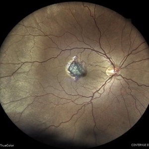
 Macula-threatening PEHCR causing a nasal visual-field defect in a patient with AMD
Apr 15 2024 by David A Reichstein, MD
(A) Ultra-widefield color fundus photograph demonstrates a PEHCR encroaching upon the temporal macula. Lipid exudation is apparent at the lesion’s anterior and inferior border. Subretinal hemorrhage is apparent at the lesion’s inferior border. Drusen are apparent in the macula. An unrelated, small choroidal nevus is apparent in the inferior fundus. (B) Ultra-widefield FA taken in early stage demonstrates hypofluorescence within the lesion consistent with blockage by possible sub-RPE or subretinal heme. (C) Ultra-widefield fundus photograph taken 6 months following the initiation of monthly anti-VEGF therapy demonstrates considerable reduction in the size of the lesion and resolution of the subretinal hemorrhage and lipid exudation. (D) Ultra-widefield fundus photograph taken 1 year after presentation where a treat-and-extend approach was performed for the most recent 6 months. The lesion had almost completely resolved.
Condition/keywords: peripheral exudative hemorrhagic chorioretinopathy (PEHCR)
-
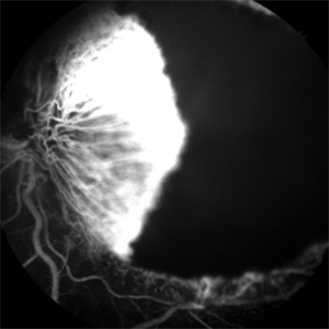
 Large PEHCR causing an exudative inferior detachment in a patient with AMD
Apr 15 2024 by David A Reichstein, MD
(A) Ultra-widefield color fundus photograph demonstrates a temporal PEHCR causing minimal intra- and subretinal hemorrhage along with lipid exudation. There is an associated inferior detachment due to the dependent nature of the exudation. Note the lipid exudation at the posterior edge of the detachment indicating chronicity of the lesion. Drusen in the macula are also appreciated. (B) Ultra-widefield FA in early stage demonstrates hypofluorescence temporally and inferiorly. (C) Ultra-widefield color fundus photograph taken after 1 year of monthly anti-VEGF therapy demonstrates resolution of the exudative detachment and resultant chorioretinal scarring.
Condition/keywords: peripheral exudative hemorrhagic chorioretinopathy (PEHCR)
-
 Choroidal Tear-Multimodal imaging
Sep 10 2023 by Maneesh M Bapaye, MD, MBA
Multimodal imaging for a 26 years old patient with history of blunt trauma. Color fundus photo shows choroidal tear through foveal center with subretinal haem, autofluorescence image shows hypoAF demarkating margins and extent of the tear while the OCT B-Scan through foveal center shows tear in choroid, BM and RPE along with elevated EZ parallel to fovea with underlying hyper reflectivity s/o hemorrhage
Photographer: Maneesh Bapaye
Condition/keywords: blunt trauma, choroidal tear, subretinal hemorrhage
-
 Active CNVM
Jul 12 2023 by Harsh Vardhan Singh, MS
55-year male with left eye sub-retinal hemorrhage due to Active CNVM, Colour fundus photograph of left eye subretinal hemorrhage due to Active CNVM; Red-free image of left eye sub-retinal hemorrhage due to Active CNVM
Photographer: Harsh Vardhan Singh
Condition/keywords: choroidal neovascular membrane (CNVM), CNVM, subretinal hemorrhage
-
 Active CNVM
Jul 12 2023 by Harsh Vardhan Singh, MS
55-year male with left eye sub-retinal hemorrhage due to Active CNVM, Colour fundus photograph of left eye subretinal hemorrhage due to Active CNVM
Photographer: Harsh Vardhan Singh
Condition/keywords: choroidal neovascular membrane (CNVM), CNVM, subretinal hemorrhage
-
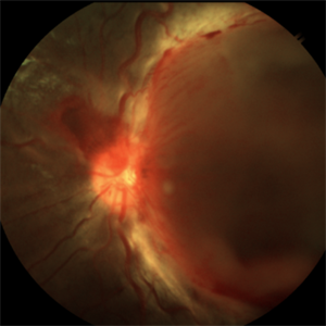
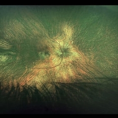
 PEHCR (Peripheral Exudative Hemorrhagic Chorioretinopathy)
May 12 2023 by Niloofar Piri, MD
Ultrawide fundus photograph of the left eye demonstrating extensive peripheral hemorrhagic exudative detachment in a 79 yo Caucasian female with prior history of non-exudative AMD. Recent diagnosis of Acute myeloid leukemia with low platelet count which might have contributed to the above presentatuon. Please note the temporal subretinal hemorrhage as well as RPE atrophy and hyperplasia in the macula.
Photographer: Rocio Bentivegna, MD, Saint Louis University; Jessica Maddox, COA, Saint Louis University
Condition/keywords: peripheral exudative hemorrhagic chorioretinopathy (PEHCR)
-
 Optos Picture 1 month after PPV with sub retinal tpa
Apr 27 2023 by Jesus Lozano, MD
Optos Image POM1 after Vitrectomy + Subretinal tPA do to subretinal hemorrhage at the macula. Visual Improvement to 6/24.
Imaging device: Optos
Condition/keywords: subretinal hemorrhage, vitrectomy
-
 SUB-RETINAL HEMORRHAGE WITH RETINITIS PIGMENTOSA
Feb 28 2023 by Akansha Sharma
COLOUR FUNDUS PHOTOGRAPH OF A 77 YEAR OLD MALE WITH SUBRETINAL HEMORRHAGE IN A CASE OF RETINITIS PIGMENTOSA
Photographer: Dr. Urmil Shah, Dr. Denish Patel, Dr. Akansha Sharma, Bharati Eye Hospital, Ahmedabad, Gujarat
Condition/keywords: retinitis pigmentosa, subretinal hemorrhage
-
 SUB-RETINAL HEMORRHAGE WITH CHOROIDAL RUPTURE
Feb 28 2023 by Akansha Sharma
COLOUR FUNDUS PHOTOGRAPH OF A 45 YEAR OLD MALE WITH SUBRETINAL HEMORRHAGE WITH CHOROIDAL RUPTURE
Photographer: Dr. Urmil Shah, Dr. Denish Patel, Dr. Akansha Sharma, Bharati Eye Hospital, Ahmedabad, Gujarat
Condition/keywords: subretinal hemorrhage
-
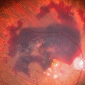
 Subretinal Hemorrhage
Feb 28 2023 by Akansha Sharma
Color fundus photograph of an 84-year old male with subretinal hemorrhage associated with areas of scarring.
Photographer: Dr. Urmil Shah, Dr. Denish Patel, Dr. Akansha Sharma, Bharati Eye Hospital, Ahmedabad, Gujarat
Condition/keywords: choroidal neovascularization (CNV), subretinal hemorrhage
-
 Lady in a dress
Feb 9 2023 by Shelby Helton
Fluorescein Angiography on a 67-year-old male with significant RPE changes secondary to a severe subretinal hemorrhage that required a vitrectomy with subretinal TPA in 2013.
Photographer: Shelby Helton
Imaging device: Heidelberg Spectralis
Condition/keywords: wet age-related macular degeneration (wet AMD)
-
 Polypoidal choroidal vasculopathy
Nov 21 2022 by T. P . VIGNESH, MBBS,MS
SD-OCT of a 60 year old woman revealing a large PED , multilobular PED and subretinal hemorrhage .
Photographer: Priyanka
Imaging device: Heidelberg Spectralis
Condition/keywords: polypoidal choroidal vasculopathy (PCV)
-
 Choroidal Melanoma
Nov 3 2022 by pedro fernandes souza neto
Enucleation specimen of Choroidal Melanoma: anterior chamber is closed. Total secondary retinal detachment with subretinal serous fluid and some subretinal hemorrhages are present.
Photographer: Eduardo Marback, Federal University of Bahia, Brazil
Condition/keywords: enucleation
 Loading…