 Initializing download.
-
 By David A Reichstein, MD
Tennessee Retina, PC - Uploaded on Apr 15, 2024.
- Last modified by Joshua Friedman on Apr 15, 2024.
- Rating
- Appears in
- Miscellaneous
- Condition/keywords
- peripheral exudative hemorrhagic chorioretinopathy (PEHCR)
- Description
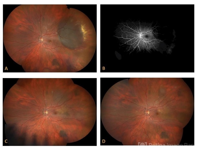
- (A) Ultra-widefield color fundus photograph demonstrates a PEHCR encroaching upon the temporal macula. Lipid exudation is apparent at the lesion’s anterior and inferior border. Subretinal hemorrhage is apparent at the lesion’s inferior border. Drusen are apparent in the macula. An unrelated, small choroidal nevus is apparent in the inferior fundus. (B) Ultra-widefield FA taken in early stage demonstrates hypofluorescence within the lesion consistent with blockage by possible sub-RPE or subretinal heme. (C) Ultra-widefield fundus photograph taken 6 months following the initiation of monthly anti-VEGF therapy demonstrates considerable reduction in the size of the lesion and resolution of the subretinal hemorrhage and lipid exudation. (D) Ultra-widefield fundus photograph taken 1 year after presentation where a treat-and-extend approach was performed for the most recent 6 months. The lesion had almost completely resolved.


")


