Search results (27 results)
-
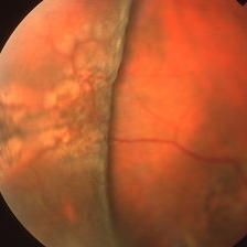
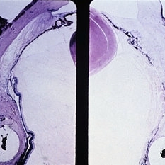
 Scleral Buckling
Apr 21 2025 by Gustavo Uriel Fonseca Aguirre
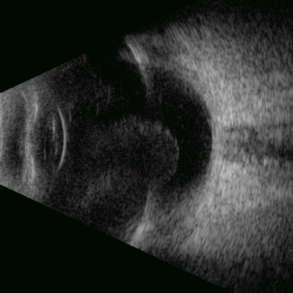
This B-mode axial ultrasound scan shows an eye with a scleral buckle in place for previous rhegmatogenous retinal detachment. The image demonstrates the characteristic indentation of the ocular wall at the buckle site, with proper retinal reattachment.
Photographer: Gustavo U. Fonseca Aguirre, Hospital Conde de Valenciana, Ciudad de México
Condition/keywords: scleral buckling
-
 Coats Disease
Mar 30 2024 by Karen Flores Guevara
Fundus photograph of a 9-year-old child with coats´ disease history of Scleral Buckling due to Retinal Detachment.
Photographer: Diana Elizabeth-Arellano-Acosta-MD Pediatric Retina,Asociación para Evitar la Ceguera en México IAP. México
Condition/keywords: Coats' disease
-
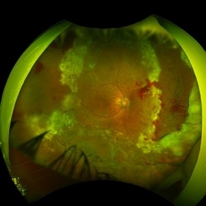
 Proliferative vitreoretinopathy in complex retinal redetachment
Dec 6 2023 by Ma. Guadalupe Perez Guevara
A 58-year-old female underwent phacovitrectomy + scleral buckling by retinal detachment. Silicon oil was used as endo tamponade. In the post-surgical period, the formation of a subretinal PRV is observed with a significant circumferential contraction that generates an inferior redetachment. It was decided to undergo a vitreous cavity lavage to remove membranes + 180° retinectomy.
Photographer: Ma. Guadalupe Pérez-Guevara. Fundación Hospital Nuestra Señora de la Luz I.A.P
Imaging device: Optos
Condition/keywords: PVR, retina surgery complications
-
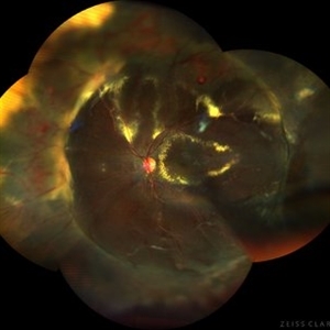
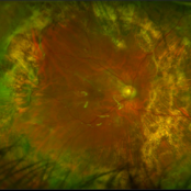
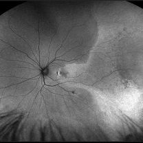
 Fully reattached retina following scleral buckling with a 360 encircling band and supero temporal buckle in a 25 year old woman.
Nov 9 2023 by Jesus Lozano, MD
Optos image of a 25 year old woman with a fully reattached retina following scleral buckling with a 360 encircling band and supero temporal buckle
Photographer: Mihaela Shlomi, Hillel Yaffe Medical Center. Israel.
Imaging device: Optos
Condition/keywords: scleral buckle
-
 Scleral Buckling IOL Drop
Aug 6 2023 by Dr.Sheetal Divate
A 27 year old female with an old history of trauma and operated with scleral buckling and cataract surgery in the past came recently with complaints of DOV . Findings noted where IOL drop, inferior retinal detachment and old scleral buckle indent.
Photographer: Dr.Sheetal Divate
Imaging device: Optos Advance
Condition/keywords: dislocated intraocular lens (IOL), Retinal Detachment, scleral buckle
-
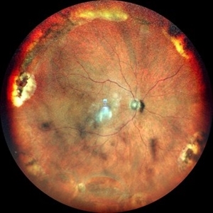 RETINAL BREAKS
Nov 21 2022 by Akansha Sharma
COLOUR FUNDUS PHOTOGRAPH OF A 60 YEAR OLD MALE PATIENT WITH RETINAL BREAK STATUS POST SCLERAL BUCKLING 25 YEARS AGO
Photographer: Dr. Akansha Sharma-Retina Foundation, Ahmedabad
Condition/keywords: retinal break, scleral buckle
-
 Retinal Detachment Following Scleral Buckling, Retinectomy, Laser, and Oil
Jan 31 2022 by Ahmad B. Tarabishy, MD
Ultra wide-field fundus photograph of a 55-year-old gentleman who is 4 days after surgery with scleral buckling, pars plana vitrectomy, perfluoron tamponade, membrane peeling, direct fluid-PFO-oil exchange, nasal and temporal retinectomies, and endolaser photocoagulation. Visual acuity was 20/150 under oil.
Photographer: Megan McLandsborough, Lakeland Eye Clinic
Imaging device: Optos California UWF Camera
Condition/keywords: endolaser, Membrane Peel, PPV, proliferative retinopathy, proliferative vitreoretinopathy (PVR), Retinal Detachment, retinal detachment with retinal defect, scleral buckle, submacular perfluorocarbon liquid (PFO)
-
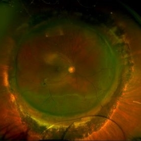
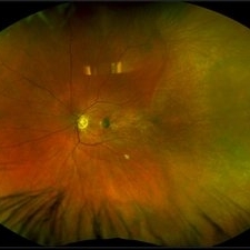
 Status Post Scleral Buckling
Jan 26 2022 by Akansha Sharma
Wide field fundus photograph of a 55-year-old male with a scleral buckle in situ post scleral buckling performed 15 years back.
Photographer: Dr. Akansha Sharma - Retina Foundation, Ahmedabad
Condition/keywords: color wide field, encircling scleral buckle
-
 Scleral Buckle with 360 Degree Laser
Sep 22 2021 by Ahmad B. Tarabishy, MD
56 year-old male with a history of retinal detachment OD, complicated by recurrent RD with PVR and cataract. He underwent multiple surgical procedures that included scleral buckling using a 3mm silicone sponge, membrane peel, inferior retinectomy, endolaser, lensectomy, and secondary IOL placement. Current VA is 20/60 OD.
Photographer: Michelle Howarth, Lakeland Eye Clinic
Imaging device: Optos P200TDx
Condition/keywords: encircling scleral buckle, proliferative vitreoretinopathy (PVR)
-
 Dexamethasone Implant
Jul 3 2021 by Gerardo Rivera Arroyo
42-year-old male, operated on for vitrectomy plus scleral buckling plus silicone plus dexamethasone implant for inferior retinal detachment with PVR.
Photographer: Rosa Elizabeth Moreno Anda, MD, Hospital Central Militar CDMX
Condition/keywords: dexamethasone implant, retina surgery, vitrectomy
-
 Ultra-Widefield Montage of Reattached Retina and Subretinal Fluid Blebs Following Scleral Buckling Surgery
Jun 1 2021 by Kushal S Delhiwala, MBBS, MS, FMRF,FICO, FAICO
Ultra-widefield fundus photograph of left eye of a 29-year-old male who underwent scleral buckling surgery for retinal detachment.279 silicone tire and 240 band was used. Fundus shows reattached retina with adequate buckle indentation and subretinal fluid blebs along inferior arcade and nasal to disc.
Photographer: Kushal Delhiwala, Netralaya superspeciality eye hospital, Ahmedabad, Gujarat,India
Imaging device: Optos Daytona
Condition/keywords: cryotherapy, external drainage, scleral band, scleral buckle, silicone band, silicone tire
-
 Submacular PFO
Feb 20 2020 by Kevin J. Blinder, MD, FASRS
This is a 53-year-old gentleman that was referred to us for a second opinion with an inoperable RD with PVR after 3 failed attempts. We performed a PPV, membranectomy, scleral buckling procedure, with silicone oil injection. This case did not require PFO. You can imagine our surprise when we discovered submacular PFO postoperatively. It is very difficult to see the PFO on the Optos. The infrared shows it clearly, with confirmation of the submacular space on the SD-OCT.
Photographer: Jarrod Wehmeier, The Retina Institute; St. Louis, MO
Imaging device: optos
Condition/keywords: submacular perfluorocarbon liquid (PFO)
-
 Submacular PFO
Feb 20 2020 by Kevin J. Blinder, MD, FASRS
This is a 53-year-old gentleman that was referred to us for a second opinion with an inoperable RD with PVR after 3 failed attempts. We performed a PPV, membranectomy, scleral buckling procedure, with silicone oil injection. This case did not require PFO. You can imagine our surprise when we discovered submacular PFO postoperatively. It is very difficult to see the PFO on the Optos. The infrared shows it clearly, with confirmation of the submacular space on the SD-OCT.
Photographer: Jarrod Wehmeier, The Retina Institute; St. Louis, MO
Imaging device: Heidelberg Spectralis
Condition/keywords: submacular perfluorocarbon liquid (PFO)
-
 Macula Off Retinal Detachment with CNV
Nov 11 2019 by Olivia Rainey
Ultra-wide field pseudocolor photograph of a 42-year-old female with a long-standing, macula-off retinal detachment affecting her left eye. Patient was unaware of vision loss until testing her visual acuity and she denied seeing flashing lights. Patient decided to proceed with scleral buckling. The CNV is potentially secondary the retinal detachment, but may be myopic related or idiopathic. The CNV appears fibrotic and inactive. The patient was warned that this will absolutely limit how much vision she recovers once the retina is reattached.
Photographer: Olivia Rainey
Imaging device: Optos California
Condition/keywords: choroidal neovascularization (CNV), left eye, montage, Optos, pseudocolor, retinal detachment of the macula, ultra-wide field imaging
-
 Macula Off Retinal Detachment with CNV
Nov 11 2019 by Olivia Rainey
Ultra-wide field pseudocolor photograph of a 42-year-old female with a long-standing, macula-off retinal detachment affecting her left eye. Patient was unaware of vision loss until testing her visual acuity and she denied seeing flashing lights. Patient decided to proceed with scleral buckling. The CNV is potentially secondary the retinal detachment, but may be myopic related or idiopathic. The CNV appears fibrotic and inactive. The patient was warned that this will absolutely limit how much vision she recovers once the retina is reattached.
Photographer: Olivia Rainey
Imaging device: Optos California
Condition/keywords: choroidal neovascularization (CNV), chronic retinal detachment, fundus autofluorescence (FAF), left eye, montage, Optos, retinal detachment of the macula, ultra-wide field imaging
-
 Bullous Retinoschisis status post Scleral Buckle
Apr 2 2019 by Gary R. Cook, MD, FACS
39-year-old white male 2 weeks status post scleral buckling for bullous retinoschisis temporally OD threatening the macula
Imaging device: Topcon VT-50
Condition/keywords: bullous retinoschisis, scleral buckle
-
 Macular Hemorrhage After Scleral Buckling
Apr 2 2019 by Gary R. Cook, MD, FACS
Partially dehemoglobinized macular hemorrhage in a 45-year-old white female following a scleral buckling procedure 4 weeks earlier; the hemorrhage was a complication of external drainage of the SRF; V.A. = 20/200
Imaging device: Topcon VT-50
Condition/keywords: dehemoglobinized hemorrhage, macular hemorrhage, retina surgery complications
-
 Macular Hemorrhage After Scleral Buckling
Apr 2 2019 by Gary R. Cook, MD, FACS
45-year-old white female with a macular hemorrhage from external drainage of SRF following a scleral buckling procedure for repair of a rhegmatogenous retinal detachment; V.A. = 20/200
Imaging device: Topcon VT-50
Condition/keywords: macular hemorrhage, retina surgery complications
-
 Pigment Fallout After Scleral Buckle
Apr 2 2019 by Gary R. Cook, MD, FACS
Another view of pigment fallout in a 49-year-old white male following a scleral buckling procedure; V.A. = 20/40
Imaging device: Topcon VT-50
Condition/keywords: pigment fallout, retina surgery complications
-
 Pigment Fallout After Scleral Buckle
Apr 2 2019 by Gary R. Cook, MD, FACS
49-year-old white male with pigment fallout from retinal cryopexy following a scleral buckling procedure; V.A. = 20/40
Imaging device: Topcon VT-50
Condition/keywords: pigment fallout, retina surgery complications
-
 Choroidal Detachment
Mar 26 2019 by Gary R. Cook, MD, FACS
Post-op choroidal detachment s/p scleral buckling procedure.
Imaging device: Topcon VT-50
Condition/keywords: choroidal detachment, serous choroidal detachment
-
 Choroidal Detachment
Mar 26 2019 by Gary R. Cook, MD, FACS
Post-op choroidal detachments status post scleral buckling procedure.
Imaging device: Topcon VT-50
Condition/keywords: choroidal detachment, serous choroidal detachment
-
 Slide 9-75
Feb 26 2019 by Lancaster Course in Ophthalmology
Ischemic necrosis of iris and ciliary body (right) in eye following scleral buckling procedure with the use of a polyethylene tube.
Condition/keywords: ciliary, retinal necrosis
-
 Macula sparing Superior Rhegmatogenous retinal detachment
Feb 15 2018 by Kushal S Delhiwala, MBBS, MS, FMRF,FICO, FAICO
60- year-old phakic female presenting with sudden onset floaters and curtain like shadow in inferior field of vision in right eye, having undergone scleral buckling surgery in left eye before 2 years. Color fundus photograph montage of right eye showing fresh superior rhegmatogenous retinal detachmnent sparing macula and well above superior arcade.
Photographer: Dr Kushal Delhiwala, Netralaya superspeciality eye hospital ,Ahmedabad
Imaging device: Zeiss Visucam 500
Condition/keywords: macula sparring
-
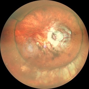
 Buckle intrusion with Retinal detachment
Feb 8 2018 by Manish Nagpal, MD, FRCS (UK), FASRS
Patient operated on 10 years back for a scleral buckling surgery presented with decreased vision and had a superonasal retinal detachment along with intrusion of the scleral buckle.
Photographer: Mehul Prajapati
Condition/keywords: acute retinal detachment, retinal break, scleral buckle
 Loading…