Search results (138 results)
-
 Folded Macula
Oct 13 2025 by Malvika Singh
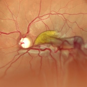
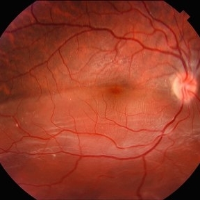
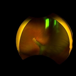
Fundus photograph of a 25 year old male showing retinal detachment with macula off with retinal folds at the macula.
Photographer: Dr Malvika Singh, Retina Foundation, Ahmedabad, India
Imaging device: Mirante SLO/OCT
Condition/keywords: retinal detachment
-
 Idiopathic Choroidal Neovascularization
Sep 30 2025 by César Adrián Gomez Valdivia, MD
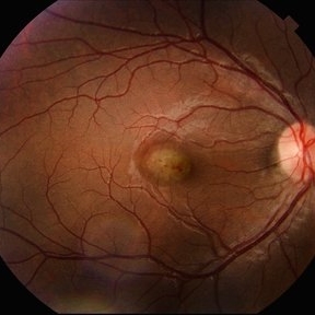
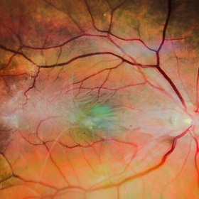
At the foveal area, there is a yellowish-greenish elevated lesion with indistinct borders, corresponding to a subfoveal choroidal neovascular membrane (CNV). There are subtle overlying changes including mild retinal pigment epithelium (RPE) disruption, and small hemorrhagic spots suggesting active leakage. Surrounding the lesion, there are faint retinal folds or striae, likely due to localized subretinal fibrosis or traction.
Photographer: @eyemissu2
Imaging device: TOPCON TRX
Condition/keywords: Idiopathic Choroidal Neovascularization
-
 Hot-Dog Like Choroidal Detachment
Aug 19 2025 by Gustavo Uriel Fonseca Aguirre
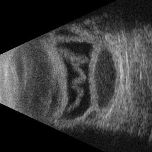
This B-mode transverse ultrasound scan reveals a bullous hemorrhagic choroidal detachment with an associated closed-funnel retinal detachment featuring central retinal folds. The choroidal detachment demonstrates convex, lobular elevation with heterogeneous internal reflectivity due to blood accumulation.
Photographer: Gustavo U. Fonseca Aguirre, Hospital Conde de Valenciana, Ciudad de México
Condition/keywords: hemorrhagic choroidal detachment
-
 Retinal Detachment
Jun 5 2025 by César Adrián Gómez Valdivia, MD
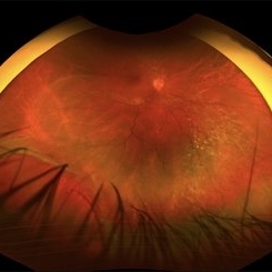
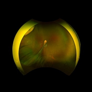
Fundus Photograph of a 19 year-old male patient with a RRD due to a Retinal Dialysis. Subretinal fluid and retinal folding can be appreciated.
Photographer: @eyemissu2
Imaging device: OPTOS
Condition/keywords: retinal detachment
-
 Retinal Detachment
Jun 5 2025 by César Adrián Gómez Valdivia, MD
Fundus Photograph of a 19 year-old male patient with a RRD due to a Retinal Dialysis. Subretinal fluid and retinal folding can be appreciated.
Photographer: @eyemissu2
Imaging device: TOPCON TRC-50DX
Condition/keywords: retinal detachment
-
 Advanced Proliferative Diabetic Retinopathy
Apr 9 2025 by Gustavo Uriel Fonseca Aguirre
B-mode ultrasound of a patient with long-standing poorly controlled diabetes demonstrates characteristic findings of advanced proliferative diabetic retinopathy. The examination reveals moderate vitreous hemorrhage appearing as diffuse hyperechoic opacities throughout the vitreous cavity, along with a posterior hyaloid membrane densely infiltrated by hemorrhagic material, showing irregular thickening and increased reflectivity. A mild subhyaloid hemorrhage is visible as a subtle hyphema-like space anterior to the retinal surface. The study documents a total tractional retinal detachment, evidenced by rigid retinal folds with clear insertion points of vitreous strands, accompanied by a significant subretinal hemorrhage seen as a prominent hyperechoic collection beneath the elevated retina. These findings collectively illustrate the severe vitreoretinal interface pathology characteristic of end-stage diabetic eye disease, with predominant tractional components and distinct echographic stratification of hemorrhagic layers - from anterior vitreous involvement to deeper subretinal blood accumulation.
Photographer: Gustavo U. Fonseca Aguirre, Hospital Conde de Valenciana, Ciudad de México
Condition/keywords: diabetic retinopathy, tractional retinal detachment, Vitreous hemorrhage
-
 Comets in the Eye (Retinopathy of Prematurity)
Apr 8 2025 by KANWALJEET HARJOT MADAN, M.S. (Ophthalmology); FAICO (Vitreous - Retina)
This is the fundus picture of right eye (RE) of a 4 years female child presented with outward deviation of right eye. Her parents also complained of diminution of vision in both eyes. On examination, her best corrected vision in RE was hand movements close to face and was 20/80 in LE. Posterior segment exam revealed presence of macular scar in RE and presence of dry retinal fold with dragging of retinal vessels. LE fundus revealed presence of nasal drag of optic disc. Parents gave history of untreated ROP as an infant. Retinopathy of Prematurity (ROP) is a Vaso proliferative disorder of Retina occurring in premature infants. Advances in neonatal care and ROP treatment has led these babies to live longer with this disease.
Photographer: Dr. Kanwaljeet Harjot Madan, Thind Eye Hospital, Jalandhar City (Punjab) INDIA.
Imaging device: Zeiss Fundus Camera
Condition/keywords: Retinopathy of Prematurity, Vaso proliferative disorder
-
 Comets in the Eye (Retinopathy of Prematurity)
Apr 8 2025 by KANWALJEET HARJOT MADAN, M.S. (Ophthalmology); FAICO (Vitreous - Retina)
This is the fundus picture of right eye (RE) of a 4 years female child presented with outward deviation of right eye. Her parents also complained of diminution of vision in both eyes. On examination, her best corrected vision in RE was hand movements close to face and was 20/80 in LE. Posterior segment exam revealed presence of macular scar in RE and presence of dry retinal fold with dragging of retinal vessels. LE fundus revealed presence of nasal drag of optic disc. Parents gave history of untreated ROP as an infant. Retinopathy of Prematurity (ROP) is a Vaso proliferative disorder of Retina occurring in premature infants. Advances in neonatal care and ROP treatment has led these babies to live longer with this disease.
Photographer: Dr. Kanwaljeet Harjot Madan, Thind Eye Hospital, Jalandhar City (Punjab) INDIA.
Imaging device: Zeiss Fundus Camera
Condition/keywords: Retinopathy of Prematurity
-
 Retinal Fold in Posterior Microphthalmos
Mar 1 2025 by Hemanth Murthy, MBBS, MD, FASRS
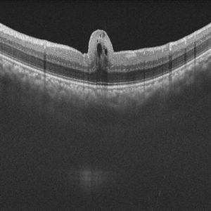
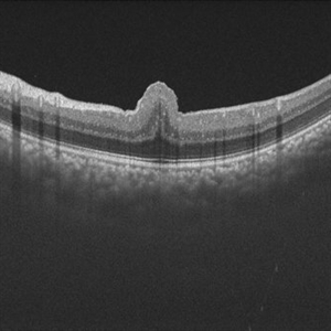
Swept source OCT image of left eye of 34 year male patient with high hypermetropia(+14). BCVA 20/20 in right eye and 20/60 in left eye. Anterior segment was normal. There is loss of foveal pit with omega shaped elevation of inner retinal layers.
Photographer: Mr Veda Vyas
Condition/keywords: posterior microphthalmos
-
 Retinal Fold in Posterior Microphthalmos
Mar 1 2025 by Hemanth Murthy, MBBS, MD, FASRS
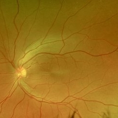
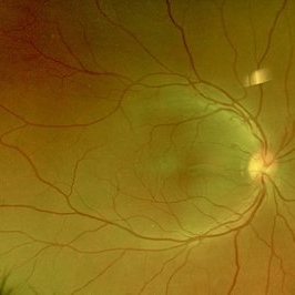
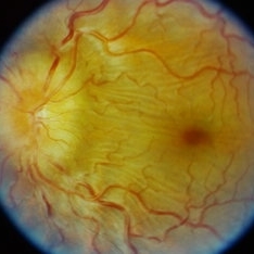
Fundus photo of left eye of 34 year male patient with high hypermetropia(+14). BCVA 20/20 in right eye and 20/60 in left eye. Anterior segment was normal. There is loss of foveal pit with omega shaped elevation of inner retinal layers.
Photographer: Mr Veda Vyas
Condition/keywords: posterior microphthalmos
-
 Retinal Fold in Posterior Microphthalmos
Mar 1 2025 by Hemanth Murthy, MBBS, MD, FASRS
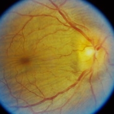
Fundus photo of Right eye of 34 year male patient with high hypermetropia(+14). BCVA 20/20 in right eye and 20/60 in left eye. Anterior segment was normal. There is loss of foveal pit with omega shaped elevation of inner retinal layers.
Photographer: Mr Veda Vyas
Condition/keywords: posterior microphthalmos
-
 Retinal Fold in Posterior Microphthalmos
Mar 1 2025 by Hemanth Murthy, MBBS, MD, FASRS
Swept source OCT image of Right eye of 34 year male patient with high hypermetropia(+14). BCVA 20/20 in right eye and 20/60 in left eye. Anterior segment was normal. There is loss of foveal pit with omega shaped elevation of inner retinal layers.
Photographer: Mr Veda Vyas
Condition/keywords: posterior microphthalmos
-
 A Classic Case of Retinal Ora Serrata Imaging
Jan 16 2025 by yuan duo
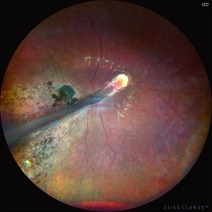
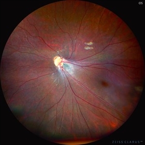
A 5-year-old girl, born full-term with no history of systemic disease, presented with poor vision since early childhood and underwent fundus examination. Anterior segments of both eyes showed no significant abnormalities. Fundus examination revealed retinal folds extending from the optic disc to the temporal peripheral retina, with blood vessels coursing through the folds (A, B). Avascular zones were observed in the peripheral retina, and the ora serrata’s boundaries were clearly visible, displaying dentate processes and bays (C, D). Retinal pigmentation was evident. Genetic testing confirmed the final diagnosis of bilateral Familial Exudative Vitreoretinopathy (FEVR).
Condition/keywords: Retinal Ora Serrata
-
 Familial Exudative Vitreoretinopathy
Jan 16 2025 by yuan duo
A 5-year-old girl, born full-term with no history of systemic disease, presented with poor vision since early childhood and underwent fundus examination. Anterior segments of both eyes showed no significant abnormalities. Fundus examination revealed retinal folds extending from the optic disc to the temporal peripheral retina, with blood vessels coursing through the folds (A, B). Avascular zones were observed in the peripheral retina, and the ora serrata’s boundaries were clearly visible, displaying dentate processes and bays (C, D). Retinal pigmentation was evident. Genetic testing confirmed the final diagnosis of bilateral Familial Exudative Vitreoretinopathy (FEVR).
Condition/keywords: Retinal Ora Serrata
-
 ERM
ERM
Jan 9 2025 by Richa Chaudhary, Mbbs,ms
52 year old male presented with idipathic ERM, with pucker showing, retinal folds. Planned for surgical removal of the same.
Condition/keywords: ERM
-
 Venolymphatic Mass With Disc Edema
Dec 5 2024 by Tejaswita Verma
Fundus picture of a 26 year old male who presented with right eye abaxial proptosis, MRI confirmed venolymphatic mass inferomedial in location located near the optic disc with disc edema , nasal elevation ,retinal folds. Vision was 6/18 . He was planned for intralesional bleomycin injection.
Photographer: DR. TEJASWITA VERMA
Imaging device: MIRANTE
Condition/keywords: disc edema, intraorbital mass, proptosis
-
 Venolymphatic Mass with Retinal Folds
Nov 25 2024 by Tejaswita Verma
Fundus picture of a 26 year old male who presented with right eye abaxial proptosis, MRI confirmed venolymphatic mass inferomedial in location located near the optic disc with disc edema , nasal elevation ,retinal folds. Vision was 6/18 . He was planned for intralesional bleomycin injection.
Photographer: DR. TEJASWITA VERMA
Imaging device: MIRANTE
Condition/keywords: disc edema, intraorbital mass, proptosis, retinal folds
-
 Giant Retinal Tear With Retinal Fold
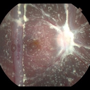
Jun 13 2024 by Anand Temkar
Intraoperative still of a 34 year old male showing giant retinal tear with retinal fold.
Photographer: Dr.Anand Temkar- Retina Foundation, Ahmedabad
Condition/keywords: giant retinal tear, GRT, retinal fold
-
 Fish Hook Eye Trauma
Jun 12 2024 by Miguel Brito, MD, FASRS
Fundus photograph of a 15-year-old boy post cataract aspiration, pars plana vitrectomy, suprachoroidal drainage, and retinal reattachment surgery secondary to traumatic endophthalmitis.
Photographer: Miguel Brito
Condition/keywords: endophthalmitis, PFCL, Retinal detachment under Silicon Oil, retinal fold
-
 Proliferative Vitreoretinopathy
Jun 9 2024 by Marcelo Zas, MD PhD
We present a case of a 20-year-old patient who underwent surgery for congenital cataract when he was born and 20 years after he developed a retinal detachment with proliferative vitreoretinopathy. Proliferative vitreoretinopathy (PVR), a major complication of rhegmatogenous retinal detachment (RRD), is an abnormal process whereby proliferative, contractile cellular membranes form in the vitreous and on both sides of the retina, resulting in tractional retinal detachment with fixed retinal folds. PVR arises in an estimated 5-10% of RRD cases, and therefore represents a major complication of retinal detachment. The best treatment of PVR is its prevention. Clinical factors associated with increased risk of PVR include: • Chronic RRD • 2 o more horseshoe retinal tears and RRD exposing three-disc diameters or more of RPE • RD associated with giant retinal • RD associated with choroidal detachment • Ocular Trauma • RRD associated with vitreous hemorrhage • Aphakia and RRD • Failure of previous surgery or multiple retinal surgeries • Aggressive retinitis, etc.
Photographer: Luciano Scorsetti MD
Condition/keywords: proliferative vitreoretinopathy (PVR)
-
 Retinal Fold
Sep 26 2023 by Mauricio Bayram-Suverza, MD
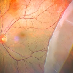
A 38-year-old man underwent vitrectomy in the left eye due to a giant tear in the upper retina. SF6 gas was used as endotamponade. During the post-surgical check-up, it was identified that the patient developed a full-thickness retinal fold due to retinal slippage during fluid-air exchange. As the fold was away from the macular area, it was decided to observe the patient. Three weeks after the surgery, his best-corrected visual acuity was 20/30.
Photographer: Mauricio Bayram-Suverza, Fundación Hospital Nuestra Señora de la Luz
Imaging device: TRC-50DX
Condition/keywords: giant retinal tear, retina surgery complications, Retinal slippage, vitreoretinal surgery
-
 Posterior Scleritis
Sep 12 2023 by Ben Serar
Fundus photograph of LE showing Disc edema with Choroidal folds in a case of Posterior Scleritis
Condition/keywords: chorioretinal folds, disc edema, posterior scleritis
-
 Posterior Scleritis
Sep 12 2023 by Ben Serar
Fundus photograph of RE showing Disc edema with Choroidal folds in a case of Posterior Scleritis.
Condition/keywords: chorioretinal folds, disc edema, Posterior scleritis
 Loading…