Search results (100 results)
-
 Hereditary Retinal Dystrophy
Feb 27 2025 by Kimberly Wakester
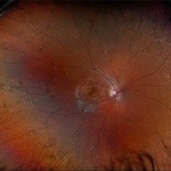
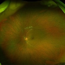
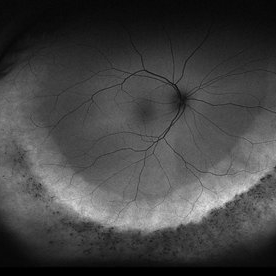
Optomap RGB image of a 7-year-old girl with Hereditary retinal dystrophy. Biological mother is a CHM gene carrier and biological father is diagnosed with RP. Patient had genetic testing and was also confirmed to be a CHM gene carrier and also has the TTC21B gene. There is linear pigmentary changes on clinical exam and fundus photos. Atypical appearance of Retinitis Pigmentosa. Patient will continue follow up care with repeat imaging.
Photographer: Kimberly Wakester, COA
Imaging device: Optos California
Condition/keywords: CHM gene, hereditary retinal dystrophy, linear pigmentary changes
-
 Astrocytic Hamartoma
Feb 27 2025 by Daniel Davis, OCT-C
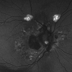
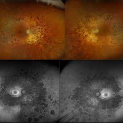
Fundus autofluorescence photo of 55-year-old female with astrocytic hamartoma in association with tuberous sclerosis. No treatment options available, benign. Other findings include; Posterior Vitreous Detachment, Vitreous Hemorrhage, Hereditary Retinal Dystrophy, Vitreous Opacities, Hypertensive Retinopathy.
Photographer: Daniel Davis, OCT-C
Imaging device: Optos California
Condition/keywords: astrocytic hamartoma, fundus autofluorescence (FAF)
-
 Astrocytic Hamartoma
Feb 27 2025 by Daniel Davis, OCT-C
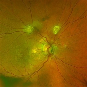
Color fundus photo of 55-year-old female with Astrocytic Hamartoma in association with tuberous sclerosis. No treatment options available, benign. Other findings include; Posterior Vitreous Detachment, Vitreous Hemorrhage, Hereditary Retinal Dystrophy, Vitreous Opacities, Hypertensive Retinopathy.
Photographer: Daniel Davis, OCT-C
Imaging device: Optos California
Condition/keywords: color fundus photograph
-
 Drusen
Jan 2 2025 by Angela Rico
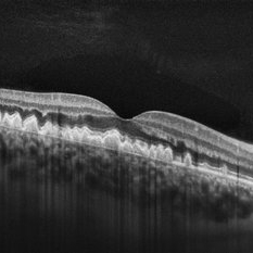
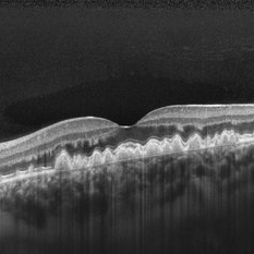
HD 1 line Raster OD in 33 y/o F with h/o Hereditary Retinal Dystrophy OU
Photographer: Angela Rico M.D.
Condition/keywords: drusen
-
 Drusen
Jan 2 2025 by Angela Rico
HD 1 line Raster OS in 33 y/o F with h/o Hereditary Retinal Dystrophy OU
Photographer: Angela Rico M.D.
Condition/keywords: drusen
-
 Asteroid Hyalosis in Retinitis Pigmentosa
Dec 9 2024 by Mauricio Bayram-Suverza, MD
A 54 year-old male patient presented with asteroid hyalosis. Retinal examination revealed the presence of bone spicules, primarily located in the mid-periphery. Genetic testing identified a pathogenic variant in the RHO gene.
Photographer: Mauricio Bayram-Suverza, Casey Eye Institute, OHSU.
Imaging device: Optos California
Condition/keywords: Asteroid hyalosis, retinal dystrophy, Retinitis Pigmentosa, vitreous
-
 Fundus Punctata Albescens
Oct 13 2024 by Brandon I Fram, MD, BS
6 year-old with inherited fleck retinal dystrophy
Condition/keywords: fleck dystrophy, fleck retinopathy, fundus punctata albescens, retinitis punctata albescens
-
 Leber´s Congenital Amaurosis
Sep 6 2024 by Mauricio Bayram-Suverza, MD
13-year-old female patient with severe nyctalopia, photophobia, and reduced peripheral vision. CRB1-related Leber´s Congenital Amaurosis. The ultra-widefield pseudocolor image shows attenuated arterioles and diffuse nummular pigmentation with important atrophy.
Photographer: Mauricio Bayram-Suverza, Casey Eye Institute, OHSU.
Imaging device: Optos California
Condition/keywords: genetic testing, Leber's congenital amaurosis, nyctalopia, retinal dystrophy
-
 Pericentral Retinitis Pigmentosa
Sep 6 2024 by Mauricio Bayram-Suverza, MD
A 65-year-old male patient reports experiencing bilateral blind spots that have gradually intensified over time. Genetic testing was unrevealing. The fundus autofluorescence image shows a hypoautofluorescent ring in the posterior pole, especially nasal to the nerve and along arcades.
Photographer: Mauricio Bayram-Suverza, Casey Eye Institute, OHSU.
Imaging device: Optos California
Condition/keywords: fundus autofluorescence (FAF), inherited retinal disease, nyctalopia, retinal dystrophy, retinitis pigmentosa
-
 Familial Dominant Drusen
Mar 28 2024 by Houda Brarou
Familial Dominant Drusen is a genetically inherited retinal dystrophy and thought to represent an early-onset variant of age related macular degeneration. The gene responsible is EFEMP1 and inherited in autosomal dominant manner with variable expressivity. It is represented with multiple radially elongated small drusen in early stages and in later stages they become larger and more confluent. Geographic atrophy occurs in advanced stages.
Photographer: Houda Braou , Mohammed V military hospital of Rabat
Imaging device: TOPCON DRI OCT Triton Plus
Condition/keywords: FAMILIAL DOMINANT DRUSEN
-
 Usher's Syndrome
Feb 6 2024 by Taylor J Slingsby, MD
54-year-old woman with bilateral, symmetric RPE degeneration. Her vision was 20/20 vision in each eye and she reported no family history of inherited retinal dystrophy. Genetic testing was positive for two pathogenic variants in ARSG, associated with autosomal recessive Usher syndrome. She also had one pathogenic (low penetrance) variant and one variant of unknown significance in ABCA4 as well as one likely pathogenic variant in PDE6A.
Photographer: Taylor Slingsby, MD, Slingsby & Huot Eye Associates, Rapid City, SD
Imaging device: Optos ultra-widefield Autofluorescence retinal imaging
Condition/keywords: inherited retinal disease, retinitis pigmentosa, usher's syndrome
-
 Retinitis Pigmentosa
Nov 7 2023 by Jolee Rodriguez
Bilateral fundus photography and fundus autofluorescence imaging of a 62-year-old male with Retinitis Pigmentosa. Patient reported visual field defects and dark adapting issues. Patient's vision at the time images were taken were sc20/20 of the right eye and sc20/25 of the left eye. Dr. Sutherland determined that based on the patient's lack of family history, the most likely route of inheritance is autosomal recessive.
Photographer: Jolee Rodriguez
Imaging device: Optos California RGB
Condition/keywords: autofluorescence imaging, fundus photography, hereditary retinal dystrophy, Optos, OPTOS CALIFORNIA RGB, retinitis pigmentosa, ultra-wide field imaging, Ultra-wide field retinal imaging, ultra-widefield image
-
 Retinitis Pigmentosa Associated with Asteroid Hyalosis
Jul 21 2023 by Mohammadkarim Johari
Fundus photograph of an 43-year-old lady with pigmentary retinal dystrophy and asteroid hyalosis, also shadow of posterior subcapsular cataract is obvious.
Photographer: Mohammadkarim Johari, Shiraz university of medical science
Condition/keywords: asteroid hyalosis, pigmentary retinal dystrophy, retinitis pigmentosa (RP) dystrophy
-
 Cone-Rod Dystrophy
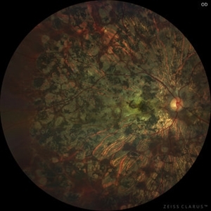
Jul 20 2023 by Harsh Vardhan Singh, MS
52-year-old male with a advanced stage of cone-rod dystrophy
Photographer: Harsh Vardhan Singh, AIIMS, Guwahati
Imaging device: Zeiss Clarus 700
Condition/keywords: cone dystrophy, Cone-Rod Dystrophy, pigmentary retinal dystrophy, retinal dystrophy
-
 Stargardt's Disease
May 5 2023 by Virginia Gebhart
51-year-old male with bilateral central retinal dystrophy consistent with Stargardt disease. No significant progression of central atrophy, and VA has remained stable at 20/150 since 2012
Photographer: Virginia Gebhart, Retina Consultants of Carolina
Imaging device: Topcon TRC 50DX
Condition/keywords: Stargardt disease
-
 Rod Cone dystrophy
Nov 29 2022 by Niloofar Piri, MD
Fundus autofluorescence of the left eye in a 58 yo male with rod cone dystrophy. He presented with night blindness and peripheral vision loss since youth and recent decrease in central vision for the past 10 years. Notice multiple coin shaped hypoautofluorescent pacthes within central 20 degrees which are coalescing centrally. (fundus photo uploaded separately) He has one pathogenic variants of both CEP290 and PRPH2 genes.
Photographer: Sean Kelso, Saint Louis University
Condition/keywords: hereditary retinal degeneration, hereditary retinal dystrophy, rod cone dystrophy
-
 Rod Cone dystrophy
Nov 29 2022 by Niloofar Piri, MD
Fundus photograph of the left eye in a 58 yo male with rod cone dystrophy. He presented with night blindness and peripheral vision loss since youth and recent decrease in central vision for the past 10 years. Notice waxy pallor of the nerve, severe arterial narrowing and chorioretinal atrophy mainly around the arcades as well as posterior pole along with RPE hyperplastic changes and atrophy. RPE atrophy in midperiphery has coin shaped appearance. FAF has characteristic appearance (uploaded separately) He has one pathogenic variants of both CEP290 and PRPH2 genes.
Photographer: Sean Kelso, Saint Louis University
Condition/keywords: hereditary retinal deg, hereditary retinal dystrophy, Rod cone dystrophy
-
 Choroideremia
Sep 21 2022 by Zach Seim
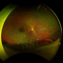
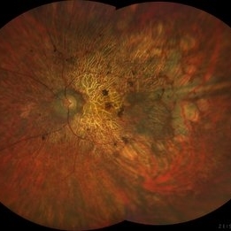
Ultra-widefield fundus photo of a 74 year old male presenting with severe vision loss beginning at age 55. Patient sought a second opinion with our office and was diagnosed with Choroideremia. Patient denies hearing loss, heart problems, balance issues, polydactyly, kidney problems, and dental problems. Patient reports that nobody in the family had blindness. Choroideremia is an X-linked chorioretinal dystrophy characterized by the diffuse, progressive degeneration of the retinal pigment epithelium (RPE), photoreceptors and choriocapillaris. It is caused by a mutation in the CHM gene.
Photographer: Zach Seim
Imaging device: Optos California
Condition/keywords: choroideremia, hereditary choroidal atrophy, hereditary retinal dystrophy, left eye, light perception, low vision, Optos, pseudocolor, ultra-wide field imaging
-
 Choroideremia
Sep 21 2022 by Zach Seim
Ultra-widefield fundus photo of a 74 year old male presenting with severe vision loss beginning at age 55. Patient sought a second opinion with our office and was diagnosed with Choroideremia. Patient denies hearing loss, heart problems, balance issues, polydactyly, kidney problems, and dental problems. Patient reports that nobody in the family had blindness. Choroideremia is an X-linked chorioretinal dystrophy characterized by the diffuse, progressive degeneration of the retinal pigment epithelium (RPE), photoreceptors and choriocapillaris. It is caused by a mutation in the CHM gene.
Photographer: Zach Seim
Imaging device: Optos California
Condition/keywords: choroideremia, hereditary choroidal atrophy, hereditary retinal dystrophy, Optos, pseudocolor, ultra-wide field imaging
-
 Rod cone dystrophy autofluorescence
Sep 19 2022 by Kenneth Fong
34 year old male with colour blindness and loss of visual field
Condition/keywords: retinal dystrophy
-
 Unilateral Macular Coloboma
Jul 29 2021 by Mihir Trivedi
Fundus examination of a 35-year-old man with focal areas of altered retinal pigment epithelium and subretinal yellowish lesion in the foveal area in the right eye. Left eye showed a punched out circumscribed lesion in the center of the macula with thin foveal roof suggestive probably of the internal limiting membrane. Macular coloboma is characterized by a sharply defined, oval or rounded, usually unilateral, atrophic lesions of varying size presenting rudimentary or absent retina, choroid and sclera located at the macula leading to decreased vision in the central area of the fundus. It can be associated with retinal dystrophy in the fellow eye, as was the case in our patient.
Photographer: Priyanshi Kambodi, RNC Eye Hospital, Valsad
Condition/keywords: macular coloboma
-
 Doyne Honeycomb Retinal Dystrophy
Sep 29 2020 by Navneet Mehrotra, DNB
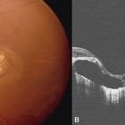
Right eye fundus photograph of a 36-year-old female with decreased vision both eyes for six months. Father also had a similar retinal disorder.
Photographer: Dr Navneet Mehrotra, Retina Care, Ahmedabad
Imaging device: TRC- NW8F
Condition/keywords: Doyne's Honeycomb, familial drusen, Malattia Leventinese
-
 Doyne Honeycomb Retinal Dystrophy
Sep 29 2020 by Navneet Mehrotra, DNB
Left eye fundus photograph of a 36-year-old female with decreased vision both eyes for six months. Father also had a similar retinal disorder.
Photographer: Dr Navneet Mehrotra
Imaging device: TRC- NW8F
Condition/keywords: Doyne's Honeycomb, drusen, Malattia Leventinese
-
 Pigmentary Retinal Dystrophy
May 5 2020 by Olivia Rainey
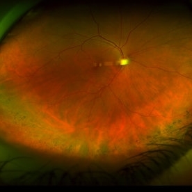
Ultra-widefield pseudocolor image of an 44-year-old male with pigmentary retinal dystrophy affecting both eyes. He presented with decreased night vision for 6 months prior to his appointment. He stated that his recovery time from transitioning from dark to light areas is reduced. He stated that his peripheral vision has never been very good for most of his life. He admits to environmental hearing loss. Patient denies family history of blin. His vision was 20/20 in both eyes. His full field ERG, visual fields were not consistent with RP. Genetic testing with ID Your IRD and annual follow up has been recommended.
Photographer: Olivia Rainey, OCT-C, COA
Imaging device: Optos California
Condition/keywords: inferior retina, Optos, pigmentary retinal dystrophy, pseudocolor, ultra-wide field imaging
-
 Pigmentary Retinal Dystrophy
May 5 2020 by Olivia Rainey
Ultra-widefield fundus autofluorescence image of an 44-year-old male with pigmentary retinal dystrophy affecting both eyes. He presented with decreased night vision for 6 months prior to his appointment. He stated that his recovery time from transitioning from dark to light areas is reduced. He stated that his peripheral vision has never been very good for most of his life. He admits to environmental hearing loss. Patient denies family history of blin. His vision was 20/20 in both eyes. His full field ERG, visual fields were not consistent with RP. Genetic testing with ID Your IRD and annual follow up has been recommended.
Photographer: Olivia Rainey, OCT-C, COA
Imaging device: Optos California
Condition/keywords: fundus autofluorescence (FAF), hyperautofluorescence, hypoautofluorescence, inferior retina, Optos, pigment, ultra-wide field imaging
 Loading…