Search results (50 results)
-
 Eye Finally Got the Ring... But the Retina Was Too Detached to Care
Nov 5 2025 by SHRADDHA RAJ SHRIVASTAVA
Left Eye B-scan ultrasound of a patient with old retinal detachment shows open funnel shaped hyperechoic membranous echoes, with high amplitude spikes on A-scan and a poor after-movement on dynamic B-scan, suggestive of retinal detachment. We can see a round echogenicity in sub-retinal location, with clear contents within, suggestive of a retinal cyst. This B-scan image is indicative of a long-standing chronic retinal detachment with secondary retinal cyst.
Photographer: Dr. Shraddha Raj Shrivastava
Condition/keywords: B scan ultrasound, chronic retinal detachment, OLD RD, open funnel RD, retinal cyst
-
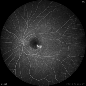
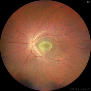
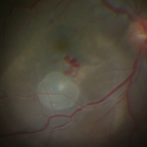
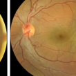
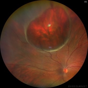 Giant Retinal Cyst
Sep 20 2025 by JORGE SOBERANES
Fundus photograph of a 45-year-old-man with a large cyst on the nasal superior side of the retina. The patient had a history of a pneumatic retinopexy two years ago and the cyst has been there since that.
Photographer: Dr. Jorge Soberanes, Asociación para Evitar la Ceguera en México (APEC), UNAM
Condition/keywords: abnormal retina, pneumatic retinopexy, retinal cyst
-
 Diabetic Macular Edema
Feb 12 2025 by Kimberly Wakester
Horizontal OCT scan of a 63-year-old woman with diabetic macular edema in the right eye. When reviewing the scan, one of the intraretinal cyst (IRC) appears heart shaped. A fun scan to see just a few day's before Valentine's day.
Photographer: Kimberly Wakester, COA
Imaging device: Heidelberg
Condition/keywords: diabetic macular edema, intraretinal cyst
-
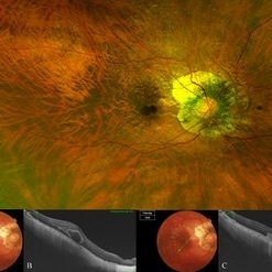
 CSR with Fibrin-FFA
Jan 29 2025 by Vishal Agrawal, MD, FRCS,FACS,FASRS
A 31-year-old female was referred with a diagnosis of subretinal cysticercosis. BCVA was 20/200 OS. OCT showed a large subfoveal bacillary layer detachment (BALAD) without any scolex. FFA revealed a smoke-stack appearance. A final diagnosis of CSR with Fibrin was made and was managed conservatively. BCVA at final visit was 20/20.
Photographer: Dr Ayushi Gupta
Imaging device: Clarus 700
Condition/keywords: central serous chorioretinopathy (CSCR)
-
 CSR with Fibrin
Jan 28 2025 by Vishal Agrawal, MD, FRCS,FACS,FASRS
A 31-year-old female was referred with a diagnosis of subretinal cysticercosis. BCVA was 20/200 OS. OCT showed a large subfoveal bacillary layer detachment (BALAD) without any scolex. FFA revealed a smoke-stack appearance. A final diagnosis of CSR with Fibrin was made and was managed conservatively. BCVA at final visit was 20/20.
Photographer: Dr Ayushi Gupta
Imaging device: Clarus 700
Condition/keywords: central serous chorioretinopathy (CSCR)
-
 Symmetric Retinal Cysts
Aug 22 2024 by Neeket R. Patel, MD
Ultrasonography of a 55-year-old woman with chronic, symmetric retinal cysts.
Condition/keywords: Retinal Cysts
-
 OCT en face of a 360 retinotomy for closed funnel combined retinal detachment
Jan 1 2023 by Malek Yassine, MD
Swept Source OCT en face at deep capillary plexus, shows foveal and parafoveal intraretinal cysts corresponding to macular edema under silicon oil
Imaging device: Topcon Triton DRI-OCT
Condition/keywords: combined retinal detachment, OCT EN FACE
-
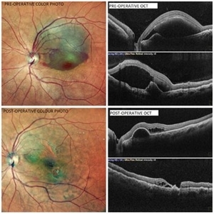 Subretinal Bleed
Jul 12 2022 by Akansha Sharma
A 60 YEAR OLD FEMALE PRESENTED WITH COUNTING FINGERS VISION FROM A SUB-RETINAL HEMORRHAGE AT THE MACULA. OCT SHOWS VARIABLE SUB -RETINAL FLUID. PARS PLANA VITRECTOMY WITH DRAINAGE OF THE SUB-RETINAL BLOOD WAS PERFORMED. POST-OPERATIVE OCT SHOWS NO SUB-RETINAL FLUID WITH VARIABLE OUTER RETINAL CYSTIC CHANGES AND VISUAL ACUITY IMPROVING TO 20/120.
Photographer: Dr. Akansha Sharma-Retina Foundation, Ahmedabad
Condition/keywords: subretinal hemorrhage, subretinal blood
-
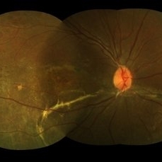
 Chronic retinal detachment changes
Apr 29 2022 by Otakar Dušek, M.D. Ph.D.
Colour fundus photo of 22-year-old woman with bulous retinal detachment number 5-9, old demarcation lines and inferotemporal periheral secondary retinal cyst.
Photographer: Otakar Dušek, Charles University, Prague
Imaging device: Zeiss Clarus
Condition/keywords: chronic retinal detachment, demarcation line, peripheral retinal cyst
-
 Macular fold posterior microphthalmos OS
Apr 24 2022 by Mariam Cernichiaro-Espinosa, MD
Macular OCT of 6-year-old girl with posterior microphthalmos, showing macular fold and one intraretinal cyst OS.
Photographer: Mariam Cernichiaro-Espinosa, Asociación para Evitar la Ceguera, I.A.P. Mexico City, Mexico.
Imaging device: Zeiss Clarus
Condition/keywords: posterior microphthalmos
-
 Macular fold posterior microphthalmos OD
Apr 24 2022 by Mariam Cernichiaro-Espinosa, MD
Macular OCT of 6-year-old girl with posterior microphthalmos, showing macular fold and intraretinal cysts OD.
Photographer: Mariam Cernichiaro-Espinosa, Asociación para Evitar la Ceguera, I.A.P. Mexico City, Mexico.
Imaging device: Zeiss Clarus
Condition/keywords: posterior microphthalmos
-
 Intraretinal cysts
Nov 15 2021 by Marcelo Zas, MD PhD
Left eye from a young patient with a chronic rhegmatogenous retinal detachment presenting intraretinal cysts.
Photographer: Zas Marcelo MD PhD
Condition/keywords: chronic retinal detachment, intraretinal cyst
-
 Intraretinal cysts
Nov 15 2021 by Marcelo Zas, MD PhD
Left eye from a young patient with a chronic rhegmatogenous retinal detachment presenting intraretinal cysts.
Photographer: Zas Marcelo MD PhD
Condition/keywords: chronic retinal detachment, intraretinal cyst
-
 Post Retinal Reattachment Surgery Epiretinal Membrane
Sep 14 2021 by Ogugua Ndubuisi Okonkwo, MD, FRCS (Edin), FASRS
Postoperative optical coherence tomography (OCT) of the right eye in a 65-year-old male who had retinal reattachment surgery for a macular hole retinal detachment. This OCT scan shows epiretinal membrane and intraretinal cystic fluid spaces.
Photographer: Oreoluwa Olabode , Eye Foundation Hospital, Lagos.
Imaging device: Optovue Avanti RTVue.
Condition/keywords: epiretinal membrane (ERM), macular hole retinal detachment, Retinal Reattachment surgery
-
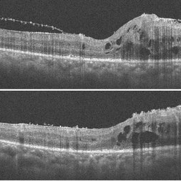
 Bow-Tie Macular Hemorrhage With Cyst- Atypical Presentation of Myopic Choroidal Neovascularization
Mar 26 2021 by RUSHIK PATEL
The image of right eye of 51-year-old lady with high myopia show " Bow-Tie" macular hemorrhage (A). Optical coherence tomography (B) scan passing through hemorrhage showed intraretinal cystic lesion. During the course of intravitreal anti-VEGF injection treatment, the lesion converted into typical myopic choroidal neovascularization (C).
Photographer: Rushik Patel, Netralaya Super Speciality Eye Hospital
Condition/keywords: cyst, macular hemorrhage, myopic choroidal neovascularization (CNV)
-
 Subretinal Cysticercus Cyst -Intraoperative Picture
Mar 5 2021 by Vishal Agrawal, MD, FRCS,FACS,FASRS
24-year-old male presented with DOV in right for the past 1 month. On examination live cyst noted at macula with inflammatory exudation at the posterior pole.
Photographer: Vishal Agrawal MD,FRCS
Imaging device: SONY PMW-10 MD HD
Condition/keywords: cyst, cysticercosis, macula lesion
-
 Macrocyst in the Fovea
Feb 2 2021 by Peter J Belin, MD
36-year-old male with a white cataract and a chronic total retinal detachment for 1 year presented with a recurrent PVR detachment after primary repair 2 weeks prior. This OCT- EDI demonstrates a large retinal cyst through the fovea.
Photographer: Holly Cheshier, CRA, OCT-C, COT
Imaging device: Heidelberg Spectralis
Condition/keywords: chronic retinal detachment, proliferative vitreoretinopathy (PVR), retinal cyst, retinal macrocyst
-
 Spontaneously Attached Retina
Jan 15 2021 by KRISHNENDU NANDI, MS
Fundus photograph of 26-year-old man with BCVA 6/60, N24, showed spontaneous retinal reattachment with multiple retinal cyst at temporal quadrant. Subretinal gliosis resemble a mustache.
Photographer: Krishnendu Nandi, Netralayam Eye Care Centre
Condition/keywords: spontaneous retinal reattachment
-
 Intraretinal Cysts in Chronic Retinal Detachment
Dec 8 2020 by Alice Kim
B-scan ultrasound showing multiple intraretinal cysts in the setting of chronic retinal detachment and proliferative vitreoretinopathy.
Condition/keywords: chronic retinal detachment, intraretinal cyst, proliferative vitreoretinopathy (PVR)
-
 Subretinal Cysticercosis Post Vitrectomy
Sep 10 2020 by Anamika Dwivedi
Fundus photograph of a 22-year-old male, case of subretinal cysticercosis, after PPV and cyst removal showing laser scar at the site of the previous cyst.
Photographer: Dr Anamika Dwivedi
Imaging device: topcon
Condition/keywords: bilateral subretinal cysticercosis
-
 Subretinal Cysticercosis
Sep 10 2020 by Anamika Dwivedi
Fundus photograph of a 22-year-old male, who was referred from physician as a case of headache under evaluation, with a history of headache for 1 month. Fundus showing bilateral subretinal cysticercus cyst with scolex.
Photographer: Dr Anamika Dwivedi
Imaging device: topcon
Condition/keywords: bilateral subretinal cysticercosis
-
 Retinal Cyst
Aug 14 2020 by Noy Ashkenazy, MD, MS
Fundus photograph of a 13-year-old male with a chronic retinal detachment following a penetrating ocular trauma. There is a retinal cyst and proliferative vitreoretinopathy.
Photographer: Giselle DeOliveira
Imaging device: Retcam III
Condition/keywords: chronic retinal detachment, proliferative vitreoretinopathy (PVR), retinal cyst
-
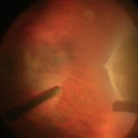
 Old RRD With Retinal Cysts and High Watermark
Apr 10 2020 by Dipak Nag, MBBS, FCPS, MSc, FRF
Intra-operative fundus picture of a 20-year-old boy showing multiple retinal cysts and high watermark in a case of old inferior retinal detachment OD.
Photographer: Dipak
Condition/keywords: high watermark, retinal cyst
-
 Rhegmatogenous Retinal Detachment With Retinal Dialysis and Intraretinal Cyst
Mar 18 2020 by Giridhar Anantharaman, MS
Optos ultra-widefield retinal imaging of the left eye of a 30-year-old lady with rhegmatogenous retinal detachment with inferotemporal retinal dialysis and a large intraretinal cyst.
Photographer: Rakesh PR, Giridhar Eye Institute, Kerala, India
Imaging device: Optos UWF Daytona plus
Condition/keywords: intraretinal cyst, retinal dialysis
-
 Ora Retinal Cyst
 Loading…