Search results (38 results)
-
 Advanced Proliferative Diabetic Retinopathy
Apr 9 2025 by Gustavo Uriel Fonseca Aguirre
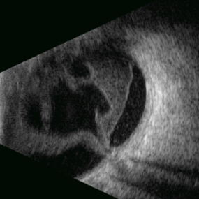
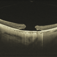
B-mode ultrasound of a patient with long-standing poorly controlled diabetes demonstrates characteristic findings of advanced proliferative diabetic retinopathy. The examination reveals moderate vitreous hemorrhage appearing as diffuse hyperechoic opacities throughout the vitreous cavity, along with a posterior hyaloid membrane densely infiltrated by hemorrhagic material, showing irregular thickening and increased reflectivity. A mild subhyaloid hemorrhage is visible as a subtle hyphema-like space anterior to the retinal surface. The study documents a total tractional retinal detachment, evidenced by rigid retinal folds with clear insertion points of vitreous strands, accompanied by a significant subretinal hemorrhage seen as a prominent hyperechoic collection beneath the elevated retina. These findings collectively illustrate the severe vitreoretinal interface pathology characteristic of end-stage diabetic eye disease, with predominant tractional components and distinct echographic stratification of hemorrhagic layers - from anterior vitreous involvement to deeper subretinal blood accumulation.
Photographer: Gustavo U. Fonseca Aguirre, Hospital Conde de Valenciana, Ciudad de México
Condition/keywords: diabetic retinopathy, tractional retinal detachment, Vitreous hemorrhage
-
 Stag Horn
Apr 8 2025 by Gustavo Uriel Fonseca Aguirre
B-mode ultrasound of a young male patient with bilateral panuveitis (currently under investigation) reveals intense vitritis with islands of preserved vitreous and partial posterior hyaloid detachment, creating a characteristic "stag horn" appearance.
Photographer: Gustavo U. Fonseca Aguirre, Hospital Conde de Valenciana, Ciudad de México
Condition/keywords: Panuveitis
-
 Removal of Hyaloid
Jul 5 2024 by Anjana Mirajkar, MS Ophthalmology
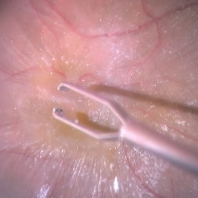
An intra operative still of hyaloid removal done with forceps.
Photographer: Dr. Anjana Mirajkar -Retina Foundation, Ahmedabad
Condition/keywords: posterior hyaloid
-
 Ocular Toxoplasmosis
Jan 5 2024 by Rolando De Leon-Barragan
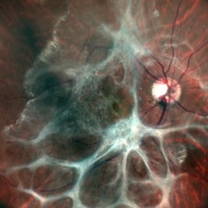
Fundus photograph of a right eye showing a thickened posterior hyaloid in a “spider web” pattern secondary to ocular toxoplasmosis.
Photographer: Rolando De Leon-Barragan, MD.
Imaging device: Nidek Mirante SLO
Condition/keywords: toxoplasmosis
-
PVD induction in a retinal detachment
Oct 24 2022 by Manish Nagpal, MD, FRCS (UK), FASRS
This video highlights the PVD induction technique in a case of retinal detachment with mobile retina, triamcinolone staining allows ease of visualizing the pvd attachment which is gradually removed from the retinal attachment using suction.
Photographer: Manish Nagpal
Condition/keywords: posterior hyaloid, PVD, triamcinolone, video, vitrectomy
-
 Staining-the-vitreous
Jan 10 2022 by Parnian Arjmand, MD, MSc, FRCSC, DABO
This is an example of staining the posterior hyaloid in an eye with no PVD with triamcinolone actinide to assist with inducing a PVD prior to repairing the macular hole.
Condition/keywords: macular hole, PVD, triamcinolone, triescence
-
 Macular Hole
Jan 3 2022 by Thirumalesh Mochi Basavaraj, MD
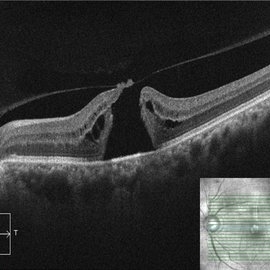
Swept source OCT of a 65-year-old patient with posterior hyaloid separation and a large macular hole with undermined edges.
Photographer: Puttaswamy Narayana Nethralaya Bangalore
Imaging device: Topcon Dri Triton
Condition/keywords: large macular hole, posterior cyclitis hyaloid separation, swept source OCT
-
Macular Hematoma Secondary Valsalva Maneuver
Oct 14 2021 by Islam bechakh
A 32-year-old man, who has presented for 02 months, a macular hematoma secondary to a Valsalva maneuver. He benefited from an attempt to open the hematoma with a Yag laser, but to no avail. We operated on and performed a 23G vitrectomy with posterior vitreous detachment, and discovered an epiretinal membrane which separated the hematoma from the posterior hyaloid. After removal of this membrane and aspiration of red blood cells and fibrin, the macula regained a normal appearance with good functional recovery.
Photographer: Islam Bechakh
Condition/keywords: epiretinal membrane (ERM), ERM, Macular hematoma, Valsalva maneuver
-
 Monocular Proliferative Diabetic Retinopathy
Sep 8 2021 by VERONICA ADRIANA ROMERO- MORALES, MD
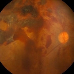
Fundus photograph of a 37-year-old woman with proliferative diabetic retinopathy and subhyaloid hemorrhage, 1 week of evolution.
Photographer: Belgica Copado Andrade
Imaging device: Cobra HD
Condition/keywords: neovascularization (NV), proliferative diabetic retinopathy (PDR), subhyaloid hemorrhage, thickening of the posterior hyaloid, vitreous blood
-
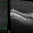
 Thickened Posterior Hyaloid in Stickler Syndrome
Feb 12 2021 by Anfisa Ayalon, MD
SD-OCT of 16-year-old male with Stickler Syndrome. Note a thickened and adherent posterior hyaloid.
Photographer: Anfisa Ayalon, MD., Meir Medical Center, Kfar Saba, Israel.
Condition/keywords: Stickler Syndrome, thickening of the posterior hyaloid
-
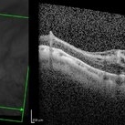
 OCT Evidence of VMT Resulting in Full Thickness Macular Hole
Dec 24 2020 by Deepak Bhojwani, MS
OCT image of a patient (with past history of focal VMT ) progressing to full thickness macular hole. Note the posterior hyaloid attachment over the torn edges of fovea.
Photographer: DEEPAK BHOJWANI
Condition/keywords: full thickness macular hole, optical coherence tomography (OCT), vitreomacular traction (VMT)
-
 Thickening of the Posterior Hyaloid
Dec 12 2020 by Anyssa Montenegro
Color fundus photograph of the right eye of a 36-year-old man showing thickening of the posterior hyaloid associated with an epiretinal membrane due to ocular toxoplasmosis.
Photographer: Anyssa Montenegro, Centro Brasileiro da Visão, Brasília-DF, Brazil
Condition/keywords: epiretinal membrane (ERM), ocular toxoplasmosis, thickening of the posterior hyaloid
-
 Thickening of the Posterior Hyaloid
Dec 12 2020 by Anyssa Montenegro
Color fundus photograph of the right eye of a 36-year-old man showing thickening of the posterior hyaloid associated with an epiretinal membrane due to ocular toxoplasmosis.
Photographer: Anyssa Montenegro, Centro Brasileiro da Visão, Brasília-DF, Brazil
Condition/keywords: epiretinal membrane (ERM), fundus photograph, ocular toxoplasmosis, thickening of the posterior hyaloid
-
 Large Sub-ILM Macular Hemorrhage
Jan 15 2020 by Deepak Bhojwani, MS
Montage image of a young hypertensive gentlemen with huge macular hemorrhage. OCT is pointing the plane of heme to be sub-ILM. High reflective taut membrane (block arrow) The line arrow shows the posterior hyaloid (low reflective band).
Photographer: Deepak Bhojwani, Raghudeep Eye Hospital, Ahmedabad
Imaging device: ZEISS VISUCAM 500
Condition/keywords: hypertension, macula lesion, ruptured macroaneurysm
-
 Branch Retinal Vein Occlusion-Posterior Hyaloid Proliferation
Jun 24 2018 by Narciso F. Atienza, MD, MBA, FASRS, FPCS, FPAO.
62-year-old male patient with sudden loss of vision on the left eye for 3 days. Wider picture of the central retina showing the relationship of the non-perfused fundus to that of the retina.
Photographer: Narciso Atienza, Jr. MD MBA, Legazpi Eye Center, Cardinal Santos Medical Center
Condition/keywords: branch retinal artery occlusion (BRAO)
-
 Branch Retinal Vein Occlusion-Posterior Hyaloid Proliferation
Dec 20 2017 by Narciso F. Atienza, MD, MBA, FASRS, FPCS, FPAO.
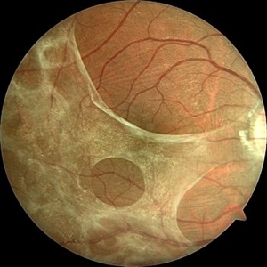
72-year-old female with ischemic branch retinal vein occlusion. Neovascular proliferation on the posterior hyaloid. Background shows ischemic BRVO with florid neovascularization emanating from the disc.
Photographer: Narciso Atienza, Jr. MD, MBA
Imaging device: Topcon
Condition/keywords: branch retinal vein occlusion (BRVO)
-
 Branch Retinal Vein Occlusion-Posterior Hyaloid Proliferation
Dec 20 2017 by Narciso F. Atienza, MD, MBA, FASRS, FPCS, FPAO.
72-year-old female with ischemic branch retinal vein occlusion. Neovascular proliferation on the posterior hyaloid. Background shows ischemic BRVO with florid neovascularization emanating from the disc.
Photographer: Narciso Atienza, Jr. MD, MBA
Imaging device: Topcon
Condition/keywords: branch retinal vein occlusion (BRVO)
-
 Vitreoschisis
Jan 26 2017 by Sara Sella
70-year-old male with high myopia -18D underwent successful surgery of pars plana vitrectomy +posterior hyaloid peel (vitreoschisis) +ELX + SF6.
Photographer: Sara Sella
Condition/keywords: high myopia, vitreoschisis
-
 Vitreoschisis
Jan 26 2017 by Sara Sella
70-year-old male with high myopia -18D underwent successful surgery of pars plana vitrectomy +posterior hyaloid peel (vitreoschisis) +ELX + SF6
Photographer: Sara Sella
Condition/keywords: high myopia, vitreoschisis
-
 Triamcinilone Stained Posterior Hyaloid
Jan 25 2017 by Manish Nagpal, MD, FRCS (UK), FASRS
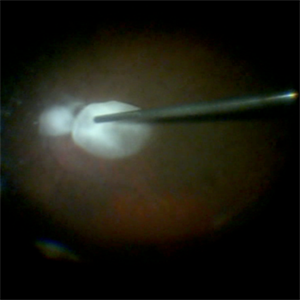
Intraoperative photo in a case of macular hole, the diffuse triamcinolone is removed and only the stained paramacular hyaloid is showing the staining. The cutter is in the process of pulling that hyaloid to release traction.
Photographer: Manish Nagpal
Imaging device: Still captured from a 3 chip HD camera on microscope
Condition/keywords: hyaloid, macular hole, stained posterior
-
 Lutein: A New Dye for Chromovitrectomy
May 16 2014 by Mauricio Maia, MD, PhD
This video shows a new dye for vitreoretinal surgery comprised of soluble lutein/zeaxanthin 1% and brilliant blue 0.025 %. The green dye was deposited on the posterior pole; vigorous dye flushing into the vitreous cavity was unnecessary. The dye indirectly shows the posterior hyaloid by deposition of the golden lutein crystals. The ILM stained greenish-blue; No evidence of toxicity was observed.
Photographer: Mauricio Maia, Federal University of São Paulo
Condition/keywords: chromovitrectomy, internal limiting membrane (ILM) peeling, lutein
 Loading…