Search results (50 results)
-
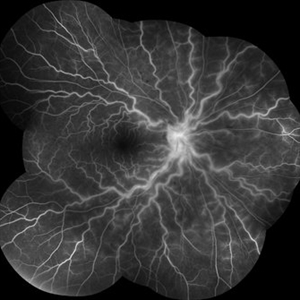 Fluorescein Angiography Papillophlebitis Salauno
Sep 3 2025 by Pablo Angel Garcia Uribe
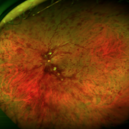
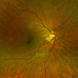
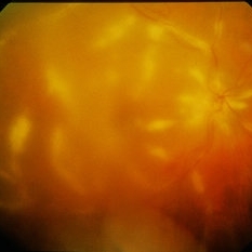
In the arteriovenous phase, fluorescein angiography demonstrated venous engorgement and tortuosity, with relative incompetence of the venous walls leading to mild leakage. Optic disc staining with late leakage was also observed. There was no evidence of significant capillary non-perfusion, and only subtle perivenous leakage was noted. The foveal region remained spared.
Photographer: Optom. Marilyn Alvarez Monroy, Clínica Oftalmológica Salauno
Imaging device: Visucam 524, Carl Zeiss Meditec AG, Jena, Germany
Condition/keywords: FA late phase leakage, retina
-
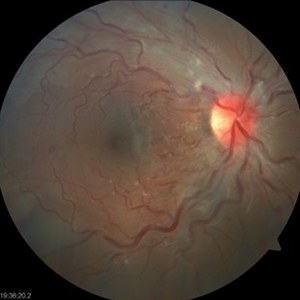 Papillophlebitis Salauno
Sep 3 2025 by Pablo Angel Garcia Uribe
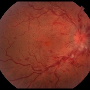
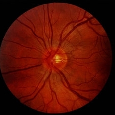
Fundus photograph of a 24-year-old woman, previously healthy, with a history of recreational inhaled cannabis use, presented with a 24-hour history of photopsias and mild decrease in visual acuity, associated with a subtle relative central scotoma in the right eye. On ophthalmic examination, the anterior segment of both eyes was unremarkable. Best-corrected visual acuity was slightly reduced in the right eye and normal in the left. Fundus biomicroscopy of the right eye revealed moderate disc edema with hyperemia and well-defined margins, accompanied by venous engorgement and tortuosity, predominantly affecting the venules. No retinal hemorrhages were observed. Additionally, retinal thickening was noted along the temporal arcades, with apparent foveal sparing. The left eye showed no pathological findings. Based on the patient’s age, the acute onset of symptoms, the fundoscopic features, and the absence of systemic risk factors, the clinical presentation was consistent with papillophlebitis.
Photographer: Clínica Oftalmológica Salauno
Imaging device: Visucam 524, Carl Zeiss Meditec AG, Jena, Germany
Condition/keywords: papillophlebitis
-
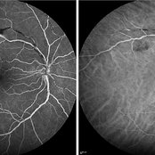 Late FA/ICG at 4 Minutes of Atypical ANCA Associated Retinal Vasculitis
Nov 13 2024 by Deepak Sambhara, MD
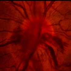
Fluorescein and Indocyanine Green Angiography of a 49-year-old male with high ANA titer, atypical ANCA positivity, who presented to clinic with 1 month of vision loss. Exam revealed anterior chamber cell, mild vitreous cell, sclerotic vessels along arterioles. Late FA/ICG at 4 minutes demonstrates absent arteriole fill with venular periphlebitis.
Photographer: Killian Roberts, Micaela Hertz; Eye Clinic of Wisconsin
Imaging device: Heidelberg Spectralis
Condition/keywords: A-ANCA, autoimmune vasculitis, fluorescein angiogram (FA), indocyanine green (ICG) angiography, retinal vasculitis
-
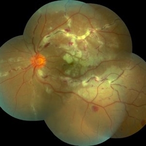 Eales Disease
May 23 2021 by Katia Delalibera Pacheco, MD
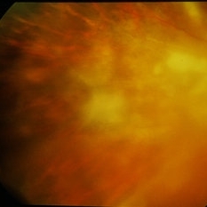
Color fundus photograph of the left eye of a 37-year-old man with Eales disease. Note the peripheral to mid-peripheral periphlebitis in multiple quadrants concurrently. Venous dilation and perivascular exudate can be observed. We can also note the demarcation between perfused and nonperfused retina.
Photographer: CBV- Eye Hospital, Brasilia, DF, Brazil
Condition/keywords: Eales disease, occlusive retinal vasculitis
-
 Eales Disease
May 23 2021 by Katia Delalibera Pacheco, MD
Color fundus photograph of the left eye of a 37-year-old man with Eales disease. Note the peripheral to mid-peripheral periphlebitis in multiple quadrants concurrently. Venous dilation and perivascular exudate can be observed. We can also note the demarcation between perfused and nonperfused retina.
Photographer: CBV- Eye Hospital Brasilia, DF, Brazil
Condition/keywords: Eales disease
-
 Central Retinal Vein Occlusion (CRVO) Associated with Papillophlebitis
Apr 16 2021 by Gabriel Costa Andrade, PhD
Fundus photograph of an 38-year-old woman with Central retinal vein occlusion (CRVO) associated with papillophlebitis.
Photographer: Dr Gabriel Andrade
Condition/keywords: central retinal vein occlusion (CRVO), ischemic CRVO
-
 CRVO with papillophlebitis
Oct 22 2020 by Gabriel Costa Andrade, PhD
Fundus photograph of an 43-year-old man with CRVO and papillophlebitis.
Photographer: Gabriel Andrade
Condition/keywords: central retinal vein occlusion (CRVO)
-
 Papillophlebitis
Dec 23 2019 by Stephanie Burke
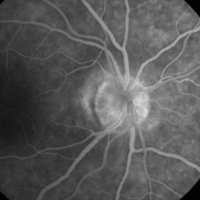
Fluorescein angiogram, mid-phase image of a 46-year-old woman with papillophlebitis.
Photographer: Stephanie Burke, CRA, OCT-C
Imaging device: Zeiss 450 Plus
Condition/keywords: optic disc edema, papillophlebitis
-
 Slide 9-90
Feb 26 2019 by Lancaster Course in Ophthalmology
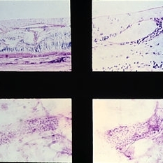
lrvine-Gass syndrome. Note presence of cystoid macular edema (upper left), retinal phlebitis in routine section (upper right), and trypsin digestion (lower views).
Condition/keywords: cystoid macular edema (CME), lrvine-Gass syndrome, phlebitis
-
 Roth Spots - Bacterial Endocarditis
Dec 3 2017 by John S. King, MD
Initial presentation; 29-year-old white female denied ivdu p/c with acute scotoma due to the sub-ILM foveal heme. She did have some roth spots in both eyes. There was a focal area of periphlebitis just superior to the fovea OD. Work up for roth spots and retinal vasculitis initiated. She did have a low grade fever that she attributed to a urinary tract infection being treated by her PCP.
Imaging device: Optos
Condition/keywords: sub-inner limiting membrane hemorrhage, white centered retinal hemorrhage (Roth Spot)
-
 Idiopathic Papillophlebitis- Fluorescein Angiography
Apr 5 2017 by Linda A Cernichiaro- Espinosa, MD
18-year-old otherwise healthy female with sudden visual loss on the left eye.
Photographer: Linda A Cernichiaro MD
Imaging device: Optos Daytona
Condition/keywords: cystoid macular edema (CME), papilledema, venous tortuosity
-
 Papillophlebitis
Jan 5 2015 by H. Michael Lambert, MD
Acute appearance with optic nerve hemorrhage and congestion.
Condition/keywords: papillophlebitis
-
 Papillophlebitis
Jan 5 2015 by H. Michael Lambert, MD
Resolving optic nerve hemorrhage and congestion, 6 weeks.
Condition/keywords: papillophlebitis
-
 Papillophlebitis
Jan 5 2015 by H. Michael Lambert, MD
Acute appearance with optic nerve hemorrhage and congestion.
Condition/keywords: papillophlebitis
-
 Papillophlebitis
-
 Papillophlebitis
Jan 5 2015 by H. Michael Lambert, MD
Acute appearance with optic nerve hemorrhage and congestion.
Condition/keywords: papillophlebitis
-
 Papillophlebitis
Jan 5 2015 by H. Michael Lambert, MD
Resolving optic nerve hemorrhage and congestion, 6 weeks.
Condition/keywords: papillophlebitis
-
 CMV Retinitis / Amazing Periphlebitis
Dec 9 2014 by David Callanan, MD
CMV retinitis / amazing periphlebitis.
Condition/keywords: CMV retinitis, periphlebitis
-
 CMV Retinitis / Amazing Periphlebitis
Dec 9 2014 by David Callanan, MD
CMV retinitis / amazing periphlebitis.
Condition/keywords: CMV retinitis, periphlebitis
-
 CMV Retinitis / Amazing Periphlebitis
Dec 9 2014 by David Callanan, MD
CMV retinitis / amazing periphlebitis.
Condition/keywords: CMV retinitis, periphlebitis
 Loading…