 Initializing download.
-
 By Katia Delalibera Pacheco, MD
- Uploaded on May 23, 2021.
- Last modified by Caroline Bozell on May 25, 2021.
- Rating
- Appears in
- Miscellaneous
- Condition/keywords
- Eales disease, occlusive retinal vasculitis
- Photographer
- CBV- Eye Hospital, Brasilia, DF, Brazil
- Imaging device
- Fundus camera
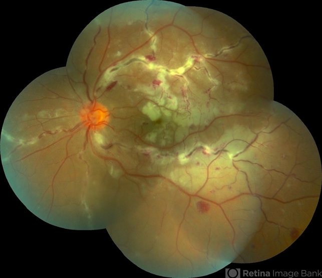
- Description
- Color fundus photograph of the left eye of a 37-year-old man with Eales disease. Note the peripheral to mid-peripheral periphlebitis in multiple quadrants concurrently. Venous dilation and perivascular exudate can be observed. We can also note the demarcation between perfused and nonperfused retina.


