Search results (98 results)
-
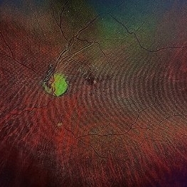
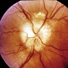
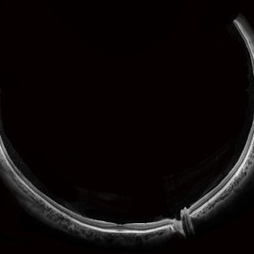
 Venous Loop
Feb 20 2024 by Soobien Lee
A 77-year-old male with a history of bilateral optic neuropathy from bilateral optic nerve sheath meningiomas S/P radiation/proton-beam therapies. Presented with radiation retinopathy OS and a known venous loop OS.
Photographer: Gavin Bragdon, Elman Retina Group
Imaging device: Optos Ultra-Widefield Fluorescein Angiography
Condition/keywords: fluorescein angiogram (FA), Optos, radiation retinopathy, retinal vascular disease, venous loop
-
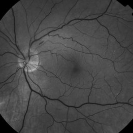
 Venous Loop
Feb 20 2024 by Soobien Lee
A 77-year-old male with a history of bilateral optic neuropathy from bilateral optic nerve sheath meningiomas S/P radiation/proton-beam therapies. Presented with radiation retinopathy OS and a known venous loop OS.
Photographer: Gavin Bragdon, Elman Retina Group
Imaging device: Optos Ultra-Widefield Imaging
Condition/keywords: Optos, OPTOS CALIFORNIA, radiation retinopathy, retinal vascular disease, venous loop
-
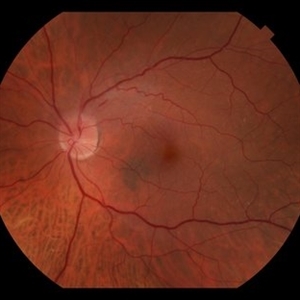
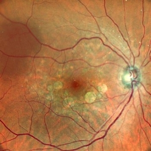 HEREDITARY MACULAR DEGENERATION WITH ETHAMBUTOL TOXICITY
Nov 1 2022 by Akansha Sharma
COLOUR FUNDUS PHOTOGRAPH OF A 45 YEAR OLD MALE WITH HEREDITARY MACULAR DEGENERATION WITH ETHAMBUTOL TOXICITY
Photographer: Dr. Akansha Sharma-Retina Foundation, Ahmedabad
Condition/keywords: drug toxicity, hereditary retinal degeneration, toxic optic neuropathy
-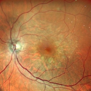
 Leukemic optic neuropathy
Oct 28 2022 by pedro fernandes souza neto
Fundus photograph of an 18-year-old woman with Leukemic optic neuropathy.
Photographer: Pedro Fernandes, Universidade Federal da Bahia
Condition/keywords: Leukemic optic neuropathy
-
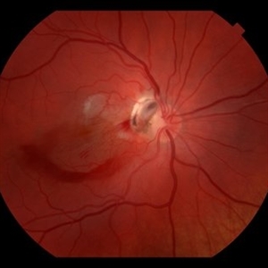
 Pseudo Foster Kennedy Syndrome
Oct 13 2022 by Aditya S Kelkar, MS, FRCS, FASRS,FRCOphth
Colour fundus photograph of a 44-year-old man showing bilateral small discs with optic atrophy on the right eye and disc edema on the left eye resulting from consecutive NAAION in both eyes.
Photographer: Dr Sukanya Mondal, National Institute of Ophthalmology, Pune. India
Imaging device: Zeiss Clarus 500
Condition/keywords: ischemic optic neuropathy, optic atrophy, optic disc edema
-
 Leukemic optic nerve infiltration
Mar 31 2022 by Franco Benvenuto, MD
Fundus photograph of a 21-year-old otherwise healthy male presenting with acute onset of blurry vision in his both eyes for one week. Acute promyelocytic leukemia was confirmed by bone marrow study. The fundus exam showed bilateral optic nerve infiltration: Leukemic optic neuropathy.
Photographer: Franco Benvenuto, Universidad de Buenos Aires, Argentina; Universidad de Guadalajara, México
Condition/keywords: acute leukemia, infiltration of the optic nerve
-
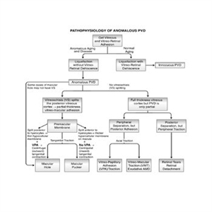 Pathophysiology of Anomalous PVD
Sep 1 2020 by J. Sebag, MD, FACS, FRCOphth, FARVO
This unifying concept of vitreo-retinopathies hypothesizes that the pathogenesis of several vitreoretinal diseases that were previously considered very disparate, are actually all manifestations of the same underlying pathophysiology – anomalous PVD. Note that vitreo-papillary adhesion (VPA) and traction can cause primary optic neuropathy, but might also play a role in facilitating/promoting cell migration and proliferation during pathologic neovascularization of the optic disc. Further, VPA seems to alter the vector of tangential forces exerted by a membrane, in some cases full-thickness posterior vitreous cortex and in some cases the outer layer of the posterior vitreous cortex left attached to the macula after vitreoschisis. While not all cases of macular holes have vitreoschisis, they feature vitreomacular adhesion and traction almost always with VPA. [From Sebag J: Anomalous PVD – a unifying concept in vitreo-retinal diseases. Graefe’s Arch Clin Exp Ophthalmol 2004;242:690-8 and Sebag J, Niemeyer M, Koss M: Anomalous PVD and vitreoschisis. In: Vitreous – in Health & Disease (J. Sebag, ed.) Springer, New York, 2014, pg. 252; image © Springer Nature, reprinted with permission]
Condition/keywords: pathology, peripheral vascular disease (PVD)
-
 Traumatic Giant Retinal Tear Associated Retinal Detachment
Nov 9 2019 by Luis J Haddock, MD
This wide field fundus photograph of the left eye shows a traumatic giant retinal tear associated with total retinal detachment. The image shows the torn superior retina folded over the macula with the underside of the retina visible. There is associated peripheral choroidal detachment due to hypotony from giant retinal tear. This patient has history of spondyloepithelial dysplasia with dwarfism and presented with vision loss after a recent blunt trauma with elbow to the eye.
Imaging device: Optos
Condition/keywords: giant retinal tear, traumatic optic neuropathy
-
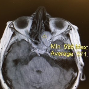
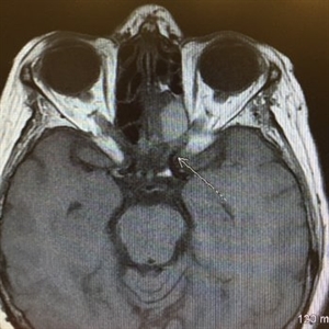 Acute Compressive Optic Neuropathy
Jun 1 2019 by John S. King, MD
84-year-old white female with acute loss of vision in the left eye one day ago was sent here after going to the ED per primary eye provider. She described vision loss as a grey curtain that became total darkness. She had left sided temporal tenderness and some left sided neck pain. In the ED the cardiac work-up was u/r, the ESR and CRP were normal, and the CTH showed some non-specific opacification in the L ethmoid sinus. Acuity was HM OS with RAPD, normal EOMs, no proptosis or ptosis, posteriorly no SVPs were noted; the optic discs were pink and flat; no emboli or retinal whitening present; some bear tracks located nasally (see photo). She was referred to Dr. Doyle, who ordered an MRI, which showed a large mucocele with bony erosion into the left orbit, along with some ON enhancement possibly from compression (see images). She was operated that night and later recovered to 20/40 in that eye with a residual, inferior arcuate scotoma.
Condition/keywords: bear tracks, optic neuropathy
-
 Acute Optic Neuropathy Due to Large Mucocele
Jun 1 2019 by John S. King, MD
84-year-old white female with acute loss of vision in the left eye one day ago was sent here after going to the ED per primary eye provider. She described vision loss as a grey curtain that became total darkness. She had left sided temporal tenderness and some left sided neck pain. In the ED the cardiac work-up was u/r, the ESR and CRP were normal, and the CTH showed some non-specific opacification in the L ethmoid sinus. Acuity was HM OS with RAPD, normal EOMs, no proptosis or ptosis, posteriorly no SVPs were noted; the optic discs were pink and flat; no emboli or retinal whitening present; some bear tracks located nasally (see photo). She was referred to Dr. Doyle, who ordered an MRI, which showed a large mucocele with bony erosion into the left orbit, along with some ON enhancement possibly from compression (see Images). She was operated that night and later recovered to 20/40 in that eye with a residual, inferior arcuate scotoma.
Condition/keywords: bear tracks, optic neuropathy
-
 Acute Optic Neuropathy Due to Large Mucocele (Incidental Bear Tracks)
Jun 1 2019 by John S. King, MD
84-year-old white female with acute loss of vision in the left eye one day ago was sent here after going to the ED per primary eye provider. She described vision loss as a grey curtain that became total darkness. She had left sided temporal tenderness and some left sided neck pain. In the ED the cardiac work-up was u/r, the ESR and CRP were normal, and the CTH showed some non-specific opacification in the L ethmoid sinus. Acuity was HM OS with RAPD, normal EOMs, no proptosis or ptosis, posteriorly no SVPs were noted; the optic discs were pink and flat; no emboli or retinal whitening present; some bear tracks located nasally (see photo). She was referred to Dr. Doyle, who ordered an MRI, which showed a large mucocele with bony erosion into the left orbit, along with some ON enhancement possibly from compression (see images). She was operated that night and later recovered to 20/40 in that eye with a residual, inferior arcuate scotoma.
Photographer: Karin Aletter
Imaging device: Topcon 50
Condition/keywords: bear tracks, optic neuropathy
-
 Radiation Retinopathy
Apr 2 2019 by Gary R. Cook, MD, FACS
White male with radiation retinopathy OS; he also had glaucoma and radiation-induced optic atrophy present in this eye
Condition/keywords: radiation optic neuropathy, radiation retinopathy, retinal hemorrhage
-
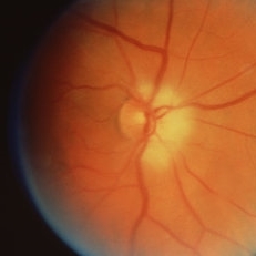 Anterior Ischemic Optic Neuropathy
Mar 26 2019 by Gary R. Cook, MD, FACS
Anterior ischemic optic neuropathy OD.
Condition/keywords: anterior ischemic optic neuropathy
-
 Slide 11-24
Feb 26 2019 by Lancaster Course in Ophthalmology
Atherosclerosis of a posterior ciliary artery in a patient with clinical signs of ischemic optic neuropathy (Verhoeff-Van Gieson stain xllO). (Scheie Eye Institute, No. 5368.)
Condition/keywords: atherosclerosis, ciliary
-
 Slide 11-23
Feb 26 2019 by Lancaster Course in Ophthalmology
Ischemic optic neuropathy. In the same eye, 3 years later, the patient developed edema of the lower pole of the disk, together with a superior altitudinal field defect.
Condition/keywords: ischemic optic neuropathy
-
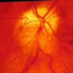
 Slide 11-22
Feb 26 2019 by Lancaster Course in Ophthalmology
Ischemic optic neuropathy. Sudden onset of segmental edema of the upper pole of the disk and adjacent retina accompanied by an inferior altitudinal field defect.
Condition/keywords: ischemic optic neuropathy
-
 NAION, ERM
Sep 7 2018 by John S. King, MD
70-year-old white male with background history of fovea involving RD repaired with PPV 5 months ago, and history of HTN. Some recent photopsias, mild scotoma. Focal ERM superiorly and swollen superior part of the optic disc.
Photographer: Macey
Imaging device: Topcon
Condition/keywords: drusen, epiretinal membrane (ERM), ischemic optic neuropathy
-
 NAION, ERM
Sep 7 2018 by John S. King, MD
70-year-old white male with background history of fovea involving RD repaired with PPV 5 months ago, and history of HTN. Some recent photopsias, mild scotoma. Focal ERM superiorly and swollen superior part of the optic disc.
Photographer: Macey
Imaging device: Topcan
Condition/keywords: drusen, epiretinal membrane (ERM), ischemic optic neuropathy
-
 Partial Optic Disc Avulsion with Optic Disc Pit
Jul 1 2018 by John S. King, MD
16-year-old with acute loss of vision after blunt finger injury to eye while playing football. Five days post-injury. Vision HM. Decreasing heme and retinal whitening.
Imaging device: Optos
Condition/keywords: traumatic optic neuropathy
-
 Partial Optic Disc Avulsion with Optic Disc Pit
Jul 1 2018 by John S. King, MD
16-year-old with acute loss of vision after blunt finger injury to eye while playing football. He was seen in ED and this is the appearance the next day. Vitreous heme, subhyaloid heme,
Condition/keywords: traumatic optic neuropathy
-
 Partial Optic Disc Avulsion with Optic Disc Pit
Jul 1 2018 by John S. King, MD
16-year-old with acute loss of vision after blunt finger injury to eye while playing football. This photo is three weeks post-injury. Vision HM.
Photographer: Maisee Yang
Imaging device: Topcon
Condition/keywords: epiretinal membrane (ERM), optic disc pit, optic nerve head avulsion, traumatic optic neuropathy
-
 Partial Optic Disc Avulsion with Optic Disc Pit
Jul 1 2018 by John S. King, MD
16-year-old with acute loss of vision after blunt finger injury to eye while playing football. This photo is three weeks post-injury. Vision HM. Retinal striae with subhyaloid heme. Decreased retinal whitening. Peripapillary heme clearing, and temporal optic disc avulsion with optic disc pit can be seen.
Photographer: Maisee Yang
Imaging device: Topcon
Condition/keywords: epiretinal membrane (ERM), optic nerve head avulsion, optic nerve pit, traumatic optic neuropathy
-
 Wide-Field-OCT-montage
Jan 8 2018 by Netan Choudhry, MD, FRCS(C) FASRS
This is an SD-OCT montage image of a 55 year old male with optic neuropathy representing a wide-field OCT spanning 130 degrees.
Photographer: John Golding, Vitreous Retina Macula Specialists of Toronto
Imaging device: Heidelberg Spectralis OCT system
Condition/keywords: wide angle imaging
-
 Traumatic Optic Nerve Avulsion
Jul 16 2015 by Mehul A Shah
A 21-year-old male presented to outdoor with history of blunt trauma and loss of vision on examination we found anterior segment to be normal and posterior segment had this picture.
Photographer: Mehul Shah, Drashti Netralaya
Imaging device: FF450 plus zeiss
Condition/keywords: blunt trauma, optic neuropathy
 Loading…