Search results (883 results)
-
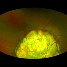
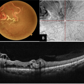
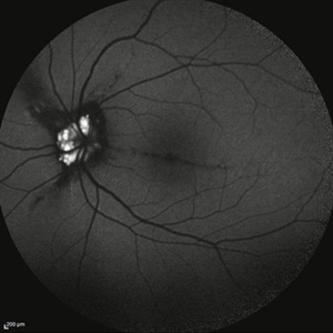
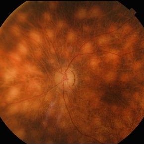 Birdshot Retinochoroidopathy
Jun 18 2025 by César Adrián Gómez Valdivia, MD
Fundus photograph of a 86 YO female patient diagnosed with Birdshot Retinochoroidopathy. Characteristically multifocal cream-colored or yellow-orange, oval or round lesions that emerge from around the optic nerve can be appreciated.
Photographer: @eyemissu2
Imaging device: TOPCON TRC-50DX
Condition/keywords: Birdshot Retinochoroidopathy
-
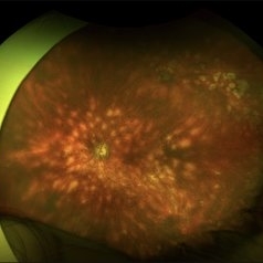 Birdshot Retinochoroidopathy
Jun 18 2025 by César Adrián Gómez Valdivia, MD
Fundus photograph of a 86 YO female patient diagnosed with Birdshot Retinochoroidopathy. Characteristically multifocal cream-colored or yellow-orange, oval or round lesions that emerge from around the optic nerve can be appreciated.
Photographer: @eyemissu2
Imaging device: California ICG OPTOS
Condition/keywords: Birdshot Retinochoroidopathy
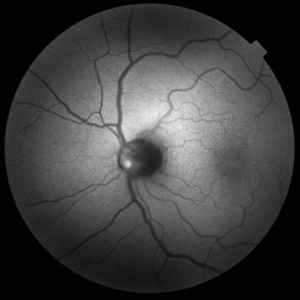
-
Central Retinal Artery Pulsations
May 27 2025 by Malvika Singh
Fundus video showing pulsations of the central retinal artery at the excavated optic nerve head.
Condition/keywords: Central retinal artery, Excavated Disc, Fundus Video
-
 Macular Star
May 27 2025 by César Adrián Gómez Valdivia, MD
Macular Star found in a 31 year-old male patient with suspected Cat Scratch Disease. Typical intraocular presentations include neuroretinitis with optic nerve edema, macular star formation, and discrete white retinal or choroidal lesions. Findings were unilateral.
Photographer: @eyemissu2
Imaging device: TOPCON TRC-50DX
Condition/keywords: macular star
-
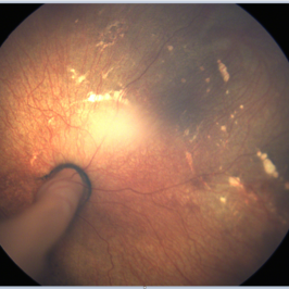
 Optic Nerve Metastasis
May 26 2025 by César Adrián Gómez Valdivia, MD
Fundus photograph of a 62 year-old woman with breast cancer history presented to the ER with decreased visual acuity. Optic nerve metastasis were found. Findings were bilateral.
Photographer: @eyemissu2
Imaging device: California ICG OPTOS
Condition/keywords: nerve, Optic, retinal metastasis
-
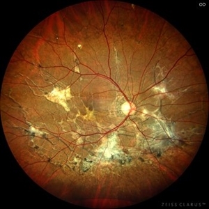
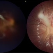
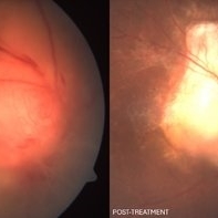 Optic Disc Granuloma
May 7 2025 by Aayesha - Khanum, MBBS. D.N.B
A 45-year-old male presented with diminished vision for one month. His Mantoux test was negative, but as steroids worsened the condition, quantiferon TB was advised and it returned positive. He was started on anti-tuberculosis treatment (ATT). Oral steroids were reintroduced after one week of ATT. Optic disc granulomas can arise from direct invasion of the optic nerve or may represent hypersensitivity reaction to tuberculous antigens. The pathogenesis involves infiltration of immune cells, leading to formation of a granulomatous structure that disrupts normal architecture and function of the optic disc. Steroids with ATT facilitated regression of granulomatous lesion.
Photographer: Ms. Krishna Jeyanthi
Imaging device: Zeiss Clarus 500
Condition/keywords: Tuberculosis
-
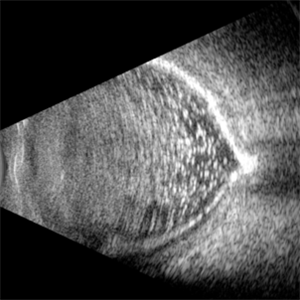
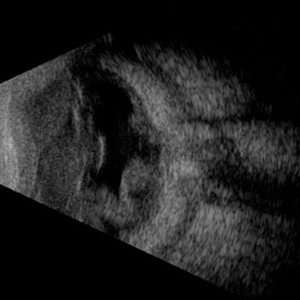 Traumatic T
May 5 2025 by Gustavo Uriel Fonseca Aguirre
This B-mode axial ultrasound scan reveals vitreous hemorrhage, a folded retinal detachment, and sub-Tenon’s fluid extending into the optic nerve sheath, forming the characteristic 'T-sign.' These findings are consistent with severe posterior segment trauma secondary to blunt ocular injury.
Photographer: Gustavo U. Fonseca Aguirre, Hospital Conde de Valenciana, Ciudad de México
Condition/keywords: blunt trauma, retinal detachment, T sign
-
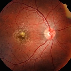
 Optic Nerve Melanocytoma
May 4 2025 by KANWALJEET HARJOT MADAN, M.S. (Ophthalmology); FAICO (Vitreous - Retina)
This is a fundus picture of a young 42-year male who visited for a routine eye exam. His BCVA was 20/20 in both eyes. Anterior segment examination was normal. His left eye showed grey-black pigmentation at the infero-nasal margin of the optic disc. Fundus of the right eye was normal. The patient was diagnosed to have optic disc melanocytoma on multimodal imaging and was advised regular follow-up. Optic nerve melanocytoma is typically a benign tumor made up of melanocytes and melanin. It can grow, but rarely transforms into a malignancy. Patients with Optic Nerve Melanocytoma should be periodically examined for evidence of growth, loss of visual field and optic nerve compression.
Photographer: Dr. Kanwaljeet Harjot Madan, Thind Eye Hospital, Jalandhar City (Punjab) INDIA.
Imaging device: Zeiss Fundus Camera
Condition/keywords: melanocytoma, melanoma, optic nerve
-
 Central Posterior Granuloma
Apr 18 2025 by Chellarani Kumarasamy, MD
Fundus photo showed central posterior granuloma on the optic nerve
Condition/keywords: toxocara granuloma
-
 Myelinated Nerve Fibers
Apr 18 2025 by DR Rohit Gupta
The **myelinated nerve fibers of the optic disc** (also known as **medullated nerve fibers**) are retinal nerve fibers that retain their myelin sheath as they pass through the optic nerve head. Normally, retinal nerve fibers are unmyelinated to allow for light transparency, but in some cases, myelination extends anteriorly into the retina, appearing as a striking white, feathery patch on the optic disc or peripapillary retina. ### **Key Features:** 1. **Appearance:** - Dense, white, striated patches with feathery edges. - Typically located at the superior or inferior pole of the optic disc. - May obscure retinal vessels underneath. 2. **Clinical Significance:** - Usually **benign** and asymptomatic. - **Congenital** (present at birth or early childhood). - Rarely associated with **visual field defects** (e.g., scotomas corresponding to the area of myelination). - Occasionally linked with **high myopia** or **amblyopia** if extensive. 3. **Pathophysiology:** - Failure of oligodendrocytes or Schwann cells to stop myelination at the lamina cribrosa. - Normally, myelination stops at the optic nerve head, but in this condition, it extends into the retina. 4. **Diagnosis:** - **Fundoscopy:** Classic white, feathery appearance. - **Optical Coherence Tomography (OCT):** Shows thickened retinal nerve fiber layer (RNFL). - **Visual Field Testing:** May detect defects if large. 5. **Differential Diagnosis:** - Optic disc edema - Cotton wool spots - Retinoblastoma (rarely, but must be ruled out in children) 6. **Management:** - No treatment required if asymptomatic. - Monitor for amblyopia in children. - Rare cases with significant visual impairment may need further evaluation. ### **Fun Fact:** Myelinated nerve fibers are seen in **~0.5-1%** of the population and are usually an incidental finding.
Photographer: Dr Rohit gupta
Imaging device: Samsung S21
Condition/keywords: Medulated Nerve fibre, Medullated Nerve fibres, myelinated nerve fibers, Myelinated Nerve Fibres, optic disc drusen
-
 Calcification of the Retina
Apr 7 2025 by Gustavo Uriel Fonseca Aguirre
B-mode ultrasound of a vitrectomized eye reveals emulsified silicone oil in the vitreous cavity, retinal detachment, and calcification of the retina and optic nerve head.
Photographer: Gustavo U. Fonseca Aguirre, Hospital Conde de Valenciana, Ciudad de México
Condition/keywords: calcification, Retina detachment, vitrectomy
-
 Shunt Vessels
Apr 1 2025 by Korey Starkey
62-year-old patient presented with stable CRVO in the left eye. FA performed that day shows delayed AV transit is present, this has compensated with shunt vessels at the disc. However there is no evidence of active leakage. OS vision 20/25.
Photographer: Korey Starkey
Imaging device: Topcon
Condition/keywords: central retinal vein occlusion (CRVO), fundus photograph, optic nerve, shunts vessels, Topcon
-
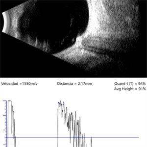 Melanocytoma
Mar 25 2025 by Gustavo Uriel Fonseca Aguirre
Longitudinal B-scan echogram shows mildly elevated lesion overlying surface of optic nerve. A-scan shows regular internal structure and high reflectivity of lesion.
Photographer: Gustavo U. Fonseca Aguirre, Hospital Conde de Valenciana, Ciudad de México
Condition/keywords: Melanocytoma
-
 Suspicious Nevus
Jan 15 2025 by Virginia Gebhart
14 year female with suspicious nevus located adjacent to the optic nerve. Questionable orange pigment present and worsening SRF compared to previous photos/OCT. RPE atrophy also present from previous fluid. No elevation. Will continue observation. BCVA 20/25
Photographer: Virginia Gebhart, Retina Consultants of Carolina
Imaging device: Topcon 50DX
Condition/keywords: choroidal nevus, nevus
-
 Coloboma
Dec 12 2024 by DANNA ALEXANDRA ALVIRDE AYALA
Ultra-wide field image of a 30-year-old man with coloboma.
Photographer: Danna Alexandra Alvirde-Ayala, Hospital Militar de Especialidades Oftalmológicas, Ciudad de México, México
Imaging device: Optos ultra-widefield
Condition/keywords: coloboma, optic nerve, retina
-
 Persistent Fetal Vasculature
Oct 10 2024 by Philip Conkling, MD
Fundus photograph of infant with persistent fetal vasculature demonstrating large stalk emanating from the optic nerve head.
Condition/keywords: persistent fetal vasculature (PFV), persistent hyperplastic primary vitreous (PHPV)
-
 Optic Nerve Head Avulsion
Sep 24 2024 by Gustavo Uriel Fonseca Aguirre
A 14-year-old male with a history of blunt ocular trauma in the right eye presented partial avulsion of the optic nerve head and submacular hemorrhage that was managed with neumatic displacement.
Photographer: Gustavo U. Fonseca Aguirre, Fundación Hospital Nuestra Señora de la Luz, Ciudad de México
Condition/keywords: optic nerve head avulsion
-
 Multimodal Imaging of a Type 3 Retinal Racemose Hemangioma
Sep 8 2024 by Maria Antonia Orrego
We present the case of a 33 year-old woman with visual loss of her left eye since childhood. Fundus examination revealed a retinal arteriovenous malformation with vessels originating from the optic nerve and extending to the fovea and equator, corresponding to a type 3 retinal racemose hemangioma (A). Infrared reflectance imaging confirmed findings described in funduscopy (B). Spectral domain optical coherence tomography shows dilated vessels in the internal and external retinal layers and adjacent intraretinal fluid (C).
Photographer: Dr. Maria Antonia Orrego V, Universidad CES, Clinica Clofán, Medellín, Colombia
Imaging device: Optovue Solix
Condition/keywords: arteriovenous malformation, multimodal imaging, racemose hemangioma, retinal arteriovenous malformations
-
 Pseudoxanthoma Elasticum Associated Angioid Streaks
Aug 18 2024 by KANWALJEET HARJOT MADAN, M.S. (Ophthalmology); FAICO (Vitreous - Retina)
This is fundus photograph of a young 31 years male patient depicting Angioid streaks emanating from optic nerve towards the periphery and subretinal fibrosis. There is peau de orange appearance temporal to fovea with Salmon Spots in periphery. He was diagnosed to have Pseudoxanthoma Elasticum.
Photographer: Dr. Kanwaljeet Harjot Madan, M.S. (Ophthalmologist) Fellow in Vitrous & Retina. Thind Eye Hospital, Jalandhar City. Punjab. India
Imaging device: Zeiss Clarus
Condition/keywords: Angioid Streaks, fundus photograph, pseudoxanthoma elasticum (PXE)
-
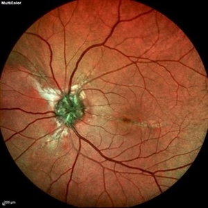 Optic Nerve Head Drusen With Angiod Streaks in Hyperphosphatemic Familial Tumoral Calcinosis
Aug 8 2024 by Hemanth Murthy, MBBS, MD, FASRS
Multicolor image of left eye of 53 year female patient with decreased vision in left eye. Patient gives history of multiple joint swellings with multiple dental procedures due to calcification of the roots. She had type2 MNV demonstrated on OCT and OCTA. Her blood reports showed elevated serum phosphorus (6.4 mg/dl) with normal serum calcium, vitamin D and parathyroid hormone. Her fibroblast growth factor 23 was markedly elevated(>1500RU/ml).
Photographer: Mr Veda Vyas
Condition/keywords: Optic disc drusen and Angiod streaks
-
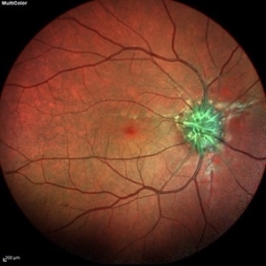 Optic Nerve Head Drusen With Angiod Streaks in Hyperphosphatemic Familial Tumoral Calcinosis
Aug 8 2024 by Hemanth Murthy, MBBS, MD, FASRS
Multicolor image of right eye of 53 year female patient with decreased vision in left eye. Patient gives history of multiple joint swellings with multiple dental procedures due to calcification of the roots. She showed type2 MNV on OCT and OCTA. Her blood reports showed elevated serum phosphorus (6.4 mg/dl) with normal serum calcium, vitamin D and parathyroid hormone. Her fibroblast growth factor 23 was markedly elevated(>1500RU/ml).
Photographer: Mr Veda Vyas
Condition/keywords: Optic disc drusen and Angiod streaks
-
 Optic Nerve Head Drusen With Angiod Streaks in Phosphatemic Familial Tumoral Calcinosis
Aug 8 2024 by Hemanth Murthy, MBBS, MD, FASRS
Autofluorescence image of left eye of 53 year female patient with decreased vision in left eye. Patient gives history of multiple joint swellings with multiple dental procedures due to calcification of the roots. She showed type 2 MNV in left eye on OCT and OCTA. Her blood reports showed elevated serum phosphorus (6.4 mg/dl) with normal serum calcium, vitamin D and parathyroid hormone. Her fibroblast growth factor 23 was markedly elevated(>1500RU/ml).
Photographer: Mr Veda Vyas
Condition/keywords: Optic disc drusen and Angiod streaks
-
 Posterior-PFV
Jul 27 2024 by Gokcen Deniz Gulpinar Ikiz
7 Year old girl presented with blurred vision on the left eye, with intermittent esotopia. She had been followed conservatively for intermittent esotropia on the left eye, recently advised for patching of the right eye. The vision is 1.0 on the right eye and 0.4 (Snellen) on the left eye. Anterior segment is natural bilaterally, except 20 PD esotropia on the left eye, with alternation and fixation. Refraction was +0.25 +0.25 x180 and +1.00-1.50 x60 on the right and left eyes respectively. Dilated fundus examination was natural on the right eye. However, there was a fibrotic stalk originating from the optic nerve head extending to the vitreous, terminating in the middle of the vitreous cavity, in a spider web configuration. Which also causes nasal dragging of the macula, leading to partial shallow detachment of the fovea nasally. Vitrectomy is advised for the left eye, with lens preserving approach, to preserve the current functional potential and the anatomy of the globe in long term.
Photographer: Gokcen Deniz Gulpinar Ikiz, Special Eye Clinic
Condition/keywords: amblyopia, posterior PFV, vitrectomy
-
 Fluorescein Angiography Montage
Jun 21 2024 by BENITO VERGARA, MD
Montage of an angiography with fluorescein from the left eye of a 32 year-old male with diabetic retinopathy previously treated with panretinal photocoagulation, that shows leakage at optic nerve and upper nasal arcade.
Photographer: Benito Vergara, Asociación Para Evitar la Ceguera en México.
Imaging device: Zeiss Clarus 700
Condition/keywords: Angiography Montage, angiography with fluorescein, diabetic retinopathy, FA montage, fluorescein angiogram (FA), peripheral scars
-
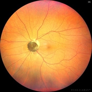
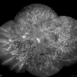
 Autofluorescence in Optic Nerve Head Drusen
May 28 2024 by Nishikant J Borse, MS, FMRF, FASRS
65-year-old female was referred for disc edema. An Autofluorescence Imaging was done which showed the autofluorescence of the optic nerve head drusen.
Photographer: Dr Nishikant Borse , Insight eye Clinic , Mumbai
Imaging device: Topcon Triton
Condition/keywords: Autofluorescence imaging of Optic Disc Drusen
 Loading…