Search results (69 results)
-
 The Halloween Smile
Mar 27 2025 by Shrishti mishra
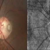
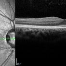
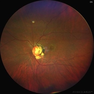
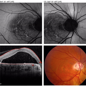
A 73 year old male with Le optic disc pit . On color fundus photo a single pit can be noted whereas on oct enface is- os interface 2 optic disc pits are noted which resembles a halloween smile .
Photographer: Mr Sudhakar
Imaging device: Zeiss cirrus6000
Condition/keywords: OCT, oct en face, optic disc pit
-
 Left Eye Optical Coherence Tomography Showing Optic Disc Pit
Nov 9 2024 by Anand Temkar
Left Eye Optical Coherence Tomography of a 48 years old male patient showing Optic Disc Pit.
Photographer: Dr.Anand Temkar- Retina Foundation, Ahmedabad
Imaging device: Mirante
Condition/keywords: optic disc pit, Optic pit
-
 Optic Disc Pit With Macular Scar
Jun 24 2024 by Akansha Sharma
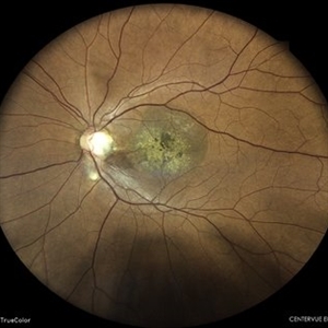
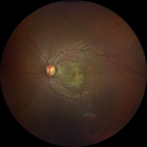
Color fundus photograph of a 42 year old male with optic disc pit with macular scar.
Photographer: Dr. Akansha Sharma, Bharati Eye Hospital
Condition/keywords: macular scar, Optic disc pit
-
 ILM Peeling in Case of Optic Disc Pit Maculopathy
Jun 14 2024 by Tejaswita Verma
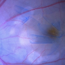
Intraoperative still of a 38 year old male post initiation of ILM peeling in a case of optic disc pit maculopathy.
Photographer: DR. TEJASWITA VERMA
Condition/keywords: intraoperative, optic pit
-
 Optic Disc Pit
May 17 2024 by T. P . VIGNESH, MBBS,MS
SD-OCT of the right eye of a 26 year man revealing Optic disc pit .
Photographer: Sivanath
Imaging device: Heidelberg Spectralis
Condition/keywords: Optic disc pit
-
 Optic Disc Pit OCT
Aug 1 2023 by Aditya S Kelkar, MS, FRCS, FASRS,FRCOphth
Optical Coherence Tomography of an 21 year old male with a Optic Disc Pit.
Photographer: Dr. Ajinkya Rawale. National institute of Ophthalmology, Pune, India.
Imaging device: Zeiss Plex
Condition/keywords: congenital optic nerve pit
-
 Optic Disc Pit Associated with Multilayered Retinoschisis
Apr 26 2023 by Shaleen Arora
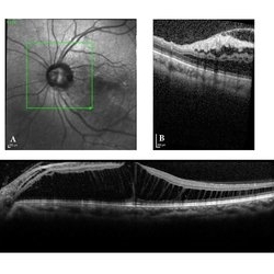
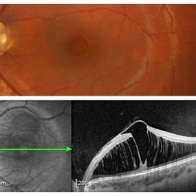
46-year-old female with an optic nerve pit in the left eye (A). OCT reveals retinoschisis involving multiple retinal layers due to intraretinal fluid tracking from the nerve pit (B). Muller cell processes maintain the architecture of individual retinal layers in the region of retinoschisis (C).
Photographer: George Washington University, Department of Ophthalmology
Condition/keywords: maculopathy, optic disc pit, optic pit
-
 Optic disc pit
Apr 14 2023 by T. P . VIGNESH, MBBS,MS
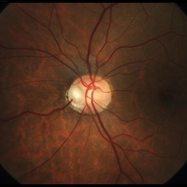
Fundus photograph of an 32-year-old woman with optic disc pit and macular RPE atrophy .
Photographer: Bharathi S
Imaging device: ZEISS CLARUS
Condition/keywords: Optic disc pit
-
 Nasal Optic Disc Pit
May 3 2022 by Bernardo Araújo
Asymptomatic patient. 41-year-old woman.
Photographer: Bernardo Araújo, Retina Clinic, São Paulo, SP, Brazil.
Condition/keywords: nasal, optic disc
-
 Optic disc pit
Mar 21 2022 by T. P . VIGNESH, MBBS,MS
Fundus photo of Left eye of a 55 year male patient revealing optic disc pit with temporal barrage laser marks and foveal schisis with RPE atrophic changes.
Photographer: Bharathi Singaravel
Imaging device: Zeiss Clarus
Condition/keywords: Optic disc pit
-
 Optic Disc Pit
Nov 8 2021 by Michael Grinton
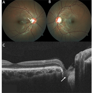
Optic disc pits are rare congenital abnormalities of the optic nerve head. Colour fundus image of an asymptomatic 18-year old male shows an optic disc pit in the right eye (A, white arrow); a small, grey, oval shaped excavation in the temporal segment of the optic disc. These pits are usually unilateral (B shows normal colour fundus of left eye) and asymptomatic. Imaging with optical coherence tomography (C) shows the optic disc pit in cross section (white arrow) and normal macular structure. In some patients with the condition, fluid can accumulate underneath the macular (serous macular detachment).
Condition/keywords: Optic disc pit, Optic nerve pit, Optic pit
-
 Optic Disc Pit with Coloboma (Hybrid Anomaly)
Jun 10 2021 by Janani Sreenivasan
Optic disc pit is a rare anomaly of the optic nerve head that can be associated with maculopathy leading to progressive visual deterioration. It belongs to the spectrum of congenital cavitary anomalies of the optic disc which encompasses extrapapillary cavitation, optic disc coloboma, and morning glory. Very rarely, optic disc pits are seen in combination with optic disc colobomas. Histopathologically, disc pit is defined as herniation of dysplastic retinal tissue into an excavation, rich in collagen, which can stretch into the subarachnoid space via a defect in the lamina cribrosa. Interestingly, this structural abnormality leading to a non-physiological communication between the intraocular and extraocular spaces is a common feature among all the congenital cavitary disc anomalies. Optic disc pit maculopathy is characterized by intraretinal and subretinal fluid at the area of macula. The origin of the retinal fluid remains unclear. Possible sources include the vitreous cavity, the subarachnoid space and the orbital space surrounding the dura. It has been estimated that approximately 25% to 75% of patients will develop serous detachment and/or retinoschisis of the central macula at some stage of their life. On fundus examination, ODPs typically appear as single grayish, round or oval depressions at the optic disc. Most commonly, they are detected at the inferotemporal segment of the disc, but may also be observed elsewhere, including the central area.The coexisting macular detachment can be related to lamellar or full-thickness macular holes, cystoid changes, retinal pigment epithelium atrophy and eventually to irreversible loss of vision,especially in longstanding cases. Herewith, we present a 32-years-old male patient presenting with an unusual combination of optic disc pit with maculopathy and optic disc coloboma (hybrid anomaly) in the same eye with corrected visual acuity of 3/60.
Photographer: Dr Janani Sreenivasan
Imaging device: Zeiss Cirrus HD-OCT
Condition/keywords: coloboma of optic disc, hybrid anomaly, macular detachment, optic disc, optic disc pit
-
 Optic Disc Pit with Maculopathy
Feb 25 2021 by Niloofar Piri, MD
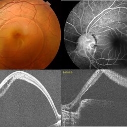
Color fundus photograph and SD OCT of a 6-year-old patient with optic disc pit associated with large retinoschisis involving the entire macula. SD OCT demonstrating large retinoschisis with ILM draping centrally giving it the appearance of pseudohole on the corresponding central area of color photo. Vision 20/80
Photographer: Lisa Breeding, St Louis University
Condition/keywords: maculopathy, optic disc
-
 Optic Pit Maculopathy
Sep 3 2020 by Ankur S. Gupta, MD, MS
36-year-old female with optic disc pit maculopathy (ODP-M) as evidenced by macular swelling on fundus photos. OCT reveals buildup of subretinal fluid spanning the entirety of the macula and FAF shows loss of RPE.
Photographer: Demi Miller, Geisinger Eye Institute
Condition/keywords: optic pit
-
 Optic Disc Pit Associated with Optic Disc Coloboma and Retinochoroidal Coloboma
Jul 22 2020 by Deepak Bhojwani, MS
Fundus photograph of a 32-year-old male showing large optic disc pit in a colobomatous optic nerve head along with isolated inferior retino-choroidal coloboma. (A rare / coincidental occurrence of multiple congenital anomalies of optic disc and retina)
Photographer: DEEPAK BHOJWANI
Condition/keywords: coloboma of choroid, coloboma of optic disc, congenital optic nerve pit
-
 Optic Disc Pit Maculopathy
Jan 28 2020 by Pierre-Henry Gabrielle, MD
Optic disc pit maculopathy of a 26-year-old man with optic disc pit maculopathy of his right eye.
Photographer: Pierre-Henry Gabrielle, Ophthalmology department, Dijon University Hospital
Imaging device: Heidelberg Spectralis
Condition/keywords: maculopathy, optic disc pit, optical coherence tomography (OCT)
-
 Optic Disc Pit Maculopathy
Jan 28 2020 by Pierre-Henry Gabrielle, MD
Optic disc pit maculopathy of a 26-year-old man with optic disc pit maculopathy of his right eye.
Photographer: Pierre-Henry Gabrielle, Ophthalmology department, Dijon University Hospital
Imaging device: Zeiss Visucam
Condition/keywords: fundus photograph, maculopathy, optic disc pit
-
 Re-Imaging Optic Disc Pit
Oct 26 2019 by Deepak Bhojwani, MS
OCT-angiography of a classic case of optic disc pit with maculopathy.
Imaging device: ZEISS CIRRUS 5000 HD-OCT WITH ANGIOPLEX
Condition/keywords: optic disc pit
-
 Optic Disc Pit
Oct 8 2019 by DIEGO TOLENTINO
OCT of a patient with optic disc pit.
Photographer: Diego Tolentino
Condition/keywords: optic disc pit, optical coherence tomography (OCT)
-
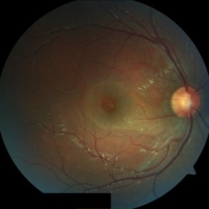
 RNFL in Right Optic Disc Pit
Jul 20 2019 by Arwa Azmeh, MD, PhD
Fundus photograph of 38-year-old healthy man with right optic disc pit, who recently noticed slightly blurred vision in right eye while closing the left eye. BCVA was 20/25 in OD and 20/20 in OS. IOP was 15mmHg OD and 14 mmHg OS. Right fundus exam showed small optic disc pit near the temporal rim of optic disc with abnormal reflex of nasal macula. Left fundus was normal. Late FA of right optic disc showed no leakage or staining of optic disc. Macular OCT showed normal foveal contour with no subretinal fluid or macular edema. There was significant reduction in RNFL thickness in the temporal sector in right eye. coloboma is clearly seen on vertical OCT scan as well as horizontal scans through right optic pit.
Photographer: Ebtisam Aljbeili, Damascus university, Almouassat university hospital
Imaging device: Heidelberg Spectralis 2
Condition/keywords: optic pit
-
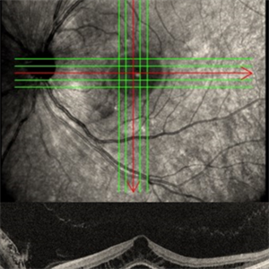
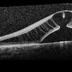
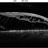
 Vertical OCT Scan Through Right Optic Disc Pit
Jul 20 2019 by Arwa Azmeh, MD, PhD
Fundus photograph of 38-year-old healthy man with right optic disc pit, who recently noticed slightly blurred vision in right eye while closing the left eye. BCVA was 20/25 in OD and 20/20 in OS. IOP was 15mmHg OD and 14 mmHg OS. Right fundus exam showed small optic disc pit near the temporal rim of optic disc with abnormal reflex of nasal macula. Left fundus was normal. Late FA of right optic disc showed no leakage or staining of optic disc. Macular OCT showed normal foveal contour with no subretinal fluid or macular edema. There was significant reduction in RNFL thickness in the temporal sector in right eye. Coloboma is clearly seen on vertical OCT scan as well as horizontal scans through right optic pit.
Photographer: Ebtisam Aljbeili, Damascus university, Almouassat university hospital
Imaging device: Heidelberg Spectralis 2
Condition/keywords: optic pit, optical coherence tomography (OCT)
-
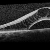
 Horizontal OCT Scan Through Right Optic Pit
Jul 20 2019 by Arwa Azmeh, MD, PhD
Fundus photograph of 38-year-old healthy man with right optic disc pit, who recently noticed slightly blurred vision in right eye while closing the left eye. BCVA was 20/25 in OD and 20/20 in OS. IOP was 15mmHg OD and 14 mmHg OS. Right fundus exam showed small optic disc pit near the temporal rim of optic disc with abnormal reflex of nasal macula. Left fundus was normal. Late FA of right optic disc showed no leakage or staining of optic disc. Macular OCT showed normal foveal contour with no subretinal fluid or macular edema. There was significant reduction in RNFL thickness in the temporal sector in right eye. Coloboma is clearly seen on vertical OCT scan as well as horizontal scans through right optic pit.
Photographer: Ebtisam Aljbeili, Damascus university, Almouassat university hospital
Imaging device: Heidelberg Spectralis 2
Condition/keywords: optic pit, optical coherence tomography (OCT)
-
 Macular OCT in Right Optic Disc Pit
Jul 20 2019 by Arwa Azmeh, MD, PhD
Fundus photograph of 38-year-old healthy man with right optic disc pit, who recently noticed slightly blurred vision in right eye while closing the left eye. BCVA was 20/25 in OD and 20/20 in OS. IOP was 15mmHg OD and 14 mmHg OS. Right fundus exam showed small optic disc pit near the temporal rim of optic disc with abnormal reflex of nasal macula. Left fundus was normal. Late FA of right optic disc showed no leakage or staining of optic disc. Macular OCT showed normal foveal contour with no subretinal fluid or macular edema. There was significant reduction in RNFL thickness in the temporal sector in right eye. Coloboma is clearly seen on vertical OCT scan as well as horizontal scans through right optic pit.
Photographer: Ebtisam Aljbeili, Damascus university, Almouassat university hospital
Imaging device: Heidelberg Spectralis 2
Condition/keywords: optic pit, optical coherence tomography (OCT)
-
 Late fluorescein Angiography of Right Optic Pit
Jul 20 2019 by Arwa Azmeh, MD, PhD
Fundus photograph of 38-year-old healthy man with right optic disc pit, who recently noticed slightly blurred vision in right eye while closing the left eye. BCVA was 20/25 in OD and 20/20 in OS. IOP was 15mmHg OD and 14 mmHg OS. Right fundus exam showed small optic disc pit near the temporal rim of optic disc with abnormal reflex of nasal macula. Left fundus was normal. Late FA of right optic disc showed no leakage or staining of optic disc. Macular OCT showed normal foveal contour with no subretinal fluid or macular edema. There was significant reduction in RNFL thickness in the temporal sector in right eye. Coloboma is clearly seen on vertical OCT scan as well as horizontal scans through right optic pit.
Photographer: Ebtisam Aljbeili, Damascus university, Almouassat university hospital
Imaging device: Heidelberg Spectralis 2
Condition/keywords: fluorescein angiogram (FA), optic pit
 Loading…