 Initializing download.
-
 By Arwa Azmeh, MD, PhD
Damascus University, Faculty of medicine
Co-author(s): Labib Alshaweesh, Damascus university, Almouassat university hospital - Uploaded on Jul 20, 2019.
- Last modified by Caroline Bozell on Jul 23, 2019.
- Rating
- Appears in
- Miscellaneous
- Condition/keywords
- optical coherence tomography (OCT), optic pit
- Photographer
- Ebtisam Aljbeili, Damascus university, Almouassat university hospital
- Imaging device
- Heidelberg Spectralis 2
- Description
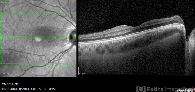
- Fundus photograph of 38-year-old healthy man with right optic disc pit, who recently noticed slightly blurred vision in right eye while closing the left eye. BCVA was 20/25 in OD and 20/20 in OS. IOP was 15mmHg OD and 14 mmHg OS. Right fundus exam showed small optic disc pit near the temporal rim of optic disc with abnormal reflex of nasal macula. Left fundus was normal. Late FA of right optic disc showed no leakage or staining of optic disc. Macular OCT showed normal foveal contour with no subretinal fluid or macular edema. There was significant reduction in RNFL thickness in the temporal sector in right eye. Coloboma is clearly seen on vertical OCT scan as well as horizontal scans through right optic pit.

")


")
")
")


