Search results (67 results)
-
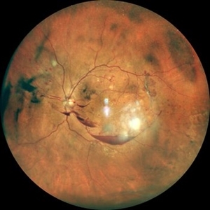
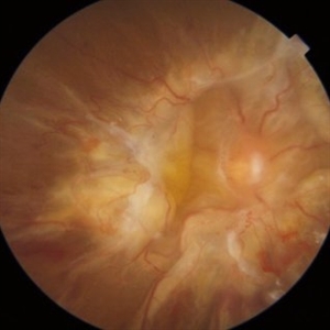
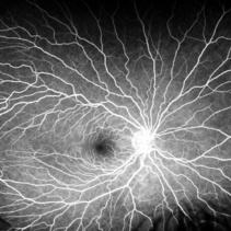
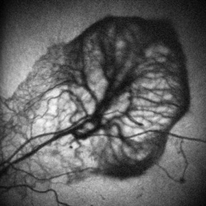
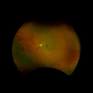 PDR with Peripheral NVEOS
Jul 23 2024 by Ashley Phillips
Fundus photo of a 62 y/o male with PDR and NVE (periphery)
Photographer: Ashley Phillips
Imaging device: Optos-California
Condition/keywords: neovascularization elsewhere (NVE), PDR
-
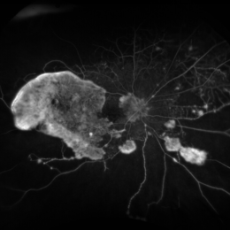
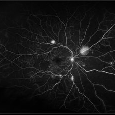
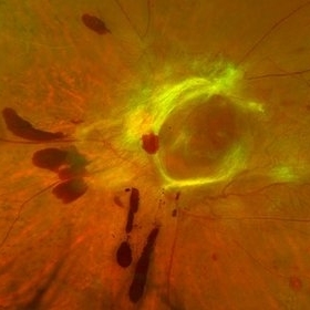
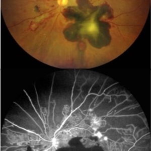 Lizard-shaped-hemorrhage
Apr 29 2023 by Saagar A Pandit, MD, MPH
49 year-old male with a history of Type 1 Diabetes Mellitus, past ocular history significant for proliferative diabetic retinopathy of both eyes. Left eye significant for a unique, "lizard-shaped" pre-retinal hemorrhage in an area of neovascularization. Note the corresponding fluorescein angiography which demonstrates blockage from hemorrhage and significant posterior non-perfusion, in addition to tufts of neovascularization which are hyperfluorescent.
Photographer: Maria Pei, Bellevue Hospital Ophthalmology Clinic, New York, NY
Condition/keywords: hemorrhage, neovascularization elsewhere (NVE), proliferative diabetic retinopathy (PDR)
-
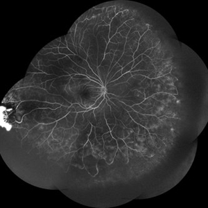
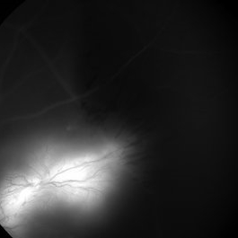
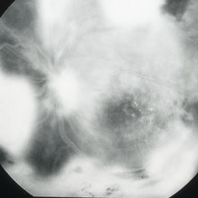
 Subhyaloid Hemorrhage with JXT and Proliferative Diabetic Retinopathy
Jan 13 2022 by ASRS Staff
Wide field photograph of 50 year-old female, known case of idiopathic juxtafoveal telangiectasia in both eyes and known diabetic, presented with subhyaloid hemorrhage and NVE.
Imaging device: Nidek Mirante
Condition/keywords: florid type PDR, JXT, neovascularization elsewhere (NVE), subhyaloid hemorrhage, ultra-wide field imaging
-
 Proliferative Diabetic Retinopathy
Oct 25 2021 by VIRAL SHAH
25 year-old patient suffering with type 1 diabetes mellitus since age 17 has developed proliferative diabetic retinopathy with extensive neovascularization elsewhere and neovascularization at disc.
Photographer: VIRAL SHAH, NETRALOK RETINA CLINIC, AHMEDABAD
Condition/keywords: neovascularization at the disc, neovascularization elsewhere (NVE), proliferative diabetic retinopathy (PDR)
-
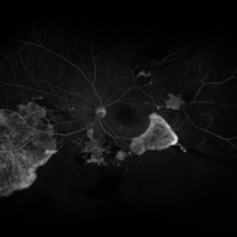
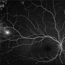
 NVE in an HIV Positive Case
Sep 26 2021 by Nivesh Gupta
FA Montage of a 22-year-old female with Neovascularization Elsewhere
Photographer: DR. NIVESH GUPTA, RETINA FOUNDATION, AHMEDABAD
Imaging device: NIDEK MIRANTE
Condition/keywords: HIV, neovascularization elsewhere (NVE)
-
 Proliferative Diabetic Retinopathy
Sep 16 2021 by Tandava Krishnan
Fundus photograph of a 60 year old poorly controlled diabetic who presented with decreased vision in left eye.
Photographer: Tandava Krishnan
Condition/keywords: fibrovascular proliferation, neovascularization elsewhere (NVE), proliferative diabetic retinopathy (PDR), subhyaloid hemorrhage
-
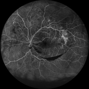 Fluorescein Angiography Neovascularization Elsewhere and Subhyaloid Hemorrhage
Aug 15 2021 by ASRS Staff
38 year-old male, presented with complaint of dark spot in vision of left eye. His vision was 6/6 in both eyes. On examination he was having subhyaloid hemorrhage and NVE in left eye.NVE was also present in RE. Patient was referred for carotid Doppler and cardiologist opinion.
Imaging device: Nidek Mirante
Condition/keywords: neovascularization elsewhere (NVE), subhyaloid hemorrhage
-
 Wide-field photo of NVE and Subhyaloid Hemorrhage
Aug 15 2021 by ASRS Staff
38 year-old male, presented with complaint of dark spot in vision of left eye. His vision was 6/6 in both eyes. On examination he was having subhyaloid hemorrhage and NVE in left eye.NVE was also present in RE. Patient was referred for carotid Doppler and cardiologist opinion.
Imaging device: Nidek Mirante
Condition/keywords: neovascularization elsewhere (NVE), subhyaloid hemorrhage, wide angle imaging
-
 Subhyaloid Hemorrhage with NVE
Aug 15 2021 by ASRS Staff
38 year-old male, presented with complaint of dark spot in vision of left eye. His vision was 6/6 in both eyes. On examination he was having subhyaloid hemorrhage and NVE in left eye. NVE was also present in RE. Patient was referred for carotid Doppler and cardiologist opinion.
Imaging device: Nidek Mirante
Condition/keywords: Central fundus, neovascularization elsewhere (NVE), subhyaloid hemorrhage
-
 Lightening In Eyes
Jul 22 2021 by Vishal Gupta, MBBS, MS
Fundus fluorescein angiogram of a neovascular frond lighting up like a silent lightening thunder in the dark night.
Photographer: Dr Vishal Gupta, INHS Asvini, Mumbai, INDIA
Imaging device: zeiss
Condition/keywords: branch retinal vein occlusion (BRVO), fluorescein angiogram (FA), neovascularization elsewhere (NVE)
-
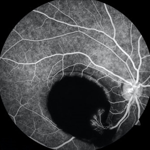
 Severe Diabetic Retinopathy, Nonperfusion and NVE
Jun 18 2021 by Kristen Wagner
Optos angiogram of severe diabetic retinopathy, nonperfusion and NVE.
Photographer: Kristen Wagner, COT Tennessee Retina Nashville TN
Imaging device: Optos
Condition/keywords: diabetic macular edema, neovascularization elsewhere (NVE), nonperfusion diabetic retinopathy, proliferative diabetic retinopathy (PDR)
-
 Severe Diabetic Retinopathy and NVE with Macular Hole
Jun 18 2021 by Kristen Wagner
Fundus photo of severe PDR with NVE and nonperfusion. The patient also has a macular hole.
Photographer: Kristen Wagner, COT Tennessee Retina Nashville TN
Imaging device: Optos
Condition/keywords: diabetic macular edema, macular hole, neovascularization elsewhere (NVE), proliferative diabetic retinopathy (PDR)
-
 Tractional Retinal Detachment
May 21 2021 by Rutul R Patel, MD Ophthalmology
Fundus photograph of a 54-year-old male patient with treatment naïve advanced proliferative diabetic retinopathy with tractional retinal detachment.
Photographer: Vidhi Bavishi
Imaging device: Topcon Maestro
Condition/keywords: neovascularization elsewhere (NVE), proliferative diabetic retinopathy (PDR), tractional retinal detachment
-
 Proliferative Diabetic Retinopathy
Jan 29 2021 by Olivia Rainey
Ultra-widefield fluorescein angiogram of a 65-year-old male with proliferative diabetic retinopathy affecting his right eye. The patient's diabetic retinopathy has progressed significantly since he was last seen in 2014. It was recommended to begin antiVEGF to control DME followed by laser treatment OU.
Photographer: Olivia Rainey, OCT-C, COA
Imaging device: Optos California
Condition/keywords: anti-VEGF, diabetes, diabetic macular edema, fluorescein angiogram (FA), fluorescein leakage, neovascularization (NV), neovascularization elsewhere (NVE), non-perfusion, Optos, proliferative diabetic retinopathy (PDR), ultra-wide field imaging
-
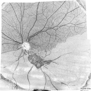
 Proliferative Sickle Cell Retinopathy
Jan 29 2021 by Olivia Rainey
Ultra-widefield fluorescein angiogram of a 24-year-old female with proliferative sickle cell retinopathy affecting her right eye. The physician's interpretation of the fluorescein shows seafan neovascularization superotemporally, AV anastomeses, and good peripheral laser. He performed scatter PRP OD on 12/2/2020 to nonperfusion in temporal far periphery. The patient's 12/2020 Hb electrophoresis came back showing Hb SC (rather than sickle cell trait). Patient was born at full term, but she reports that her mother used drugs while pregnant with the patient. The patient also mentioned that her niece has full sickle cell disease and her grandmother, mother, and sibling all have condition on the sickle cell spectrum.
Photographer: Olivia Rainey, OCT-C, COA
Imaging device: Optos California
Condition/keywords: fluorescein angiogram (FA), fluorescein leakage, neovascularization (NV), neovascularization elsewhere (NVE), Optos, sea fan, sickle cell retinopathy
-
 Proliferative Sickle Cell Retinopathy
Jan 29 2021 by Olivia Rainey
Ultra-widefield fundus photograph of a 24-year-old female with proliferative sickle cell retinopathy affecting her right eye. He performed scatter PRP OD on 12/2/2020 to nonperfusion in temporal far periphery. The patient's 12/2020 Hb electrophoresis came back showing Hb SC (rather than sickle cell trait). Patient was born at full term, but she reports that her mother used drugs while pregnant with the patient. The patient also mentioned that her niece has full sickle cell disease and her grandmother, mother, and sibling all have condition on the sickle cell spectrum.
Photographer: Olivia Rainey, OCT-C, COA
Imaging device: Optos California
Condition/keywords: fundus photograph, laser photocoagulation, neovascularization (NV), neovascularization elsewhere (NVE), Optos, pseudocolor, sea fan, sickle cell retinopathy
-
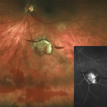
 Occlusive Retinal Vasculitis
Dec 17 2020 by Somnath Chakraborty, MD
Fundus photo montage of left eye of a 45-year-old male showing a large neovascular frond secondary to peripheral occlusive vasculitis.
Photographer: Pulak Roy
Condition/keywords: neovascularization elsewhere (NVE), peripheral retinal vasculitis, retinal vasculitis
-
 Coats' Disease
Oct 20 2020 by Anfisa Ayalon, MD
Fundus fluorescein angiography of 35-year-old female with right eye asymptomatic coats disease.
Photographer: Anfisa Ayalon, Meir Medical Center, Kfar Saba, Israel.
Imaging device: California, Optos 200 DTX
Condition/keywords: Coats' disease, neovascularization elsewhere (NVE), retina
-
 The Barren Field
Jun 27 2020 by SANDEEP KUMAR
A 59-year-old man with DM for 18 years operated for mature cataract. Post op left eye had a visual acuity of 20/80. Wide field swept source OCTA revealed gross vessel wipe out in inferior hemi quadrant with branching out neovascular frond inferior to disc with terminal loops, The patient underwent Anti VEGF injection followed by OCTA guided sectoral retinal photocoagulation.Image J software used here to generate reverse image that sharply delineates the non perfusion area.
Photographer: Sandeep Kumar
Imaging device: Optical coherence tomography system Zeiss Plex Elite 9000
Condition/keywords: hemi CRVO, neovascularization elsewhere (NVE)
-
 Sub-ILM Hemorrhage with Neovessels
Apr 30 2020 by Saurabh Deshmukh, MBBS, DNB, FVRS, MNAMS
Late arteriovenous phase FA showing a large sub-internal limiting membrane hemorrhage with overlying neovessels. This hypertensive patient presented with a visual acuity of counting fingers at 2 meters. The patient was advised intravitreal anti-VEGF injection, Nd: YAG Membranotomy, and systemic control of hypertension.
Photographer: Saurabh Deshmukh, Sri Sankaradeva Nethralaya, Guwahati, India
Imaging device: Topcon TRC-50 DX
Condition/keywords: hypertensive retinopathy, neovascularization elsewhere (NVE), subILM hemorrhage
-
 Tractional Detachment of Retina
Apr 28 2020 by Pauline T Merrill, MD, FASRS
29-year-old male with PDR and DME.
Photographer: Karen Parque, Illinois Retina Associates
Imaging device: Topcon TRC 50DX
Condition/keywords: hyperfluorescence, neovascularization elsewhere (NVE)
-
 NVE with Hemorrhages
Jan 20 2020 by Sarah Oelrich
NVE with hemorrhages
Photographer: Sarah Oelrich CRA COT OCT-C Southeastern Retina Associates
Condition/keywords: hemorrhage, neovascularization elsewhere (NVE)
-
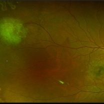
 Neovascularization Elsewhere in Proliferative Diabetic Retinopathy
Nov 16 2019 by Anfisa Ayalon, MD
Fundus autofluorescence image of neovascularization elsewhere, patient with high-risk proliferative diabetic retinopathy.
Photographer: Anfisa Ayalon, MD., Meir Medical Center, Kfar Saba, Israel.
Condition/keywords: diabetes, fundus autofluorescence (FAF), ischemia, neovascularization elsewhere (NVE), proliferative diabetic retinopathy (PDR)
-
 Wolf Jaw
Jun 23 2019 by Veronika Yehezkeli
Wolf Jaw
Imaging device: Optos
Condition/keywords: ischemia, neovascularization (NV), neovascularization elsewhere (NVE), preretinal hemorrhage, proliferative diabetic retinopathy (PDR), wolf jaw
-
 Eales Disease
Apr 1 2019 by Gary R. Cook, MD, FACS
Late-phase fluorescein angiogram image of the left eye of a 23-year-old Vietnamese female with Eales Disease showing extensive dye leakage from multiple areas of NVE and from some NVD.
Imaging device: Topcon VT-50
Condition/keywords: Eales disease, FA late phase, fluorescein angiogram (FA), neovascularization elsewhere (NVE)
 Loading…