Search results (551 results)
-
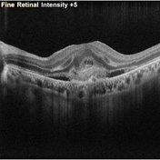
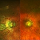
 Both Eyes OCT in Case of CNVM with Angioid Streaks
Nov 29 2024 by Anand Temkar
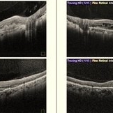
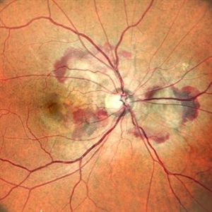
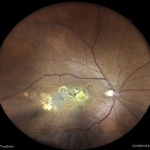
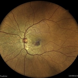
A 45 year old male came with chief complaint of blurring vision in right eyes since past 4 days. His vision is 6/12 in right eye and 6/9 in left eye. His vision was 14 mmHg in right eye and 16 mmHg in left eye. He was diagnosed with Angioid Streaks in both eyes about a year ago, then he developed choroidal neovascularization in his left eye 8 months ago, for which he received AntiVEGF injections x 3. Left eye is a stable eye now. Patient presented with right eye choroidal neovascularization in a case of Angioid Streaks on recent follow up. We have advised him right eye AntiVEGF injections x 3. In this image we can see the subretinal hyperreflective material in right eye and in left eye few cystic spaces are noted.
Photographer: Dr.Anand Temkar- Retina Foundation, Ahmedabad
Imaging device: Mirante
Condition/keywords: Angioid Streaks, choroidal neovascular membrane (CNVM)
-
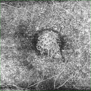
 Both Eyes Fundus Autofluorescence in Case of CNVM with Angioid Streaks
Nov 29 2024 by Anand Temkar
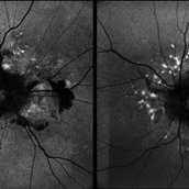
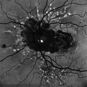
A 45 year old male came with chief complaint of blurring vision in right eyes since past 4 days. His vision is 6/12 in right eye and 6/9 in left eye. His vision was 14 mmHg in right eye and 16 mmHg in left eye. He was diagnosed with Angioid Streaks in both eyes about a year ago, then he developed choroidal neovascularization in his left eye 8 months ago, for which he received AntiVEGF injections x 3. Left eye is a stable eye now. Patient presented with right eye choroidal neovascularization in a case of Angioid Streaks on recent follow up. We have advised him right eye AntiVEGF injections x 3. In this image we can see fundus hypoautofluorescence in right eye due to hemorrhages and angioid streaks and in left eye fundus hypoautofluorescence is noted due to angioid streaks.
Photographer: Dr.Anand Temkar- Retina Foundation, Ahmedabad
Imaging device: Mirante
Condition/keywords: Angioid Streaks, choroidal neovascular membrane (CNVM), fundus autofluorescence (FAF)
-
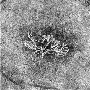
 RE OCTA (ORCC) in case of CNVM with Angioid Streaks
Nov 29 2024 by Anand Temkar
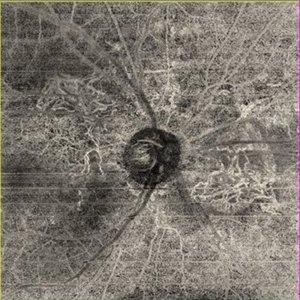
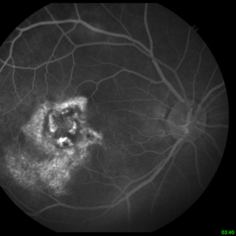
A 45 year old male came with chief complaint of blurring vision in right eyes since past 4 days. His vision is 6/12 in right eye and 6/9 in left eye. His vision was 14 mmHg in right eye and 16 mmHg in left eye. He was diagnosed with Angioid Streaks in both eyes about a year ago, then he developed choroidal neovascularization in his left eye 8 months ago, for which he received AntiVEGF injections x 3. Left eye is a stable eye now. Patient presented with right eye choroidal neovascularization in a case of Angioid Streaks on recent follow up. We have advised him right eye AntiVEGF injections x 3. In this image we can see the abnormal vessels at outer retina chorio-capillary ( ORCC ) junction in right eye.
Photographer: Dr.Anand Temkar- Retina Foundation, Ahmedabad
Imaging device: Mirante
Condition/keywords: Angioid Streaks, choroidal neovascular membrane (CNVM), OCT Angiography
-
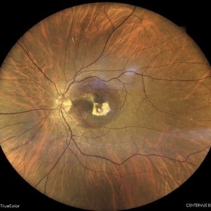
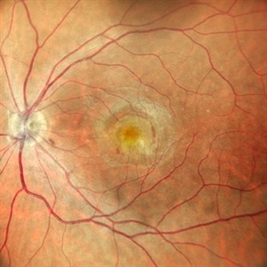
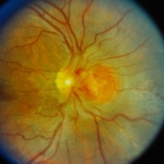
 Left Eye Color Photo With Extrafoveal CNVM (Stable) in Case of Angioid Streaks
Nov 29 2024 by Anand Temkar
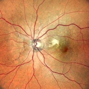
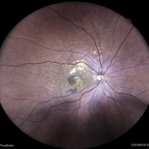
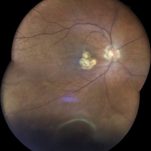
A 45 year old male came with chief complaint of blurring vision in right eyes since past 4 days. His vision is 6/12 in right eye and 6/9 in left eye. His vision was 14 mmHg in right eye and 16 mmHg in left eye. He was diagnosed with Angioid Streaks in both eyes about a year ago, then he developed choroidal neovascularization in his left eye 8 months ago, for which he received AntiVEGF injections x 3. Left eye is a stable eye now. Patient presented with right eye choroidal neovascularization in a case of Angioid Streaks on recent follow up. We have advised him right eye AntiVEGF injections x 3. In this image, the left eye color photo shows angioid streaks with extrafoveal CNVM ( stable ) ( status post antiVEGF x 3 )
Photographer: Dr.Anand Temkar- Retina Foundation, Ahmedabad
Imaging device: Mirante
Condition/keywords: Angioid Streaks, choroidal neovascular membrane (CNVM)
-
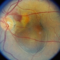
 Right Eye Color Photo With Hemorrhages in Case of CNVM With Angioid Streaks
Nov 29 2024 by Anand Temkar
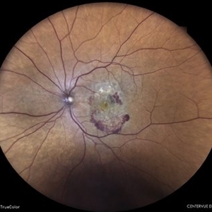
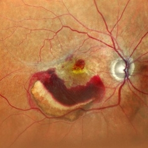
A 45 year old male came with chief complaint of blurring vision in right eyes since past 4 days. His vision is 6/12 in right eye and 6/9 in left eye. His vision was 14 mmHg in right eye and 16 mmHg in left eye. He was diagnosed with Angioid Streaks in both eyes about a year ago, then he developed choroidal neovascularization in his left eye 8 months ago, for which he received AntiVEGF injections x 3. Left eye is a stable eye now. Patient presented with right eye choroidal neovascularization in a case of Angioid Streaks on recent follow up. We have advised him right eye AntiVEGF injections x 3. In this image, the right eye color photo shows bleed from CNVM in case of angioid streaks.
Photographer: Dr.Anand Temkar- Retina Foundation, Ahmedabad
Imaging device: Mirante
Condition/keywords: Angioid Streaks, choroidal neovascular membrane (CNVM)
-
 Angioid Streaks/Optic Disc Drusen
Oct 30 2024 by JULIAN VILLARREAL, MD
FAF showing angiod streaks , optic disc drusen, and macular atrophy secondary to macular neovascular membrane.
Photographer: Julián Villarreal MD
Imaging device: Mirante
Condition/keywords: Angioid Streaks, macular atrophy, optic disc drusen
-
 Subretinal Neovascular Membrane
Jun 5 2024 by Akansha Sharma
Color fundus photograph of a 49 year old female with subretinal bleed suggestive of subretinal neovascular membrane.
Photographer: Dr. Akansha Sharma, Bharati Eye Hospital
Condition/keywords: choroidal neovascular membrane (CNVM), CNVM, SRNVM, subretinal neovascularization (SRNV), wet age-related macular degeneration (wet AMD)
-
 Subretinal Neovascular Membrane
Jun 5 2024 by Akansha Sharma
Color fundus photograph of a 94 year old female with subretinal bleed suggestive of subretinal neovascular membrane.
Photographer: Dr. Akansha Sharma, Bharati Eye Hospital
Condition/keywords: choroidal neovascular membrane (CNVM), CNVM, SRNVM, subretinal neovascularization (SRNV), wet age-related macular degeneration (wet AMD)
-
 Scarred Choroidal Neovacular Membrane
May 7 2024 by Akansha Sharma
Color fundus photograph of a 75 year old male with scarred choroidal neovascular membrane.
Photographer: Dr. Akansha Sharma, Bharati Eye Hospital
Condition/keywords: choroidal neovascular membrane (CNVM), CNVM
-
 Subretinal Neovascular Membrane
Apr 19 2024 by Akansha Sharma
Color fundus photograph of a 74 year old female with subretinal neovascular membrane with scarring below the fovea.
Photographer: Dr. Akansha Sharma, Bharati Eye Hospital
Condition/keywords: SRNVM, subretinal neovascularization (SRNV)
-
 Subretinal Neovascular Membrane with PED
Apr 17 2024 by Akansha Sharma
Color fundus photograph of a 72 year old male with ped along with subretinal bleed around it.
Photographer: Dr. Akansha Sharma, Bharati Eye Hospital
Condition/keywords: CNVM, PED, SRNVM, subretinal neovascularization (SRNV), wet age-related macular degeneration (wet AMD)
-
 Myopic Subretinal Neovascular Membrane
Apr 9 2024 by Akansha Sharma
Color fundus photograph of a 23 year old female with subretinal bleed in a case of high myopia.
Photographer: Dr. Akansha Sharma, Bharati Eye Hospital
Condition/keywords: myopic choroidal neovascularization (CNV), SRNVM, subretinal hemorrhage
-
 Macular Telangiectasia Type 2 OCTA
Mar 29 2024 by Lucy V Cobbs, M.D.
Optical coherence tomography angiography allows for 3-dimensional vessel imaging and may help detect abnormal vessels earlier than fluorescein angiography, which was historically used in diagnosis of MacTel type 2. This OCTA of the left eye of a 52-year-old male captures superficial telangiectatic macular vessels (top left) and follows them as they dive into deeper capillary layers (top right). The structural image of this OCTA (bottom right) shows the classic “right angles” of these abnormal vessels as they plunge. The outer retinal slab image (bottom left) shows a choroidal neovascular membrane, which is a rare complication of MacTel type 2.
Condition/keywords: Mac Tel type 2
-
 Macular Telangiectasia Type 2
Mar 29 2024 by Lucy V Cobbs, M.D.
Color fundus photograph of the left eye of a 70-year-old male with a disciform scar resulting from a neovascular membrane. A minority of MacTel type 2 patients develop neovascular disease, and the gold standard treatment is anti-VEGF intravitreal therapy. Without treatment, membranes may progress to severe central macular scarring. Late stages of proliferative MacTel type 2 may be confused with AMD, and a differentiating aspect is that MacTel type 2 typically lacks pigment epithelial detachments and drusen.
Condition/keywords: Mac Tel type 2
-
 Subretinal Neovascular Membrane
Mar 26 2024 by Akansha Sharma
Color fundus photograph of a 65 year old female patient with subretinal bleed suggestive of subretinal neovascular membrane.
Photographer: Dr. Akansha Sharma, Bharati Eye Hospital
Condition/keywords: CNVM, SRNVM, subretinal neovascularization (SRNV), wet age-related macular degeneration (wet AMD)
-
 Ghost of Halloween
Mar 22 2024 by Tushar Agrawal
Fundus photograph of a 57-year-old man with a macular choroidal neovascular membrane observed over time.
Condition/keywords: choroidal neovascular membrane (CNVM), macula choroidal neovascularization, wet age-related macular degeneration (wet AMD)
-
 Scarred Choroidal Neovacular Membrane With Large Inferior Horse Shoe Tear
Feb 7 2024 by Akansha Sharma
Color fundus photograph of a 73 year old male with scarred choroidal neovascular membrane with large horse shoe tear inferiorly.
Photographer: Dr. Akansha Sharma, Bharati Eye Hospital
Condition/keywords: atrophic scar, choroidal neovascular membrane (CNVM), CNVM
-
 Wet age related macular degeneration
Jan 28 2024 by Anjana Mirajkar, MS Ophthalmology
Fundus photograph of an 70 year old male with sub retinal bleed and exudation as well as scarring in case of wet age related macular degeneration.
Photographer: Dr. Anjana Mirajkar -Retina Foundation, Ahmedabad
Imaging device: Mirante-Nidek
Condition/keywords: choroidal neovascular membrane (CNVM), wet age-related macular degeneration (wet AMD)
-
 Neovascular-network-OCTA
Jan 2 2024 by Tahsin Khundkar, MD
En Face optical coherence tomography (OCT).- angiography shows a large choroidal neovascular membrane in the outer retina to choriocapillaris slab.
Photographer: Jeffrey Zeigler, Concord Eye Center
Imaging device: Zeiss
Condition/keywords: neovascular membrane, wet age-related macular degeneration (wet AMD)
-
 Subretinal Neovascular Membrane
Oct 25 2023 by Vaidehi Sathaye
OCT image of a 45 year old female with SRNVM in the LE
Photographer: Dr. Vaidehi Sathaye
Imaging device: Mirante
Condition/keywords: OCT, SRNVM
-
 Subretinal Neovascular Membrane
Oct 25 2023 by Vaidehi Sathaye
OCTA image showing Neovascular network at the macula in the ORCC slab , in the LE of a 45 year old female
Photographer: Dr. Vaidehi Sathaye
Imaging device: Mirante
Condition/keywords: OCTA, SRNVM
-
 Subretinal Neovascular Membrane
Oct 25 2023 by Vaidehi Sathaye
Fundus of a 45 year old female with SRNVM in the LE
Photographer: Dr. Vaidehi Sathaye
Imaging device: Mirante
Condition/keywords: color photo, SRNVM
-
 WED-ARMD-CNVM
Sep 29 2023 by PUSHPANJALI BADOLE
Fundus photograph of a 72 year old male with right eye choroidal neovascularization. There is a thick membrane over macula along with subretinal fibrosis in right eye. Left eye shows drusen.
Photographer: NITIN DESALE, ISHA NETRALAYA, KALYAN
Imaging device: DAYTONA OPTOS
Condition/keywords: choroidal neovascular membrane (CNVM), choroidal neovascularization (CNV)
-
 Peripapillary Choroidal Neovascular Membrane (CNVM)
Sep 26 2023 by Ben Serar
Fundus photograph showing Peripapillary Choroidal Neovascular Membrane (CNVM) with subretinal bleed and surrounding sub retinal fluid.
Condition/keywords: Peripapillary Choroidal Neovascular Membrane (CNVM)
-
 Peripapillary Choroidal Neovascular Membrane (CNVM) with Angioid Streaks
Sep 21 2023 by Ben Serar
Fundus photograph of RE showing Peripapillary Choroidal Neovascular Membrane (CNVM) nasal to disc with blurring of nasal disc margin and superficial retinal haemorrhages, with peripapillary angioid streaks.
Condition/keywords: angioid streaks, Peripapillary Choroidal Neovascular Membrane (CNVM)
 Loading…