Search results (87 results)
-
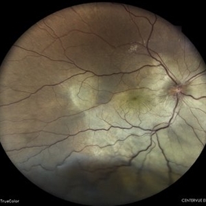
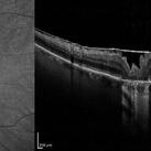
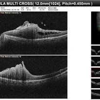
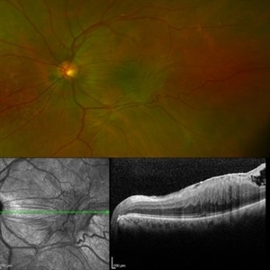
 Retinal Detachment with Multiple OCT Overlays
Jan 7 2025 by Drew Mitchell
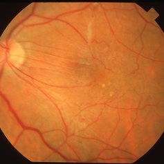
Optos 360* Color photo montage with multiple Zeiss Cirrus OCT scan overlays. Retinal Detachment with multiple breaks and a Epiretinal Membrane.
Photographer: Drew Mitchel, OCT-C
Imaging device: Optos California
Condition/keywords: ERM, macular pucker, montage, Optos, OPTOS CALIFORNIA, RD, Retinal Detachment
-
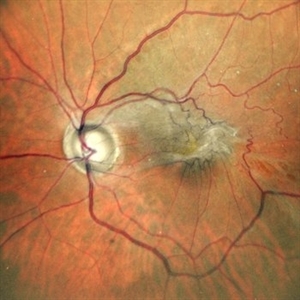
 Exudative Retinal Detachment
May 27 2024 by Akansha Sharma
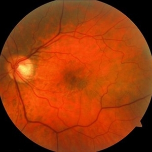
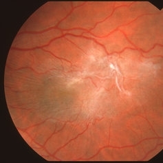
Color fundus photograph of a 38 year old male with breast carcinoma leading to intraocular metastasis as represented by an exudative retinal detachment.
Photographer: Dr. Akansha Sharma, Bharati Eye Hospital
Condition/keywords: Disc Edema, exudative detachment, macular pucker, METASTATSIS
-
 Bullseye Maculopathy
Jan 22 2024 by Kali Jend
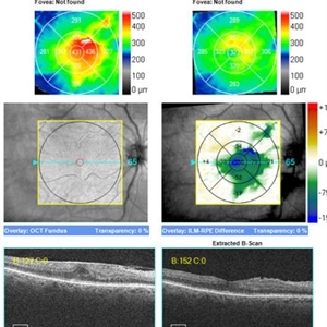
Optical coherence tomography of a 73-year-old female with Bullseye Macular Changes affecting her left eye. Patient reports having a family history of this condition and denies prior Plaquenil or Elmiron use. Compared to previous imaging, the patient's condition progressed in the left eye from 2020 to 2023. Patient has a history of fluctuating Diabetic Macular Edema and a current Epiretinal Membrane as well. Patient's vision was Ncc20/60 at the time the image was taken.
Photographer: Kali Jend
Imaging device: Heidelberg Spectralis
Condition/keywords: bullseye maculopathy, epiretinal membrane (ERM), heidelberg spectralis, left eye, macular pucker, OCT, optical coherence tomography (OCT)
-
Vitrectomy for Subsilicon ERM removal
Jan 6 2023 by Manish Nagpal, MD, FRCS (UK), FASRS
Vitrectomy for Subsilicon ERM removal. This is a case of Retinal detachment with PVR which had earlier underwent Vitrectomy with silicon oil. Now the patient was posted for Silicon oil removal and there was a macular pucker/epiretinal membrane which required removal. I prefer to remove the membrane with the oil in situ in such cases. There retinas can redetach easily if one peels the membrane directly under fluid and hence its safer to do it under oil. Here I use a 25-gauge forceps to carefully peel it tangentially. It is quite adherent but peels of leaving a few haemorrhages and folds on posterior pole. I also inject PFCL after removal of membrane to iron the posterior pole. After that Silicon oil removal is done normally as planned.
Condition/keywords: forceps, macular pucker, peeling, silicon oil, subsilicon ERM, vitrectomy
-
 Epiretinal Membrane causing Macular Pucker.
Dec 11 2022 by Anjana Mirajkar, MS Ophthalmology
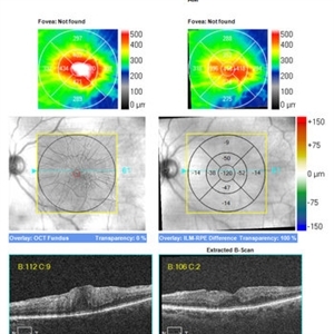
OCT of LE in a 65 year old male case of Epiretinal Membrane causing Macular Pucker.
Photographer: Dr. Anjana Mirajkar -Retina Foundation, Ahmedabad.
Condition/keywords: epiretinal membrane (ERM), macular pucker
-
 Epiretinal Membrane causing Macular Pucker.
Dec 11 2022 by Anjana Mirajkar, MS Ophthalmology
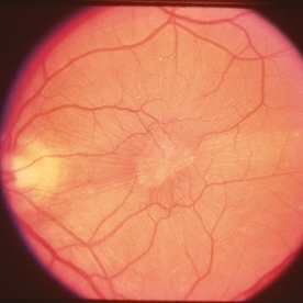
Color Photo of LE in a 65 year old male case of Epiretinal Membrane causing Macular Pucker.
Photographer: Dr. Anjana Mirajkar -Retina Foundation, Ahmedabad.
Condition/keywords: epiretinal membrane (ERM), macular pucker
-
Vitrectomy for epiretinal membrane removal
Nov 30 2022 by Manish Nagpal, MD, FRCS (UK), FASRS
Vitrectomy is carried out for epiretinal membrane removal. After doing core vitrectomy and pvd induction a 25 gauge forceps is used to pinch and peel the macular pucker. After finding a edge the forceps is gradually moved tangentially over the retinal surface to remove the membrane.
Photographer: Manish Nagpal
Condition/keywords: epiretinal membrane, forceps, macular pucker, peeling, video, vitrectomy
-
Epiretinal membrane removal
Oct 24 2022 by Manish Nagpal, MD, FRCS (UK), FASRS
This video highlights the surgical technique of tangentially removing the epiretinal membrane using a forceps
Photographer: Manish Nagpal
Condition/keywords: epiretinal membrane, ERM, macular pucker, staining, video, vitrectomy
-
Epiretinal membrane and ILM peeling
Oct 24 2022 by Manish Nagpal, MD, FRCS (UK), FASRS
This video shows the technique of peeling a epiretinal membrane after triamcinilone staining followed by ILM removal.
Photographer: Manish Nagpal
Condition/keywords: epiretinal membrane (ERM), ILM, macular pucker, staining, video, vitrectomy
-
 Epiretinal Membrane
May 14 2022 by Rinat Sutiushev
Female, born in 1961. Complains of decreased vision and distortion when reading text. The ocular fundus showed retinal surface wrinkling due to membrane contracture.
Photographer: Rinat Sutiushev
Condition/keywords: Cellophane Maculopathy, epiretinal membrane (ERM), macular pucker
-
 Spontaneous Resolution of ERM
Dec 19 2020 by John S. King, MD
78-year-old diabetic with ERM OD and mild PCO OD that was stable for a few years, and eventually improved on own. Her acuity ranged from 20/25-2040 and she did not notice significant visual issues, and we decided to monitor. On her last visit she was 20/40 with 1+ PCO, and has not noticed any visual changes.
Imaging device: Zeiss Cirrus
Condition/keywords: epiretinal membrane (ERM), macular pucker
-
 ERM Improvement Without Surgery
Dec 19 2020 by John S. King, MD
70-year-old white female with a visually significant ERM that did not want surgery. Initially 20/60 J7 OS, and a year later had improved to 20/40 J2 with significant improvement in the foveal region on OCT.
Imaging device: Zeiss Cirrus
Condition/keywords: epiretinal membrane (ERM), macular pucker
-
 Vitreoschisis
Sep 3 2020 by J. Sebag, MD, FACS, FRCOphth, FARVO
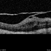
OCT of the left eye in a patient with macular pucker (see SLO image below to right) demonstrates splitting of the posterior vitreous cortex in two separate places. Tangential traction caused thickening of the underlying macula. [For histopathology see: Gupta P, Yee KMP, Garcia P, Rosen RB, Parikh J, Hageman GS, Sadun AA, Sebag J: Vitreoschisis in macular diseases. Brit J Ophthalmol 2011;95(3):376-80]
Condition/keywords: vitreoschisis
-
 Pathophysiology of Vitreoschisis
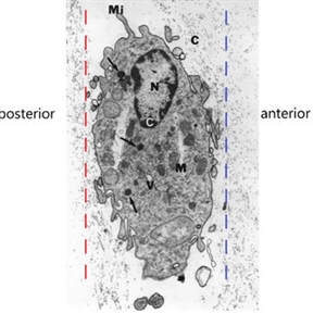
Sep 1 2020 by J. Sebag, MD, FACS, FRCOphth, FARVO
Transmission electron microscopy of human hyalocyte in situ demonstrates embedding within the dense collagen matrix of the posterior vitreous cortex. The retina was to the left (“posterior”) and the anterior segment was to the right (“anterior”). The red dashed line indicates the level of vitreoschisis split that might occur posterior to the level of the hyalocyte monolayer, leaving a thin, hypocellular membrane attached to the macula. The dashed blue line indicates the level of vitreoschisis split that might occur anterior to the level of the hyalocyte monolayer, leaving a thick, hypercellular membrane attached to the macula. The former is more likely to present as macular hole, while the latter as macular pucker (see Figure 12). Mi = microvilli; black C = collagen of posterior vitreous cortex; N = lobulated nucleus typical of mononuclear phagocytes; white C = dense marginal chromatin in nucleus; M = mitochondria; V = vacuoles; arrows = dense granule (original magnification = 11,670) [Modified from Sebag J: Anomalous PVD – a unifying concept in vitreo-retinal diseases. Graefe’s Arch Clin Exp Ophthalmol 2004;242:690-8 and Sebag J, Niemeyer M, Koss M: Anomalous PVD and vitreoschisis. In: Vitreous – in Health & Disease (J. Sebag, ed.) Springer, New York, 2014, pg. 252]
Condition/keywords: pathology, vitreoschisis
-
 Macular Pucker
Jan 7 2020 by RAFAEL REIS PEREIRA, MD
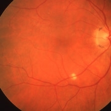
A clinical grading system was proposed by Gass in 1987 describe the different stages of the epiretinal membrane. Grade 2 Macular pucker consists of a thick fibroglial membrane that contracts and produces obscuration of underlying vessels and marked full-thickness retinal distortion. Sometimes associated with cotton-wool spots, exudates, blot hemorrhages, microaneurysms, and cystoid macular edema.
Photographer: Rafael Reis, Retina Clinic - Brazil
Condition/keywords: macular pucker
-
 Macular Pucker with Pseudohole
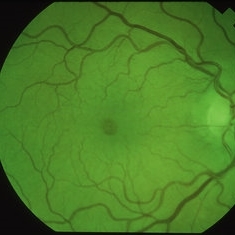
Apr 8 2019 by Gary R. Cook, MD, FACS
Red-free photograph of a 52-year-old white male with a diffuse ERM and pseudohole OD; V.A. = 20/20-1
Imaging device: Topcon VT-50
Condition/keywords: epiretinal membrane (ERM), idiopathic epiretinal membrane, macular pseudohole, macular pucker
-
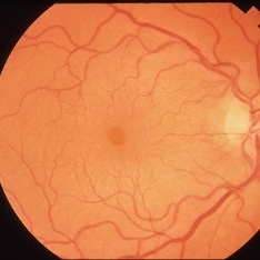 Macular Pucker with Pseudohole
Apr 8 2019 by Gary R. Cook, MD, FACS
52-year-old white male with a diffuse ERM and pseudohole OD; V.A. = 20/20-1
Imaging device: Topcon VT-50
Condition/keywords: epiretinal membrane (ERM), idiopathic epiretinal membrane, macular pseudohole, macular pucker
-
 Macular Pucker
Apr 8 2019 by Gary R. Cook, MD, FACS
64-year-old white female with an idiopathic macular pucker OS following a PVD; V.A. = 20/60
Imaging device: Topcon VT-50
Condition/keywords: epiretinal membrane (ERM), idiopathic epiretinal membrane, macular pucker
-
 Idiopathic ERM
Apr 8 2019 by Gary R. Cook, MD, FACS
22-year old white female with an idiopathic ERM OS; no history of any prior trauma; V.A. = 20/70
Imaging device: Topcon VT-50
Condition/keywords: epiretinal membrane (ERM), idiopathic epiretinal membrane, macular pucker
-
 Macular Pucker After Retinal Detachment
Apr 2 2019 by Gary R. Cook, MD, FACS
Macular pucker following retinal detachment repair OS.
Condition/keywords: epiretinal membrane (ERM), macular pucker, retina surgery complications
-
 ILM Striae fFollowing Laser PRP
Apr 2 2019 by Gary R. Cook, MD, FACS
21-year-old white female with Type 1 DM who developed ILM striae and mild macular puckering following laser P.R.P. treatment OS for PDR; V.A. = 20/40+1
Imaging device: Topcon VT-50
Condition/keywords: laser photocoagulation, laser surgery complications, macular pucker
-
 Macular Pucker Complication of Laser Photocoagulation
Apr 2 2019 by Gary R. Cook, MD, FACS
71-year-old white female with a macular pucker OD secondary to peripheral laser photocoagulation treatment.
Imaging device: Topcon VT-50
Condition/keywords: laser photocoagulation, laser surgery complications, macular pucker
-
 Histoplasmosis and Idiopathic Macular Pucker
Mar 27 2019 by Gary R. Cook, MD, FACS
62-year-old female with presumed ocular histoplasmosis and an idiopathic macular pucker in her right eye; no Histo spots or CNV present in the macula; V.A.= 20/60.
Imaging device: Topcon VT-50
Condition/keywords: epiretinal membrane (ERM), macular pucker, presumed ocular histoplasmosis syndrome (POHS)
-
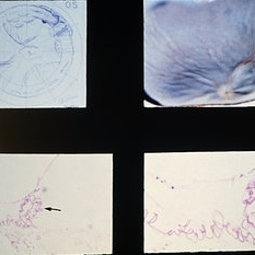 Slide 9-77
Feb 26 2019 by Lancaster Course in Ophthalmology
Case of macular pucker following retinal reattachment. There is a thin, hypocellular preretinal membrane that has contracted, producing wrinkling, detachment, and coiling (arrows) of the internal limiting membrane.
Condition/keywords: hypocellular preretinal membrane, macular pucker, retinal pigmentosa
 Loading…