Search results (48 results)
-
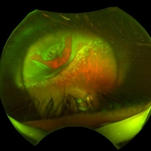
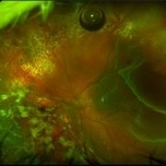 S/P Vitreo Retinal Surgery
Feb 27 2025 by Angela Rico
Patient referred to our office for Evaluation and Treatment of re- detached retina following previous repair of RD. 10% gas Bubble- Macular detachment- PVR Temporal Star fold super-temporal - Multiple irregular Tears infero nasal
Photographer: Angela Rico M.D.
Imaging device: California Optos
Condition/keywords: PVR, retinal detachment
-
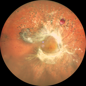
 Retinal Hemangioblastoma
Aug 10 2024 by Muhammad Amer Awan, MD, FRCSEd, FRCOphth, FRCS Glasgow, FACS, FASRS
Fundal photograph of a 21 years old female with a large and small retinal hemangioblastoma with extensive epiretinal membrane over the macula and macular detachment in the right eye.
Condition/keywords: retinal hemangioblastoma
-
 Annular Tractional Retinal Detachment
Jul 4 2024 by Hector Gabriel Moreno Solano, MD, MHA
52-year-old Hispanic female patient with a diagnosis of type II diabetes mellitus of 15 years of evolution, comes to the retina service for progressive visual loss in the right eye (single functional eye) with visual acuity of 20/100, Fundus examination reveals laser-modified proliferative diabetic retinopathy with activity + annular tractional retinal detachment with macular involvement.
Photographer: Hector Gabriel Moreno Solano, MD, MHA, HGZ #20 IMSS Puebla.
Imaging device: Mirante
Condition/keywords: macular detachment, proliferative diabetic retinopathy (PDR), tractional retinal detachment
-
 Ruptured Retinal Artery Macroaneurysm
Jun 18 2024 by KANWALJEET HARJOT MADAN, M.S. (Ophthalmology), FAICO (Vitreous - Retina)
This is a fundus photo depicting ruptured Retinal Artery Macroaneurysm (RAM) in the left eye of a 63 years old female. RAM is an acquired saccular or fusiform dilatation of the retinal arterioles that usually occur within the first three orders of bifurcation. The Superotemporal artery is the most common location. RAM may be asymptomatic or cause a number of complications such as macular edema, serous macular detachment, and hemorrhages.
Photographer: Dr Kanwaljeet Harjot Madan
Condition/keywords: Haemorrhage, macroaneurysm, retinal arteriole
-
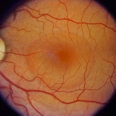
 Central Serous Chorioretinopathy (CSR)
Sep 26 2023 by Ben Serar
Fundus photograph of RE showing serous macular detachment in a case of Central Serous Chorioretinopathy.
Condition/keywords: Central Serous Chorioretinopathy (CSR)
-
 Central Serous Chorioretinopathy (CSR)
Sep 21 2023 by Ben Serar
Fundus photograph showing increased cup-disc ratio with nasalisation of vessels , with thinning of Neuroretinal rim and bayonetting of vessels in a case of Glaucomatous Optic Atrophy (GOA) Fundus photograph of LE showing serous macular detachment in a case of Central Serous Chorioretinopathy (CSR).
Condition/keywords: Central Serous Chorioretinopathy (CSR)
-
 Central Serous Chorioretinopathy (CSR)
Sep 21 2023 by Ben Serar
Fundus photograph of RE showing serous macular detachment in a case of Central Serous Chorioretinopathy.
Condition/keywords: Central Serous Chorioretinopathy (CSR)
-
 Central Serous Chorioretinopathy (CSR)
Sep 21 2023 by Ben Serar
Fundus photograph of LE showing serous macular detachment in a case of Central Serous Chorioretinopathy.
Condition/keywords: Central Serous Chorioretinopathy (CSR)
-
 Central Serous Chorioretinopathy (CSR)
Sep 14 2023 by Ben Serar
Fundus photograph of LE showing serous macular detachment in a case of Central Serous Chorioretinopathy.
Condition/keywords: Central Serous Chorioretinopathy (CSR)
-
 Central Serous Chorioretinopathy (CSR)
Sep 14 2023 by Ben Serar
Fundus photograph of LE showing serous macular detachment in a case of Central Serous Chorioretinopathy.
Condition/keywords: Central Serous Chorioretinopathy (CSR)
-
 Central Serous Chorioretinopathy (CSR)
Sep 12 2023 by Ben Serar
Fundus photograph of LE showing serous macular detachment in a case of Central Serous Chorioretinopathy
Condition/keywords: Central Serous Chorioretinopathy (CSR)
-
 Central Serous Chorioretinopathy (CSR)
Sep 12 2023 by Ben Serar
Fundus photograph of LE showing serous macular detachment in a case of Central Serous Chorioretinopathy.
Condition/keywords: Central Serous Chorioretinopathy (CSR)
-
 Central Serous Chorioretinopathy (CSR)
Sep 12 2023 by Ben Serar
Fundus photograph of RE showing serous macular detachment in a case of Central Serous Chorioretinopathy.
Condition/keywords: Central Serous Chorioretinopathy (CSR)
-
 Central Serous Chorioretinopathy (CSR)
Sep 12 2023 by Ben Serar
Fundus photograph of LE showing serous macular detachment in a case of Central Serous Chorioretinopathy.
Condition/keywords: Central Serous Chorioretinopathy (CSR)
-
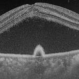 Central serous chorioretinopathy
Nov 18 2022 by T. P . VIGNESH, MBBS,MS
OCT of a 45 year old man revealing serous macular detachment with a subfoveal PED .
Photographer: Priyanka
Imaging device: Topcon Triton
Condition/keywords: central serous chorioretinopathy (CSCR)
-
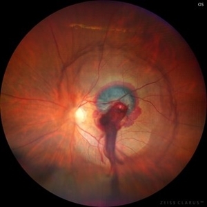
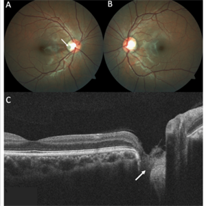 Optic Disc Pit
Nov 8 2021 by Michael Grinton
Optic disc pits are rare congenital abnormalities of the optic nerve head. Colour fundus image of an asymptomatic 18-year old male shows an optic disc pit in the right eye (A, white arrow); a small, grey, oval shaped excavation in the temporal segment of the optic disc. These pits are usually unilateral (B shows normal colour fundus of left eye) and asymptomatic. Imaging with optical coherence tomography (C) shows the optic disc pit in cross section (white arrow) and normal macular structure. In some patients with the condition, fluid can accumulate underneath the macular (serous macular detachment).
Condition/keywords: Optic disc pit, Optic nerve pit, Optic pit
-
 Optic Disc Pit with Coloboma (Hybrid Anomaly)
Jun 10 2021 by Janani Sreenivasan
Optic disc pit is a rare anomaly of the optic nerve head that can be associated with maculopathy leading to progressive visual deterioration. It belongs to the spectrum of congenital cavitary anomalies of the optic disc which encompasses extrapapillary cavitation, optic disc coloboma, and morning glory. Very rarely, optic disc pits are seen in combination with optic disc colobomas. Histopathologically, disc pit is defined as herniation of dysplastic retinal tissue into an excavation, rich in collagen, which can stretch into the subarachnoid space via a defect in the lamina cribrosa. Interestingly, this structural abnormality leading to a non-physiological communication between the intraocular and extraocular spaces is a common feature among all the congenital cavitary disc anomalies. Optic disc pit maculopathy is characterized by intraretinal and subretinal fluid at the area of macula. The origin of the retinal fluid remains unclear. Possible sources include the vitreous cavity, the subarachnoid space and the orbital space surrounding the dura. It has been estimated that approximately 25% to 75% of patients will develop serous detachment and/or retinoschisis of the central macula at some stage of their life. On fundus examination, ODPs typically appear as single grayish, round or oval depressions at the optic disc. Most commonly, they are detected at the inferotemporal segment of the disc, but may also be observed elsewhere, including the central area.The coexisting macular detachment can be related to lamellar or full-thickness macular holes, cystoid changes, retinal pigment epithelium atrophy and eventually to irreversible loss of vision,especially in longstanding cases. Herewith, we present a 32-years-old male patient presenting with an unusual combination of optic disc pit with maculopathy and optic disc coloboma (hybrid anomaly) in the same eye with corrected visual acuity of 3/60.
Photographer: Dr Janani Sreenivasan
Imaging device: Zeiss Cirrus HD-OCT
Condition/keywords: coloboma of optic disc, hybrid anomaly, macular detachment, optic disc, optic disc pit
-
 "Hang in There"
Apr 20 2021 by Tomas Minelli, MD
Fundus wide field photograph of a 50-year-old man with a macular detachment associated with a big temporal superior tear. The laser is firmly holding the progression of the tear in the 14th day post- laser. BCVA 20/20
Photographer: Livia Conci, Universtity of São Paulo
Imaging device: Optos Daytona
Condition/keywords: giant retinal tear
-
 FA 40 Seconds - Large Hemorrhage With Macular Detachment Due to AMD
Nov 7 2019 by John S. King, MD
81-year-old white female with three day history of seeing a "dark blob" nasally OD; no blood thinners; vision was 20/100- J16 with 2+NSC OD; OCT (not shown) had large SRF that included the fovea and extended out temporally. Posterior segment showed a large amount of SRF in the macula with some SRH in the inferior portion of the macula, hemorrhagic PEDs temporally with some RPE scarring and SRH in the periphery. On the FA there is blockage by the SRH and SRPE heme; there is staining peripherally; there is a wave of leakage that extends out into the macula and pools into to subretinal space.
Photographer: Brandon Peter
Condition/keywords: retinal pigment epithelium, subretinal hemorrhage, wet age-related macular degeneration (wet AMD)
-
 FA 5 min - Large Hemorrhage With Macular Detachment Due to AMD
Nov 7 2019 by John S. King, MD
81-year-old white female with three day history of seeing a "dark blob" nasally OD; no blood thinners; vision was 20/100- J16 with 2+NSC OD; OCT (not shown) had large SRF that included the fovea and extended out temporally. Posterior segment showed a large amount of SRF in the macula with some SRH in the inferior portion of the macula, hemorrhagic PEDs temporally with some RPE scarring and SRH in the periphery. On the FA there is blockage by the SRH and SRPE heme; there is staining peripherally; there is a wavbe of leakage that extends out into the macula and pools into to subretinal space.
Photographer: Brandon Peter
Condition/keywords: retinal pigment epithelium, subretinal hemorrhage, wet age-related macular degeneration (wet AMD)
-
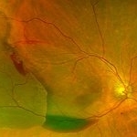 Photo of Large Hemorrhage with macular detachment due to AMD
Nov 7 2019 by John S. King, MD
81-year-old white female with three day history of seeing a "dark blob" nasally OD; no blood thinners; vision was 20/100- J16 with 2+NSC OD; OCT (not shown) had large SRF that included the fovea and extended out temporally. Posterior segment showed a large amount of SRF in the macula with some SRH in the inferior portion of the macula, hemorrhagic PEDs temporally with some RPE scarring and SRH in the periphery. On the FA there is blockage by the SRH and SRPE heme; there is staining peripherally; there is a wavbe of leakage that extends out into the macula and pools into to subretinal space. Anti-VEGF given; f/u one month.
Photographer: Brandon Peter
Condition/keywords: retinal pigment epithelium, subretinal hemorrhage, wet age-related macular degeneration (wet AMD)
-
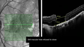 Closing the Persistent Macular Hole: Third Time’s the Charm
May 7 2019 by Srinivas Joshi, MD, FASRS
Patient was operated for macular hole with vitrectomy and conventional ILM peeling following which macular hole did not close. In second step, macular detachment induction with subretinal BSS injection using 42G needle was done but macular hole did not close. In third attempt a neurosensory retinal patch graft surgery was done and finally the macular hole was closed successfully with type 1 closure.
Condition/keywords: macular detachment, macular hole, neurosensory retinal patch graft, subretinal BSS
-
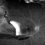
 CSR with large RPED
Mar 26 2019 by Gary R. Cook, MD, FACS
Mid-phase FA showing large RPED inferonasal to optic disc with overlying cruciate pigment figures (black lines) and neurosensory macular detachment OD.
Imaging device: Topcon VT-50
Condition/keywords: central serous retinopathy (CSR), neurosensory detachment of retina, retinal pigment epithelium (RPE) detachment
-
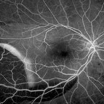
 Bilateral Central Serous Retinopathy
Mar 26 2019 by Gary R. Cook, MD, FACS
Late-phase frame of FA of 37-year-old white male with acute CSR OD showing pooling of dye beneath the small central RPED centrally, a smokestack-type leak from the RPE defect just above it, and mild late pooling of dye outlining the large neurosensory macular detachment; VA = 20/80-1.
Imaging device: Topcon VT-50
Condition/keywords: central serous retinopathy (CSR), FA late phase, FA late phase leakage, neurosensory detachment of retina
-
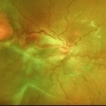
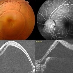
 Central Serous Retinopathy
Mar 26 2019 by Gary R. Cook, MD, FACS
43-year-old white male with acute CSR OD showing a large neurosensory macular detachment in his right eye.
Imaging device: Topcon VT-50
Condition/keywords: central serous retinopathy (CSR), neurosensory detachment of retina
 Loading…