Search results (32 results)
-
 Macular Coloboma
Jun 5 2025 by César Adrián Gómez Valdivia, MD
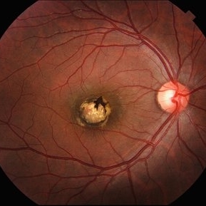
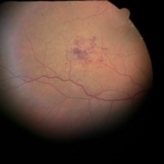
Macular Coloboma found in a 28 year-old male patient, visual acuity was 20/60. Resulting due to fusion failure of the optic fissure, colobomas are commonly found in the infero-nasal quadrant. If the retina is involved, it is reduced to glial tissue with no underlying RPE or choroid. This appears as an area of whitening often with pigment deposition at the junction of the coloboma and normal retina. Findings were bilateral.
Photographer: @eyemissu2
Imaging device: TOPCON TRC-50DX
Condition/keywords: coloboma
-
 Morning Glory Anomaly with Macular Atrophy
Oct 30 2024 by Luis Guillermo Anaya Sánchez, Ophthalmology
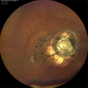
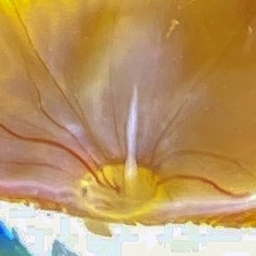
47 year old female patient, presents to evaluate low vision since birth. Fundus photography shows an enlarged disc, with radial vasculature disposition, and glial tissue; corresponding with a Morning Glory Anomaly, with macular atrophy.
Photographer: Luis Guillermo Anaya MD
Imaging device: Zeiss Clarus 700
Condition/keywords: Morning Glory Anomaly
-
 Morning glory disc anomaly-associated maculopathy: fibroglial tissue with a Mac-Off serous retinal detachment.
Jun 26 2024 by Julián Villarreal, MD
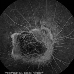
19 year old with a Morning glory disc anomaly-associated maculopathy: fibroglial tissue with a Mac-Off serous retinal detachment.
Photographer: Julián Villarreal MD
Imaging device: Mirante
Condition/keywords: fibroglial tissue, Morning Glory Anomaly, retinal detachment of the macula
-
 Morning Glory Disc Anomaly
Feb 12 2024 by NIDHI PANWAR, MD FRCS Glasgow FNB FICO
Fundus photograph of 43 year old male, hypertensive on medication, came for routine check up, and has been diagnosed to have poor vision left eye since childhood, denies any history of trauma. Vision left eye 6/18, Anterior segment normal, Fundus left eye shows excavated ,funnel-shaped optic nerve head, with central tuft of glial tissue obscuring the cup . The retinal vessels were seen emanating from the edge of disc in radial manner. In addition, the sectoral nasal retina shows localized area of hyperpigmented bony spicules like lesions. However, no history of nyctalopia or any other neurological disorder could be obtained.
Photographer: Nidhi Panwar, NMC Royal hospital, Sharjah , UAE
Imaging device: OPTOMAP
Condition/keywords: Morning Glory Anomaly, optic disc excavation
-
 Combined Pigment Epithelial and Retinal Hamartoma
Oct 3 2021 by Luiz A Zago, PhD
23-year-old male with a combined pigment epithelial and retinal hamartoma and a secondary neovascularization and evolution with a glial tissue.
Photographer: Luiz Alberto Zago Filho
Imaging device: Topcon 50IX
Condition/keywords: Combined pigment epithelial and retinal hamartoma
-
 Retinal Cavernous Hemangioma
Apr 23 2021 by Aparna Ghodake
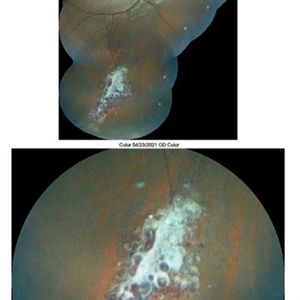
Fundus photograph of a 14-year-old girl having amblyopia was incidentally found to have numerous dark blood filled saccular aneurysms embedded in white fibroglial tissue giving it a 'bunch of grapes' appearance characteristic of retinal cavernous hemangioma.
Photographer: Dr. Aparna Ghodake, Sri Sankaradeva netralaya, Guwahati, Assam, India
Imaging device: Zeiss visucam 500
Condition/keywords: cluster of grapes, fundus photograph
-
 Bergmeister's Papilla
Sep 29 2020 by Dhaivat Shah
Bergmeister's papilla is a small tuft of glial tissue which arises from the center of the optic disc, and represents a remnant of the fetal hyaloid artery. The hyaloid artery provides nutrition to the lens during development, and runs forward to the lens from the optic disc. At birth the hyaloid artery regresses, and is normally completely regressed by the time of birth. Bergmeister's papilla is frequently observed as an incidental clinical finding if this artery has an incomplete regression posteriorly. However, in the severe forms it can be associated with cataracts, persistence of the primitive vitreous, microphthalmia, vitreous hemorrhages and sometimes tractional retinal detachment, due to contraction of the residual fibro vascular tissue. Therefore, careful monitoring of vitreous thickening in the peripapillary areas, both by examining the ocular fundus, and especially by SD-OCT, is of considerable importance. Here we have one such of a 30 year old young male who came in for a routine checkup, in whom we noted a Bergmeister’s papilla. Due to its benign nature, patient was reassured and was asked to follow up yearly.
Condition/keywords: Bergmeister's Papillae
-
 Bergmeister Papilla
Feb 20 2020 by Nisarg Joshi, MD
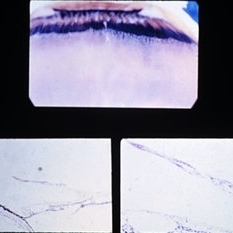
Gross pathology photo of a Bergmeister Papilla. It is a remnant of incompletely resorbed hyaloid vasculature from ocular development. This glial tissue is seen emminating from the optic nerve, which also shows glaucomatous cupping. The eye was enucleated due to a choroidal melanoma.
Photographer: Nisarg Joshi, MD, Geisinger Medical Center
Imaging device: Digital camera
Condition/keywords: Bergmeister's Papillae, hyaloid artery, persistent fetal vasculature (PFV)
-
 Slide 9-70
Feb 26 2019 by Lancaster Course in Ophthalmology
Peripheral retinal zonular traction tuft. A strand of fibroglial tissue extends anteriorly over the pars plana from the peripheral retina. A zonular fiber or condensed vitreous strand (arrow) is attached to the apex of the tuft.
Condition/keywords: fibroglial tissue, pars plana, retinal zonular traction tuft
-
 Retinal Cavernous Hemangioma
Sep 21 2018 by John S. King, MD
54 -year-old sent in with CME and Dx of BVO. She was 20/70. There are some exudates just nasal to the fovea at the edge of this photos, due to DME. There is a cluster of saccular aneurysms infero-temporally without any glial tissue overlying them.
Imaging device: Topcon
Condition/keywords: cavernous hemangioma of the retina
-
---thumb.jpg/image-square;max$300,300.ImageHandler) Accumulation Of Glial Tissue
Accumulation Of Glial Tissue
Oct 31 2013 by Maurice F. Rabb
19 year old white female with an accumulation of glial tissue.
Condition/keywords: glial tissue
-
---thumb.jpg/image-square;max$300,300.ImageHandler) Accumulation Of Glial Tissue
Accumulation Of Glial Tissue
Oct 31 2013 by Maurice F. Rabb
19 year old white female with an accumulation of glial tissue.
Condition/keywords: glial tissue
-
---thumb.jpg/image-square;max$300,300.ImageHandler) Accumulation Of Glial Tissue
Accumulation Of Glial Tissue
Oct 31 2013 by Maurice F. Rabb
19 year old white female with an accumulation of glial tissue.
Condition/keywords: glial tissue
-
---thumb.jpg/image-square;max$300,300.ImageHandler) Accumulation Of Glial Tissue
Accumulation Of Glial Tissue
Oct 31 2013 by Maurice F. Rabb
19 year old white female with an accumulation of glial tissue.
Condition/keywords: glial tissue
-
---thumb.jpg/image-square;max$300,300.ImageHandler) Accumulation Of Glial Tissue
Accumulation Of Glial Tissue
Oct 31 2013 by Maurice F. Rabb
19 year old white female with an accumulation of glial tissue.
Condition/keywords: glial tissue
-
---thumb.jpg/image-square;max$300,300.ImageHandler) Accumulation Of Glial Tissue
Accumulation Of Glial Tissue
Oct 31 2013 by Maurice F. Rabb
19 year old white female with an accumulation of glial tissue.
Condition/keywords: glial tissue
-
---thumb.jpg/image-square;max$300,300.ImageHandler) Accumulation Of Glial Tissue
Accumulation Of Glial Tissue
Oct 31 2013 by Maurice F. Rabb
19 year old white female with an accumulation of glial tissue.
Condition/keywords: glial tissue
-
---thumb.jpg/image-square;max$300,300.ImageHandler) Accumulation Of Glial Tissue
Accumulation Of Glial Tissue
Oct 31 2013 by Maurice F. Rabb
19 year old white female with an accumulation of glial tissue.
Condition/keywords: glial tissue
-
---thumb.jpg/image-square;max$300,300.ImageHandler) Accumulation Of Glial Tissue
Accumulation Of Glial Tissue
Oct 31 2013 by Maurice F. Rabb
19 year old white female with an accumulation of glial tissue.
Condition/keywords: glial tissue
-
---thumb.jpg/image-square;max$300,300.ImageHandler) Accumulation Of Glial Tissue
Accumulation Of Glial Tissue
Oct 31 2013 by Maurice F. Rabb
19 year old white female with an accumulation of glial tissue.
Condition/keywords: glial tissue
-
---thumb.jpg/image-square;max$300,300.ImageHandler) Accumulation Of Glial Tissue
Accumulation Of Glial Tissue
Oct 31 2013 by Maurice F. Rabb
19 year old white female with an accumulation of glial tissue.
Condition/keywords: glial tissue
-
---thumb.jpg/image-square;max$300,300.ImageHandler) Accumulation Of Glial Tissue
Accumulation Of Glial Tissue
Oct 31 2013 by Maurice F. Rabb
19 year old white female with an accumulation of glial tissue.
Condition/keywords: glial tissue
-
---thumb.jpg/image-square;max$300,300.ImageHandler) Accumulation Of Glial Tissue
Accumulation Of Glial Tissue
Oct 31 2013 by Maurice F. Rabb
19 year old white female with an accumulation of glial tissue.
Condition/keywords: glial tissue
-
---thumb.jpg/image-square;max$300,300.ImageHandler) Accumulation Of Glial Tissue
Accumulation Of Glial Tissue
Oct 31 2013 by Maurice F. Rabb
19 year old white female with an accumulation of glial tissue.
Condition/keywords: glial tissue
-
---thumb.jpg/image-square;max$300,300.ImageHandler) Accumulation Of Glial Tissue
Accumulation Of Glial Tissue
Oct 31 2013 by Maurice F. Rabb
19 year old white female with an accumulation of glial tissue.
Condition/keywords: glial tissue
 Loading…