 Initializing download.
-
 By John S. King, MD
Retina Associates, PA - Uploaded on Sep 21, 2018.
- Last modified by Caroline Bozell on Sep 21, 2018.
- Rating
- Appears in
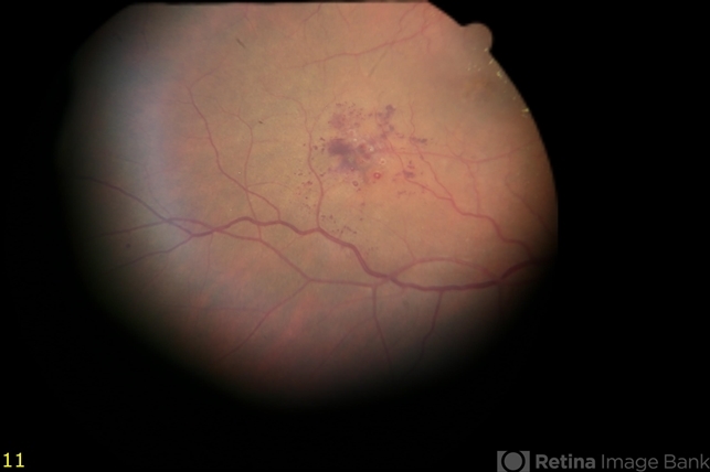
- Cavernous Hemangioma of the Retina
- Condition/keywords
- cavernous hemangioma of the retina
- Imaging device
-
Fundus camera
Topcon - Description
- 54 -year-old sent in with CME and Dx of BVO. She was 20/70. There are some exudates just nasal to the fovea at the edge of this photos, due to DME. There is a cluster of saccular aneurysms infero-temporally without any glial tissue overlying them.


---thumb.jpg/image-square;max$79,0.ImageHandler "Optic disc")


---thumb.jpg/image-square;max$79,0.ImageHandler "Cavernous Hemangioma of the Retina")


