Search results (44 results)
-
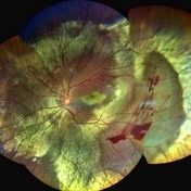
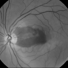
 Floral Cherry-red Spot
Dec 3 2024 by Andrea Espinosa Quintana, MD
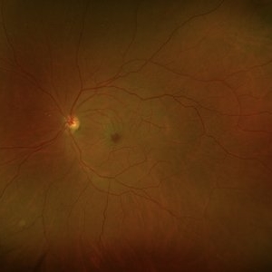
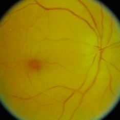
Fundus photograph of a 54 year old woman that presented Central Retinal Artery Occlusion (CRAO) with cherry-red spot in a flowery form.
Photographer: Andrea Espinosa, Fundación Hospital Nuestra Señora de la Luz
Condition/keywords: central retinal artery occlusion (CRAO), cherry red spot
-
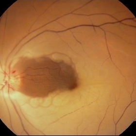
 Traumatic CRAO with Cilioretinal Artery Sparing
Sep 10 2024 by KRISHNENDU NANDI, MS
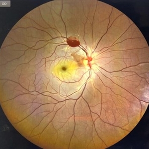
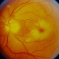
A 25 year-old male presented with dimness of vision in the right eye for the last 3 days following blunt trauma. The BCVA of the right eye was CF close to the face and left eye was 6/6, N6 in Snellen’s chart. On examination the retina showed CRAO with cherry red spot and sparing of cilioretinal artery circulation. Traumatic subhyaloid haemorrhage also noted at supero-temporal arcade.
Photographer: Dr Krishnendu Nandi
Condition/keywords: CRAO, subhyaloid hemorrhage, Trauma
-
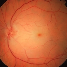
 Central Retinal Artery Occlusion-Widefield
Apr 10 2024 by Tejaswita Verma
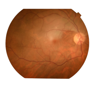
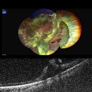
Left eye widefield color photo of 75 year old male with pale edematous retina with cherry red spot suggestive of central retinal artery occlusion.
Photographer: DR. TEJASWITA VERMA
Imaging device: MIRANTE
Condition/keywords: central retinal artery occlusion (CRAO), cherry red spot
-
 Central Retinal Artery Occlusion OCT
Apr 10 2024 by Tejaswita Verma
Left eye OCT of a 75 year old male with central retinal artery occlusion showing altered foveal contour with loss of differentiation of layers with thickening.
Photographer: DR. TEJASWITA VERMA
Imaging device: MIRANTE
Condition/keywords: central retinal artery occlusion, cherry red spot
-
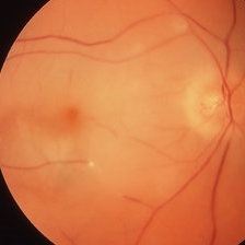
 Central Retinal Artery Occlusion
Apr 10 2024 by Tejaswita Verma
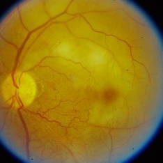
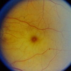
Left eye fundus photo of a 75 year old male with pale edematous retina with cherry red spot in a case of central retinal artery occlusion.
Photographer: DR. TEJASWITA VERMA
Imaging device: MIRANTE
Condition/keywords: central retinal artery occlusion (CRAO), cherry red spot
-
 CRAO
CRAO
Jan 8 2024 by ANKIT JAIN
RIGHT EYE FUNDUS IMAGE OF A 68 YEARS OLD MALE WITH SUDDEN LOSS OF VISION, WHO IS A KNOWN CASE OF HYPERTENSION FOR 15 YEARS
Photographer: Dr Ankit Jain
Condition/keywords: central retinal artery occlusion (CRAO), cherry red spot
-
 Central Retinal Artery Occlusion
Nov 16 2023 by Gabriel Costa Andrade, PhD
Fundus photograph of an 62-year-old man with retinal whitening and a cherry red spot due to Central Retinal Artery Occlusion.
Photographer: Gabriel Andrade
Condition/keywords: Central Retinal Artery Occlusion, central retinal artery occlusion (CRAO), Retina
-
 Branch Retinal Artery Occlusion (BRAO)
Sep 26 2023 by Ben Serar
Fundus photograph of LE showing retinal edema and opacification along the superotemporal arcade, with cherry red spot at the macula, in a case of Branch Retinal Artery Occlusion (BRAO).
Condition/keywords: branch retinal artery occlusion (BRAO), cherry red spot
-
 Central Retinal Artery Occlusion(CRAO)
Sep 21 2023 by Ben Serar
Fundus photograph of RE showing retinal edema and opacification with a cherry red spot at the macula in a case of Central Retinal Artery Occlusion (CRAO).
Condition/keywords: central retinal artery occlusion (CRAO), cherry red spot
-
 Central Retinal Artery Occlusion(CRAO)
Sep 14 2023 by Ben Serar
Fundus photograph of the LE showing retinal edema with opacification , with cherry red spot at the macula , in a case of Central Retinal Artery Occlusion (CRAO).
Condition/keywords: Central Retinal Artery Occlusion(CRAO), cherry red spot
-
 Cherry red spot
Sep 12 2023 by Ben Serar
Fundus Photograph of LE showing cherry red spot at the macula, with surrounding retinal edema.
Condition/keywords: cherry red spot
-
 Traumatic Retinal Tear
Jan 20 2022 by Aditya S Kelkar, MS, FRCS, FASRS,FRCOphth
Color fundus photograph of a 34-year old man's left eye, 2 hours after a tennis ball injury, showing commotio retinae with Berlin's edema and cherry red spot in the fovea along with linear retinal tears in the temporal equatorial zone. Adjoining OCT slice taken through the break shows full thickness retinal tear without any underlying choroidal rupture.
Photographer: Dr Sukanya Mondal, National Institute of Ophthalmology, Pune. India
Imaging device: Zeiss Clarus 500
Condition/keywords: Berlin's edema, cherry red spot, commotio retinae, retinal tear
-
 Traumatic Retinal Tear
Dec 5 2021 by Aditya S Kelkar, MS, FRCS, FASRS,FRCOphth
Color fundus photograph of a 34-year old man's left eye, 2 hours after a tennis ball injury, showing commotio retinae with Berlin's edema and cherry red spot in the fovea along with linear retinal tears in the temporal equatorial zone.
Photographer: Dr Sukanya Mondal. National Institute of Ophthalmology, Pune. India.
Imaging device: Zeiss Clarus 500
Condition/keywords: Berlin's edema, cherry red spot, commotio retinae, retinal tear
-
 CRAO Post COVID-19
May 21 2021 by Deepak Bhojwani, MS
Fundus Image of a 67-year old lady with history of COVID-19 disease 20 days back with elevated D-DIMER, CRP levels came to our clinic with sudden diminution of vision in her right eye since last 4 days. The fundus photo shows cherry red spot and severely attenuated vessels and pale disc suggesting classic CRAO. FFA was also done to confirm the same. We could only retrospectively attribute this artery occlusion secondary to recent past history of COVID disease since this lady had no other systemic co-morbidities.
Photographer: Deepak Bhojwani
Condition/keywords: central retinal artery occlusion (CRAO), COVID-19
-
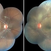 Commotio Retinae
Apr 27 2021 by Priya Rasipuram Chandrasekaran, MBBS, DO, DNB, FRCS
This is the fundus photo montage of a 11-year-old boy showing extensive commotio retinae evidenced by the absence of greyish opacification of the retina with the absence of the usual red reflex following blunt trauma to the left eye. There is no pseudo cherry red spot. The right eye has been added for comparison.
Condition/keywords: commotio retinae
-
 Acute Central Retinal Artery Occlusion with Natural Reperfusion
Mar 12 2021 by Kushal S Delhiwala, MBBS, MS, FMRF,FICO, FAICO
Fundus photographs of 33-year-old healthy male with right eye acute CRAO of 12 hours duration showing cattle trucking, extensive retinal whitening and cherry red spot (left image). Right image 18 hours later showing reduced extent of retinal whitening and absent cattle trucking, suggestive of natural restoration of perfusion.
Photographer: Kushal Delhiwala, Netralaya superspeciality eye hospital, Ahmedabad, Gujarat,India
Imaging device: Optos Daytona
Condition/keywords: cattle trucking, central retinal artery occlusion (CRAO), cherry red spot
-
 Central Retinal Artery Occlusion
Jan 22 2021 by Renata Garcia Franco, Md
65-year-old male, history of uncontrolled systemic arterial hypertension. Segmentation of blood in retinal arterioles, retinal whitening and cherry red spot.
Photographer: Fatima Hernandez, Instituto de la Retina del Bajio SC
Imaging device: Zeiss
Condition/keywords: central retinal artery occlusion (CRAO)
-
 BRAO with Cherry Red Spot
Mar 28 2020 by Sugnesh Parmar
20-year-old male presented with sudden loss of vision in his RE and on examination found to have BRAO with typical cherry red spot.
Photographer: Dr. Sugnesh Parmar, Radheshyam retina hospital, Bhavnagar, Gujarat, India
Condition/keywords: branch retinal artery occlusion (BRAO)
-
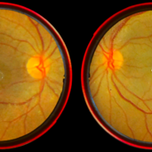 Central Retinal Artery Occlusion
Jan 13 2020 by Prithvi Chandrakanth
37-years-old male with complaints of sudden diminution of vision in the left eye for the past three days. Fundus examination revealed pale retina in the left eye with cherry red spot and normal fundus picture in right eye.
Photographer: DR.PRITHVI CHANDRAKANTH, ARAVIND EYE HOSPITAL, UDUMALPET
Imaging device: TRASH TO TREASURE RETCAM
Condition/keywords: central retinal artery occlusion (CRAO), cherry red spot, retcam, smartphone fundus photography
-
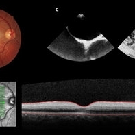 Central Retinal Artery Occlusion Leading to Patent Foramen Ovale Diagnosis
Sep 13 2019 by Patrícia José Figueiredo Lopes
A 19-year-old man presented in emergency department (ED) reporting painless blurred vision in the right eye that started one hour ago while he was doing exercise. His medical history was unremarkable. On examination, best corrected visual acuity in the right eye was counting fingers (20cm), right relative afferent pupillary defect was evident, intraocular pressure and anterior segment were normal. Dilated retinal examination revealed retinal whitening in the macular area and a cherry red spot (panel A) that became increasingly evident with time. Patient denied other systemic symptoms. Macular spectral domain optic coherence tomography showed hyperreflectivity of the inner retina (panel B). In ED, patient underwent ocular massage using a three-mirror contact lens and topical hypotensive treatment. Additionally, oral antiplatelet and hyperbaric oxygen treatment were initiated. Further investigation was performed and fluorescein angiography revealed a delay in arterial filling. Blood tests including hypercoagulation disorders investigation, plain chest radiography and electrocardiogram were unremarkable. Patent foramen ovale was diagnosed in transesophageal echocardiogram (panel C), anticoagulation therapy was promptly initiated and percutaneous closure of patent foramen ovale was done successfully a few weeks later. Final best corrected visual acuity was 20/200 and macula developed atrophy.
Photographer: Patrícia José
Condition/keywords: central retinal artery occlusion (CRAO), patent foramen ovale
-
 Central Retinal Artery Occlusion with Cilioretinal Artery Sparing
Jun 12 2019 by Unnati Vishwanath Shukla, M. S. ,DNB, FVRS FNERF, MNAMS,PhD Scholar(Retina)
A young female patient of Indian origin on oral contraceptive medication presenting with central retinal artery occlusion with cilioretinal artery sparing.
Photographer: Unnati Shukla, C.H. Nagri Eye Hospital, NHL medical college, Ahmedabad,Gujarat,India.
Condition/keywords: central retinal artery occlusion (CRAO), cherry red spot, cilioretinal sparing
-
 Central Retinal Artery Occlusion
Jun 4 2019 by Unnati Vishwanath Shukla, M. S. ,DNB, FVRS FNERF, MNAMS,PhD Scholar(Retina)
A young female patient of Indian origin on Oral Contraceptive medication presenting with Central Retinal Artery Occlusion with Cilioretinal artery Sparing.
Photographer: Unnati Shukla, C.H. Nagri Eye Hospital, NHL medical college, Ahmedabad,Gujarat,India.
Condition/keywords: central retinal artery occlusion (CRAO), cherry red spot, cilioretinal sparing, pale retina
-
 Central Retinal Artery Occlusion
Mar 26 2019 by Gary R. Cook, MD, FACS
61-year-old male patient with acute CRAO OS demonstrating a hyperemic optic disc with a couple of peripapillary hemorrhages, generalized arteriolar narrowing, a cherry-red spot in the macula, and retinal whitening surrounding the fovea; VA= LP.
Imaging device: Topcon VT-50
Condition/keywords: central retinal artery occlusion (CRAO), cherry red spot, retinal whitening
-
 Embolic Central Retinal Artery Occlusion
Mar 26 2019 by Gary R. Cook, MD, FACS
58-year-old WM with embolic CRAO demonstrating a a cherry-red spot in macula, retinal whitening around the fovea, and the embolus in a inferotemporal branch retinal arteriole; VA= HM 6''
Imaging device: Topcon VT-50
Condition/keywords: central retinal artery occlusion (CRAO), cherry red spot, embolus, retinal whitening
-
 Central Retinal Artery Occlusion With Cilioretinal Sparing
Apr 4 2018 by Soumya Venkatesh
Fundus photograph of a 23-year-old gentleman presenting with sudden loss of vision 2 days prior to presentation. He underwent all relevant investigations and found to have APLA positive. He also had dengue serology positive. On follow up, his retinal edema reduced unmasking the underlying hemorrhages( flame shaped).
Photographer: Soumya Harapanahalli Venkatesh, JSS university, Karnataka, India
Condition/keywords: central retinal artery occlusion (CRAO), cherry red spot, cilioretinal sparing, retinal ischemia
 Loading…