Search results (60 results)
-
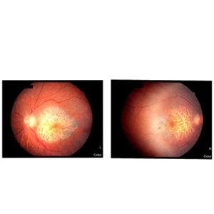
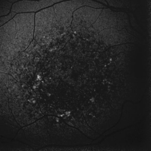
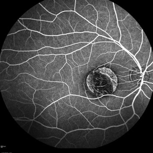
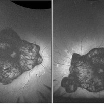 Central Areolar Choroidal Dystrophy
Aug 21 2023 by rahul saradge
54 year old female well circumscribed, bilateral and symmetrical lesion with loss of retinal and choroidal tissue in the macular area.
Photographer: Sushil Zende, Isha Netralaya
Imaging device: Optos
Condition/keywords: autofluorescence imaging, central areolar choroidal dystrophy (CACD)
-
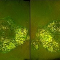 Central Areolar Choroidal Dystrophy
Aug 21 2023 by rahul saradge
54 year old female well circumscribed, bilateral and symmetrical lesion with loss of retinal and choroidal tissue in the macular area.
Photographer: Sushil Zende, Isha Netralaya
Imaging device: Optos
Condition/keywords: central areolar choroidal dystrophy (CACD)
-
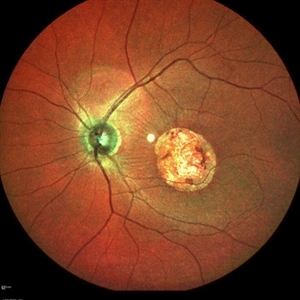
 Central Areolar Choroidal Dystrophy
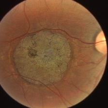
Jul 24 2023 by Mohammadkarim Johari
Fundus photograph of left eye, 45 y/o male with decreased vision bilaterally from childhood. Central areolar choroidal dystrophy (CACD) is a hereditary retinal disorder that affects the macula, resulting in progressive and usually profound visual loss. The hallmark feature of the disorder is a well-defined atrophy of the retinal pigment epithelium (RPE) and the choriocapillaris
Photographer: Mohammadkarim Johari, Shiraz university of medical science
Condition/keywords: central areolar choroidal dystrophy (CACD), choroid, dystrophy
-
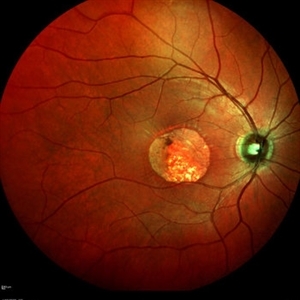
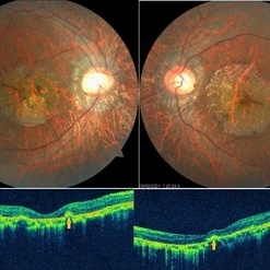 Central Areolar Choroidal Dystrophy
May 4 2021 by Priya Rasipuram Chandrasekaran, MBBS, DO, DNB, FRCS
Fundus photo of a 34-year-old male showing bilaterally symmetrical atrophy of retinal pigment epithelium (RPE) and choriocapillaris involving the fovea and highlighting the underlying large choroidal vessels. OCT macula shows atrophy of the outer retinal layers up to the external limiting membrane along with thinning of RPE and Bruch's membrane complex. Rosette - like hyperreflective structures causing retinal elevation at the border of atrophic area (yellow arrows) are seen categorizing this into stage 4 disease.
Condition/keywords: central areolar choroidal dystrophy (CACD)
-
 Central Areolar Choroidal Dystrophy
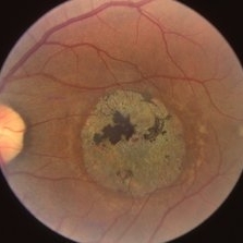
Oct 30 2020 by Mihir Trivedi
Fundus photo of a 43-year-old female with gradual onset diminution of vision in both eyes since 2-3 years. BCVA in OU was 3/60. She was diagnosed to have central areolar choroidal dystrophy(CACD). Central areolar choroidal dystrophy (CACD) is a rare inherited disease, which causes progressive profound loss of vision in patients during their fourth decade. It is characterized by atrophy of retinal pigment epithelium, photoreceptors and choriocapillaris. IT is a progressive macular dystrophy characterized by subtle, mottled depigmentation in the posterior pole in the early stages. The depigmentation area gradually enlarges until an oval or round surface of atrophy of the retinal pigmentary epithelium and choriocapillaris is formed. Drusen or flecks are absent in a typical presentation.
Photographer: Mr Ganesh Naidu
Imaging device: TOPCON DRI Triton
Condition/keywords: central areolar choroidal dystrophy (CACD)
-
 Central Areolar Choroidal Dystrophy
Apr 5 2020 by Sugnesh Parmar
65-year-old male presented with dimness of vision in both eyes over a period of time.
Photographer: Dr. Sugnesh Parmar, Radheshyam retina hospital, Bhavnagar, Gujarat, India
Condition/keywords: central areolar choroidal dystrophy (CACD)
-
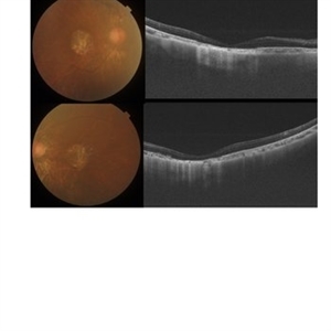
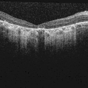
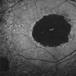
 Geographic Atrophy Secondary to Central Areolar Choroidal Dystrophy
Dec 8 2019 by Anfisa Ayalon, MD
OCT pictures of a 37-year-old male with CACD. Note atrophic changes in the outer retinal layer.
Photographer: Anfisa Ayalon,MD., Meir Medical Center, Kfar Saba, Israel.
Condition/keywords: central areolar choroidal dystrophy (CACD), geographic atrophy, optical coherence tomography (OCT)
-
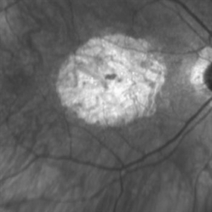
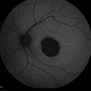
 Fundus Autofluorescence in Central Areolar Choroidal Dystrophy
Dec 8 2019 by Anfisa Ayalon, MD
Fundus autofluorescence pictures of a 37-year-old male with CACD. The patient has visual acuity of 1/18 in the right eye and 6/30 in the left eye. Full-field ERG was normal under photopic and scotopic conditions.
Photographer: Anfisa Ayalon,MD., Meir Medical Center, Kfar Saba, Israel.
Condition/keywords: central areolar choroidal dystrophy (CACD), fundus autofluorescence (FAF), hereditary retinal degeneration
-
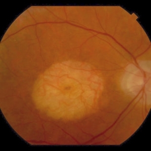
 Central Areolar Choroidal Dystrophy
Dec 8 2019 by Anfisa Ayalon, MD
Fundus photograph of a 37-year-old male with CACD. The patient has visual acuity of 1/18 in the right eye and 6/30 in the left eye. Full-field ERG was normal under photopic and scotopic conditions.
Photographer: Anfisa Ayalon,MD., Meir Medical Center, Kfar Saba, Israel.
Condition/keywords: central areolar choroidal dystrophy (CACD), geographic atrophy, hereditary retinal degeneration
-
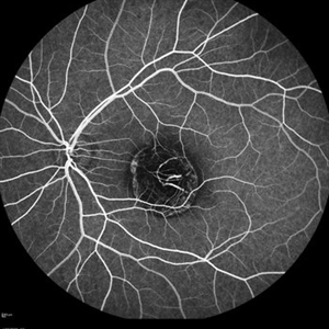
 Central Areloar Choroidal Dystrophy
Oct 1 2019 by Demetrios G. Vavvas, MD, PhD
SD-OCT 74-year-old with CACD type 2 PRPH2 mutation.
Condition/keywords: central areolar choroidal dystrophy (CACD)
-
 Central Areloar Choroidal Dystrophy
Oct 1 2019 by Demetrios G. Vavvas, MD, PhD
IR 74-year-old with CACD type 2 PRPH2 mutation.
Condition/keywords: central areolar choroidal dystrophy (CACD)
-
 Central Areloar Choroidal Dystrophy
Oct 1 2019 by Demetrios G. Vavvas, MD, PhD
Color fundus image of a 74-year-old with CACD type 2 PRPH2 mutation.
Condition/keywords: central areolar choroidal dystrophy (CACD)
-
 Central Areloar Choroidal Dystrophy
Oct 1 2019 by Demetrios G. Vavvas, MD, PhD
AF 74-year-old with CACD type 2 PRPH2 mutation.
Condition/keywords: central areolar choroidal dystrophy (CACD)
-
 Central Areloar Choroidal Dystrophy
Oct 1 2019 by Demetrios G. Vavvas, MD, PhD
AF of a 31-year-old with CACD type 2 PRPH2 mutation.
Condition/keywords: central areolar choroidal dystrophy (CACD)
-
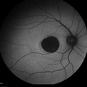
 Central Areolar Choroidal Dystrophy
Mar 26 2019 by Gary R. Cook, MD, FACS
Left eye of a patient with central areolar choroidal dystrophy.
Condition/keywords: central areolar choroidal dystrophy (CACD)
-
 Central Areolar Choroidal Dystrophy
Mar 26 2019 by Gary R. Cook, MD, FACS
Right eye of a patient with central areolar choroidal dystrophy.
Condition/keywords: central areolar choroidal dystrophy (CACD)
-
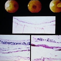
 Slide 9-79
Feb 26 2019 by Lancaster Course in Ophthalmology
Senile macular degeneration. Drusen are shown (arrows), associated with a central area of areolar atrophy (asterisks) in which there is loss of the RPE and photoreceptor cells.
Condition/keywords: central areolar choroidal dystrophy (CACD), drusen, macular degeneration, retinal pigment epithelium
-
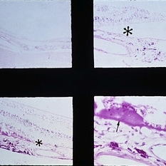
 Slide 9-44
Feb 26 2019 by Lancaster Course in Ophthalmology
Central areolar choroidal sclerosis. Macular lesions in three generations of the same family. (Upper three views courtesy of R. E. Carr, M.D. ). Histopathologic study shows loss of retinal pigment epithelium and photoreceptor cells but with a preservation of choriocapillaris in some areas. (Middle and lower views courtesy of Andrew Ferry, M.D.)
Condition/keywords: central areolar choroidal dystrophy (CACD), photoreceptor cell, retinal pigment epithelium
-
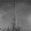
 Central Areolar Choroidal Dystrophy
Apr 14 2018 by Hamza Ahmed Shawky
Left fundus color photograph of a 35-year-old man with central areolar choroidal dystrophy, BCVA is 6/60.
Photographer: Hamza Shawky, Alferdaws eye hospital, Retina unit
Imaging device: Heidelberg Spectralis
Condition/keywords: central areolar choroidal dystrophy (CACD), hereditary retinal dystrophy, macular dystrophy, retinal dystrophy
-
 Central Areolar Choroidal Dystrophy
Apr 14 2018 by Hamza Ahmed Shawky
Left fundus autofluorescence photograph of a 35-year-old man with central areolar choroidal dystrophy, BCVA is 6/60
Photographer: Hamza Shawky, Alferdaws eye hospital, Retina unit
Imaging device: Heidelberg Spectralis
Condition/keywords: central areolar choroidal dystrophy (CACD), hereditary retinal dystrophy, macular dystrophy, retinal dystrophy
-
 Central Areolar Choroidal Dystrophy
Apr 14 2018 by Hamza Ahmed Shawky
Left fundus late FFA photograph of a 35-year-old man with central areolar choroidal dystrophy, BCVA is 6/60
Photographer: Hamza Shawky, Alferdaws eye hospital, Retina unit
Imaging device: Heidelberg Spectralis
Condition/keywords: central areolar choroidal dystrophy (CACD), hereditary retinal dystrophy, macular dystrophy, retinal dystrophy
-
 Central Areolar Choroidal Dystrophy
Apr 14 2018 by Hamza Ahmed Shawky
Right fundus color photograph of a 35-year-old man with central areolar choroidal dystrophy, BCVA is 6/60
Photographer: Hamza Shawky, Alferdaws eye hospital, Retina unit
Imaging device: Heidelberg Spectralis
Condition/keywords: central areolar choroidal dystrophy (CACD), hereditary retinal dystrophy, macular dystrophy, retinal dystrophy
-
 Central Areolar Choroidal Dystrophy
Apr 14 2018 by Hamza Ahmed Shawky
Right fundus late FFA photograph of a 35-year-old man with central areolar choroidal dystrophy, BCVA is 6/60
Photographer: Hamza Shawky, Alferdaws eye hospital, Retina unit
Imaging device: Heidelberg Spectralis
Condition/keywords: central areolar choroidal dystrophy (CACD), hereditary retinal dystrophy, macular dystrophy, retinal dystrophy
-
 Central areolar choroidal dystrophy
Apr 14 2018 by Hamza Ahmed Shawky
Right fundus autofluorescence photograph of a 35-year-old man with central areolar choroidal dystrophy, BCVA is 6/60
Photographer: Hamza Shawky, Alferdaws eye hospital, Retina unit
Imaging device: Heidelberg Spectralis
Condition/keywords: central areolar choroidal dystrophy (CACD), hereditary retinal dystrophy, macular dystrophy, retinal dystrophy
-
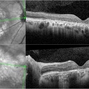
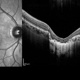 Central Areolar Choroidal Dystrophy
Apr 14 2018 by Hamza Ahmed Shawky
Right fundus OCT of a 35-year-old man with central areolar choroidal dystrophy, BCVA is 6/60
Photographer: Hamza Shawky, Alferdaws eye hospital, Retina unit
Imaging device: Heidelberg Spectralis
Condition/keywords: central areolar choroidal dystrophy (CACD), hereditary retinal dystrophy, macular dystrophy, retinal dystrophy
 Loading…