Search results (47 results)
-
 Cavernous Hemangioma
Jun 3 2023 by Alexandre Grandinetti, MD, PhD
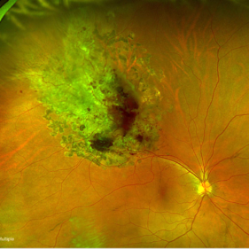
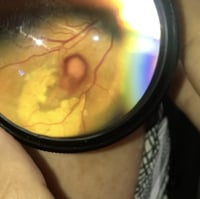
Fundus photograph of a 44-year-old woman with a cavernous hemangioma
Imaging device: Optos California
Condition/keywords: cavernous hemangioma
-
 Retinal Cavernous Hemangioma
Nov 22 2022 by Vaidehi Sathaye
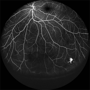
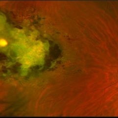
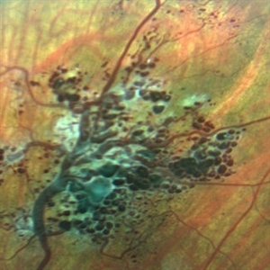
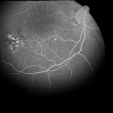
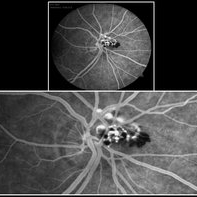
Widefield FA of LE of a 9 yr male patient with Cavernous Hemangioma appearing as a cluster of grapes
Photographer: Dr. Vaidehi Sathaye
Imaging device: Mirante
Condition/keywords: cavernous hemangioma of the retina, FA
-
 Retinal Cavernous Hemangioma
Apr 23 2021 by Aparna Ghodake
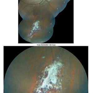
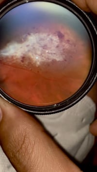
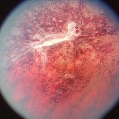
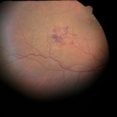
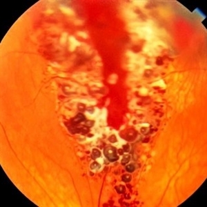
Fundus photograph of a 14-year-old girl having amblyopia was incidentally found to have numerous dark blood filled saccular aneurysms embedded in white fibroglial tissue giving it a 'bunch of grapes' appearance characteristic of retinal cavernous hemangioma.
Photographer: Dr. Aparna Ghodake, Sri Sankaradeva netralaya, Guwahati, Assam, India
Imaging device: Zeiss visucam 500
Condition/keywords: cluster of grapes, fundus photograph
-
 Retinal Cavernous Hemangioma
Nov 6 2020 by David L Kilpatrick, MD
15-year-old female with an asymptomatic retinal cavernous hemangioma.
Photographer: MS Retina Associoates
Imaging device: Optos
Condition/keywords: cavernous hemangioma of the retina
-
 Retinal Cavernous Hemangioma
Oct 22 2020 by Olivia Rainey
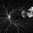
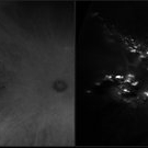
Ultra-widefield fluorescein and ICG angiogram of a 31-year-old male presenting with a retinal cavernous hemangioma affecting his left eye. Patient was 18-years-old when he was diagnosed with a retinal cavernous hemangioma. He has had a few episodes of vitreous hemorrhages since then. His vision was 20/20-1 in both eyes.
Photographer: Becca Harris
Imaging device: Optos California
Condition/keywords: cavernous hemangioma of the retina, fluorescein angiogram (FA), indocyanine green (ICG) angiography, late phase, left eye, Optos, ultra-wide field imaging
-
 Retinal Cavernous Hemangioma
Oct 22 2020 by Olivia Rainey
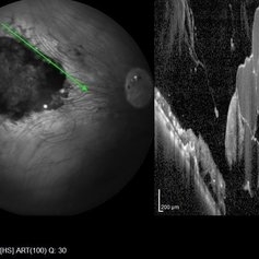
Widefield OCT of a 31-year-old male presenting with a retinal cavernous hemangioma affecting his left eye. Patient was 18-years-old when he was diagnosed with a retinal cavernous hemangioma. He has had a few episodes of vitreous hemorrhages since then. His vision was 20/20-1 in both eyes.
Photographer: Becca Harris
Imaging device: Heidelberg Spectralis
Condition/keywords: 50 degrees, cavernous hemangioma of the retina, Heidelburg Spectralis, left eye, optical coherence tomography (OCT), wide angle imaging
-
 Retinal Cavernous Hemangioma
Oct 21 2020 by Olivia Rainey
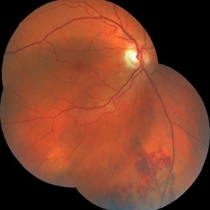
Ultra-widefield image of a 31-year-old male presenting with a Retinal Cavernous Hemangioma affecting his left eye. Patient was 18-years-old when he was diagnosed with a retinal cavernous hemangioma. He has had a few episodes of vitreous hemorrhages since then. His vision was 20/20-1 in both eyes.
Photographer: Olivia Rainey, OCT-C, COA
Imaging device: Optos California
Condition/keywords: cavernous hemangioma of the retina, color fundus photograph, fundus photograph, left eye, Optos, pseudocolor, ultra-wide field imaging
-
 Retinal Cavernous Hemangioma
May 21 2020 by Elias Khalili Pour, MD
Unilateral retinal cavernous hemangioma in a healthy pregnant woman.
Condition/keywords: cavernous hemangioma of the retina
-
 Ruptured Retinal Arterial Macroaneurysm
May 21 2020 by Elias Khalili Pour, MD
Ruptured Retinal Arterial Macroaneurysm in a patient with history of hypertension
Condition/keywords: cavernous hemangioma of the retina
-
 Multiple Cavernous Hemangioma
Jan 20 2020 by Sarah Oelrich
Multiple Cavernous Hemangioma
Photographer: Sarah Oelrich CRA COT OCT-C Southeastern Retina Associates
Condition/keywords: cavernous hemangioma of the retina
-
 Cavernous Hemangioma of Retina
Mar 27 2019 by Gary R. Cook, MD, FACS
14-year-old WM with a cavernous hemangioma of the retina OD: V.A.= 20/20.
Condition/keywords: cavernous hemangioma of the retina
-
 Slide 6-21
Feb 25 2019 by Lancaster Course in Ophthalmology
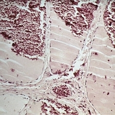
Cavernous hemangioma. Microscopic appearance. Tumor shows large, blood-filled spaces separated by endothelium-lined fibrous septa (H&E x lOl).
Condition/keywords: cavernous hemangioma of the retina, endothelium, tumor
-
 Slide 6-20
Feb 25 2019 by Lancaster Course in Ophthalmology
Cavernous hemangioma. Clinical appearance.
Condition/keywords: cavernous hemangioma of the retina
-
 Retinal Cavernous Hemangioma
Nov 30 2018 by Brenda Fallas
2-year-old female with retinal cavernous hemangioma.
Photographer: Brenda Fallas
Imaging device: Retcam 130 lens
Condition/keywords: cluster of grapes, retinal angioma
-
 Retinal Cavernous Hemangioma
Sep 21 2018 by John S. King, MD
54-year-old sent in with CME and Dx of BVO. She was 20/70. She did not have a diagnosis of diabetes at the time, but FA finding suggestive of DR, so will get her tested via her PCP. There is a cluster of saccular aneurysms infero-temporally that show some hyperFL of the top of the aneurysm.
Photographer: Kay Dalby
Imaging device: Topcon
Condition/keywords: cavernous hemangioma of the retina
-
 Retinal Cavernous Hemangioma
Sep 21 2018 by John S. King, MD
54 -year-old sent in with CME and Dx of BVO. She was 20/70. There are some exudates just nasal to the fovea at the edge of this photos, due to DME. There is a cluster of saccular aneurysms infero-temporally without any glial tissue overlying them.
Imaging device: Topcon
Condition/keywords: cavernous hemangioma of the retina
-
 Cavernous Hemangioma Fluorescein Angiography
Jul 30 2017 by Tony Tsai, MD, FASRS
15-year-old female with asymptomatic cavernous hemangioma of the retina.
Photographer: Michael Kinnison
Condition/keywords: cavernous hemangioma of the retina
-
 Cavernous Hemangioma
Jul 30 2017 by Tony Tsai, MD, FASRS
15-year-old female with asymptomatic cavernous hemangioma of the retina.
Photographer: Michael Kinnison
Condition/keywords: cavernous hemangioma of the retina
-
 Cavernous Hemangioma Color
Jul 30 2017 by Tony Tsai, MD, FASRS
15-year-old female with asymptomatic cavernous hemangioma of the retina.
Photographer: Michael Kinnison
Condition/keywords: cavernous hemangioma of the retina
-
 Cavernous Hemangioma
Oct 6 2016 by Peter W. Hadden, MBChB,FRANZCO
Fundus photo of a European male with an incidental finding of a cavernous haemangioma.
Photographer: Peter Hadden, Eye Institute, Auckland
Imaging device: Zeiss
Condition/keywords: cavernous hemangioma of the retina
-
 Cavernous Hemangioma of the Retina
Sep 11 2016 by JEFFERSON R SOUSA, Tecg.º (Biomedical Systems Technology)
A female patient, 13 years of age, with complaint of low vision in her left eye, had esotropia in this eye. In the examination of fundoscopy and color photograph, we observed a pattern of multiple formations venous aneurysm with aspects of bunches of grapes in the nasal cavity above, which is characteristic of the cavernous hemangiomas of the retina.
Photographer: JEFFERSON R SOUSA - Study Center and Ophthalmological Research Dr. Andre M V Gomes, Institute Dr. Suel Abujamra São Paulo-Brazil
Imaging device: Topcon TRC-50VT, Film, Kodak Ektachrome 160 - ASA 100 / 35mm, field of 35 degrees. Flash 100.
Condition/keywords: cavernous hemangioma of the retina, tumor
-
 Cavernous Hemangioma of the Retina
Sep 11 2016 by JEFFERSON R SOUSA, Tecg.º (Biomedical Systems Technology)
Male patient, 46-years-old, complaining of loss of the nasal field. In the examination of fluorescent angiography showed a pattern of hiperfluorescência and hipofluorescência typical of cavernous hemangioma.
Photographer: JEFFERSON R SOUSA - Study Center and Ophthalmological Research Dr. Andre M V Gomes, Institute Dr. Suel Abujamra São Paulo-Brazil
Imaging device: Zeiss / VisuCam-500 - Angulation of field photo of 45 Degrees, flash 24, digital magnification 4x
Condition/keywords: cavernous hemangioma of papilla, cavernous hemangioma of the retina, tumor
-
 Cavernous Hemangioma of Papilla
Sep 11 2016 by JEFFERSON R SOUSA, Tecg.º (Biomedical Systems Technology)
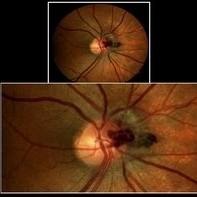
Male patient, 46-years-old, complaining of loss of the nasal field. The funduscopy revealed important vascular changes with aspects of bunch of grapes above-papillae.
Photographer: JEFFERSON R SOUSA - Study Center and Ophthalmological Research Dr. Andre M V Gomes, Institute Dr. Suel Abujamra São Paulo-Brazil
Imaging device: Zeiss / VisuCam-500 - Angulation of field photo of 45 Degrees, flash 16, digital magnification 4x
Condition/keywords: cavernous hemangioma of papilla, cavernous hemangioma of the retina, tumor
-
 Retinal Cavernous Hemangioma FA 49 Seconds
Dec 3 2015 by Daniel A. Adelberg, MD, FASRS
Fundus and fluorescein angiographic images of an 26-year-old male with a peripheral , asymptomatic retinal cavernous hemangioma.
Photographer: Robert Ramsay, COA, Southwestern Eye Center Phoenix Arizona
Condition/keywords: cavernous hemangioma of the retina
-
 Retinal Cavernous Hemangioma - FA 23 Seconds
Dec 3 2015 by Daniel A. Adelberg, MD, FASRS
Fundus and fluorescein angiographic images of an 26-year-old male with a peripheral , asymptomatic retinal cavernous hemangioma.
Photographer: Robert Ramsay, COA, Southwestern Eye Center Phoenix Arizona
Condition/keywords: cavernous hemangioma of the retina
 Loading…