Search results (102 results)
-
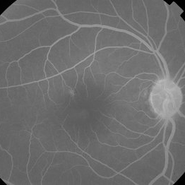
 Central Retinal Vein Occlusion associated with disc edema
Oct 19 2023 by Gabriel Costa Andrade, PhD
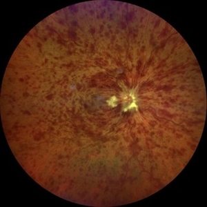
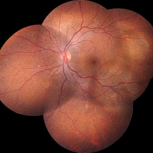
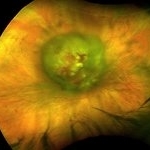
53-year-old woman with an acute CRVO. The patient has a history of breast cancer undergoing treatment with systemic chemotherapy. Notice the peripapillary cotton wool spots, superficial flame shaped hemorrhages and deeper dot and blot hemorrhages in all 4 quadrants.
Photographer: Gabriel Andrade
Condition/keywords: central retinal vein occlusion (CRVO), macular edema, Retina
-
 Breast cancer metastatic to choroid
Jul 13 2021 by Odette M. Houghton, MD
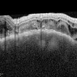
EDI-OCT image of a 59-year-old female with a choroidal tumor secondary to metastatic breast cancer.
Photographer: David Saiz COT, Mayo Clinic Arizona
Imaging device: Heidelberg Spectralis
Condition/keywords: breast cancer, choroidal metastasis, metastatic lesion
-
 Breast cancer metastatic to choroid
Jul 13 2021 by Odette M. Houghton, MD
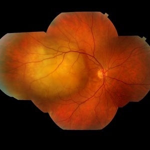
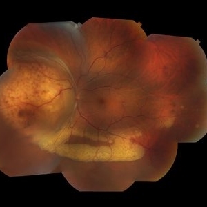
Montage photograph of a 59-year-old female with a choroidal tumor secondary to metastatic breast cancer.
Photographer: David Saiz COT, Mayo Clinic Arizona
Imaging device: Topcon
Condition/keywords: breast cancer, metastatic cancer, metastatic lesion
-
 Breast cancer metastatic to choroid
Jul 13 2021 by Odette M. Houghton, MD
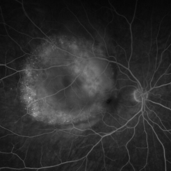
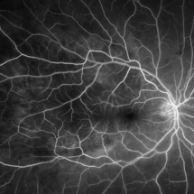
Late phase fluorescein angiogram of a 59-year-old female with a choroidal tumor secondary to metastatic breast cancer.
Photographer: David Saiz COT, Mayo Clinic Arizona
Imaging device: Optos California
Condition/keywords: breast cancer, FA late phase, metastatic cancer
-
 Breast cancer metastatic to choroid
Jul 13 2021 by Odette M. Houghton, MD
Arteriovenous phase fluorescein angiogram of a 59-year-old female with a choroidal tumor secondary to metastatic breast cancer.
Photographer: David Saiz COT, Mayo Clinic Arizona
Imaging device: Optos California
Condition/keywords: breast cancer, choroidal metastasis, metastatic lesion
-
 Breast cancer metastatic to choroid
Jul 13 2021 by Odette M. Houghton, MD
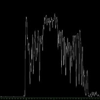
B-scan image of a 59-year-old female with a choroidal tumor secondary to metastatic breast cancer.
Photographer: Christina Carpenter COA, ROUB, OSC, Mayo Clinic Arizona
Imaging device: Ellex
Condition/keywords: B scan ultrasound, breast cancer, metastatic cancer
-
 Breast cancer metastatic to choroid
Jul 13 2021 by Odette M. Houghton, MD
A-scan image of a 59-year-old female with a choroidal tumor secondary to metastatic breast cancer.
Photographer: Christina Carpenter COA, ROUB, OSC, Mayo Clinic Arizona
Imaging device: Ellex
Condition/keywords: a-scan image, breast cancer, metastatic cancer
-
 Metastatic Breast Carcinoma
Jan 21 2021 by Jamin S. Brown, MD
This anterior segment photograph was taken with a smartphone camera attached to a regular Haag Streit slit lamp ocular demonstrates unusual clustering of white cells on the posterior surface of the intraocular lens. The clinical diagnosis is metastatic breast carcinoma to the vitreous, which is very rare.
Photographer: Stefanie Palmer CRA, Retina Vitreous Surgeons of CNY
Imaging device: Cell phone camera
Condition/keywords: anterior segment, breast cancer, cell phone camera, slit lamp photo
-
 Metastatic Adenocarcinoma
May 18 2020 by McGill University Health Centre
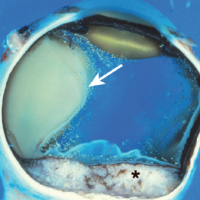
Metastatic disease is the most frequent intraocular malignant tumor. In women, the most common origin is breast cancer. In men, the most common origin is lung cancer. This pupil–optic nerve section shows a whitish tumor with several foci of necrosis (*) occupying the posterior aspect of the choroid. Note the pigment epithelium over the inner surface of the tumor. A serous retinal detachment is present (arrow) with a retinal detachment artifact overlying the tumor and normal choroid. Note the air bubble artifacts in the vitreous cavity. Another artifact, the compression of the eyeball, is present on the right side.
Condition/keywords: breast cancer, foci of necrosis, metastatic adenocarcinoma, tumor
-
 Fundus Fluorescein Angiography of Choroidal Metastases
Jan 18 2020 by Vishal Agrawal, MD, FRCS,FACS,FASRS
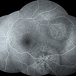
Left eye FFA montage of a 55-year-old female with choroidal metastases with the primary being breast carcinoma. The right eye had exudative retinal detachment . Note the pin point leaks at the border of the 2 lesions.
Photographer: Dr Vishal Agrawal MD,FRCS
Imaging device: Zeiss
Condition/keywords: breast cancer, FA mid phase, metastatic lesion
-
 Choroidal Metastases
Jan 18 2020 by Vishal Agrawal, MD, FRCS,FACS,FASRS
Left eye fundus montage of a 55-year-old female with choroidal metastases with the primary being breast carcinoma. The right eye had exudative retinal detachment.
Photographer: Dr Vishal Agrawal MD,FRCS
Imaging device: Zeiss
Condition/keywords: breast cancer, choroidal metastasis, metastatic lesion
-
 Tamoxifen Retinopathy with Pseudocystic Foveal Cavitation - FA Recirculation
Nov 7 2019 by John S. King, MD
50-year-old female with breast cancer referred for CME had noticed gradual decrease in vision over the last 6-12 months; pseudocystic foveal cavitation due to tamoxifen retinopathy diagnosed by Dr. Hruby. Visual acuity cc 20/360, J16 OD and 20/40, J7 OS, normal IOPs, trace NSC OU. Bilateral pseudoholes noted on posterior segment exam without any retinal crystals. No leakage or tel vessels seen on the FA. Cystic cavitary alterations seen on the OCT with a large outer hole in the left eye. Plaquenil will be d/c.
Photographer: Gretchen Harper
Condition/keywords: foveal cavitation, pseudocystic foveal cavitiation, tamoxifen maculopathy, tamoxifen retinopathy, tamoxifen toxicity
-
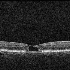 OCT OD Tamoxifen Retinopathy with Pseudocystic Foveal Cavitation in Tamoxifen Retinopathy
Nov 7 2019 by John S. King, MD
50-year-old female with breast cancer referred for CME had noticed gradual decrease in vision over the last 6-12 months; pseudocystic foveal cavitation due to tamoxifen retinopathy diagnosed by Dr. Hruby. Visual acuity cc 20/360, J16 OD and 20/40, J7 OS, normal IOPs, trace NSC OU. Bilateral pseudoholes noted on posterior segment exam without any retinal crystals. No leakage or tel vessels seen on the FA. Cystic cavitary alterations seen on the OCT with a large outer hole in the left eye. Plaquenil will be d/c.
Photographer: Gretchen Harper
Condition/keywords: foveal cavitation, pseudocystic foveal cavitiation, tamoxifen maculopathy, tamoxifen retinopathy, tamoxifen toxicity
-
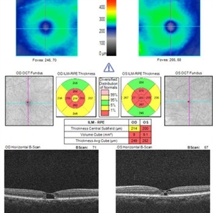 OCT OU Tamoxifen Retinopathy with Pseudocystic Foveal Cavitation
Nov 7 2019 by John S. King, MD
50-year-old female with breast cancer referred for CME had noticed gradual decrease in vision over the last 6-12 months; pseudocystic foveal cavitation due to tamoxifen retinopathy diagnosed by Dr. Hruby. Visual acuity cc 20/360, J16 OD and 20/40, J7 OS, normal IOPs, trace NSC OU. Bilateral pseudoholes noted on posterior segment exam without any retinal crystals. No leakage or tel vessels seen on the FA. Cystic cavitary alterations seen on the OCT with a large outer hole in the left eye. Plaquenil will be d/c.
Photographer: Gretchen Harper
Condition/keywords: foveal cavitation, pseudocystic foveal cavitiation, tamoxifen maculopathy, tamoxifen retinopathy, tamoxifen toxicity
-
 Tamoxifen Retinopathy Pseudocystic Foveal Cavitatio - FA Late
Nov 7 2019 by John S. King, MD
50-year-old female with breast cancer referred for CME had noticed gradual decrease in vision over the last 6-12 months; Pseudocystic foveal cavitation due to tamoxifen retinopathy diagnosed by Dr. Hruby. Visual acuity cc 20/360, J16 OD and 20/40, J7 OS, normal IOPs, trace NSC OU. Bilateral pseudoholes noted on posterior segment exam without any retinal crystals. No leakage or tel vessels seen on the FA. Cystic cavitary alterations seen on the OCT with a large outer hole in the left eye. Plaquenil will be d/c.
Photographer: Gretchen Harper
Condition/keywords: foveal cavitation, pseudocystic foveal cavitiation, tamoxifen maculopathy, tamoxifen retinopathy, tamoxifen toxicity
-
 Pseudohole OS in Tamoxifen Retinopathy with Pseudocystic Foveal Cavitation
Nov 7 2019 by John S. King, MD
50-year-old female with breast cancer referred for CME had noticed gradual decrease in vision over the last 6-12 months; pseudocystic foveal cavitation due to tamoxifen retinopathy diagnosed by Dr. Hruby. Visual acuity cc 20/360, J16 OD and 20/40, J7 OS, normal IOPs, trace NSC OU. Bilateral pseudoholes noted on posterior segment exam without any retinal crystals. No leakage or tel vessels seen on the FA. Cystic cavitary alterations seen on the OCT with a large outer hole in the left eye. Plaquenil will be d/c.
Photographer: Gretchen Harper
Condition/keywords: foveal cavitation, pseudocystic foveal cavitiation, tamoxifen maculopathy, tamoxifen retinopathy, tamoxifen toxicity
-
 Tamoxifen Retinopathy with a Large atrophic, near full-thickness macular hole OD (Pseudocystic Foveal Cavitation)
Nov 7 2019 by John S. King, MD
50-year-old female with breast cancer referred for CME had noticed gradual decrease in vision over the last 6-12 months; pseudocystic foveal cavitation due to tamoxifen retinopathy diagnosed by Dr. Hruby. Visual acuity cc 20/360, J16 OD and 20/40, J7 OS, normal IOPs, trace NSC OU. Bilateral pseudoholes noted on posterior segment exam without any retinal crystals. No leakage or tel vessels seen on the FA. Cystic cavitary alterations seen on the OCT with a large outer hole in the left eye. Plaquenil will be d/c.
Photographer: Gretchen Harper
Condition/keywords: foveal cavitation, pseudocystic foveal cavitiation, tamoxifen maculopathy, tamoxifen retinopathy, tamoxifen toxicity
-
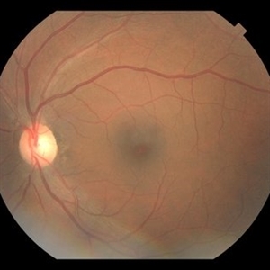
 Choroidal Metastasis from Breast Cancer
Oct 1 2019 by John S. King, MD
60-year-old white female with four year history of breast cancer associated with metastases to many organs including the CNS, was sent her to r/o melanoma, found on routine exam. Visual acuity was HM; there was NSC/PSC; there was a unilateral, large choroidal lesion in the posterior pole that was yellow, well circumscribed, with plateau configuration associated with SRF adn heme.
Photographer: Kay Dalby
Imaging device: Optos CA
Condition/keywords: breast cancer, choroidal lesions, choroidal metastasis
-
 Choroidal Metastasis from Breast Cancer
Oct 1 2019 by John S. King, MD
60-year-old white female with four year history of breast cancer associated with metastasis to many organs including the CNS, was sent her to r/o melanoma, found on routine exam. Visual acuity was HM; there was NSC/PSC; there was a unilateral, large choroidal lesion in the posterior pole that was yellow, well circumscribed, with plateau configuration associated with SRF adn heme.
Photographer: Kay Dalby
Imaging device: Optos CA
Condition/keywords: breast cancer, choroidal lesions, choroidal metastasis
-
 Metastatic Cancer
Mar 26 2019 by Gary R. Cook, MD, FACS
64-year-old WF with metastatic breast carcinoma OD s/p radiation treatment; VA improved to 20/25.
Imaging device: Topcon VT-50
Condition/keywords: breast cancer, breast carcinoma, choroidal metastasis, metastatic lesion
-
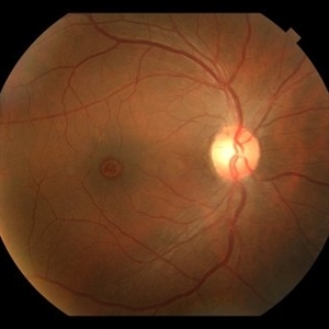
 Metastatic Breast Carcinoma
Mar 26 2019 by Gary R. Cook, MD, FACS
64-year-old white female with metastatic breast carcinoma lesion superior to optic disc OD; VA= 20/70-1.
Imaging device: Topcon VT-50
Condition/keywords: breast cancer, breast carcinoma, choroidal metastasis, metastatic cancer, metastatic lesion
-
 Choroidal Metastasis
Mar 26 2019 by Gary R. Cook, MD, FACS
Fluorescein angiogram image of the choroidal metastasis secondary to breast carcinoma OD.
Imaging device: Topcon VT-50
Condition/keywords: breast cancer, breast carcinoma, choroidal metastasis, fluorescein angiogram (FA), metastatic lesion
-
 Choroidal Metastasis
Mar 26 2019 by Gary R. Cook, MD, FACS
Choroidal metastasis secondary to breast carcinoma.
Imaging device: Topcon VT-50
Condition/keywords: breast cancer, breast carcinoma, choroidal metastasis, metastatic lesion
-
 Metastatic Breast Carcinoma
Mar 26 2019 by Gary R. Cook, MD, FACS
55-year-old white female with metastatic breast carcinoma OD; V.A. = 20/60.
Imaging device: Topcon VT-50
Condition/keywords: breast cancer, breast carcinoma, choroidal metastasis, metastatic lesion
-
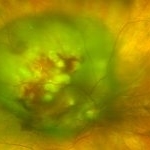
 Ocular Metastasis of Breast Cancer
Mar 13 2018 by Olivia Rainey
Color fundus montage of a 45-year-old female presenting with ocular metastasis affecting her left eye. She had been treated for pneumonia, had progressive lumbar back pain, and a 29 pound weight loss recently. She reported that she had a breast lump and a mammogram, but had not been provided the results. After she was sent to oncology, it was confirmed that she has widely metastatic breast cancer and tumors throughout the brain. The oncology team felt she could not wait weeks for her chemotherapy to start and consequently decided to do whole brain radiation and treat the affected eye just posterior to the lens.
Photographer: Olivia Rainey
Imaging device: Topcon DX50
Condition/keywords: choroidal metastasis, color fundus photograph, exudative detachment, left eye, lipid exudation, montage
 Loading…