Search results (2962 results)
-
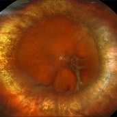
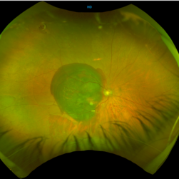 Sub retinal Bleed
Apr 23 2025 by Anjana Mirajkar, MS Ophthalmology
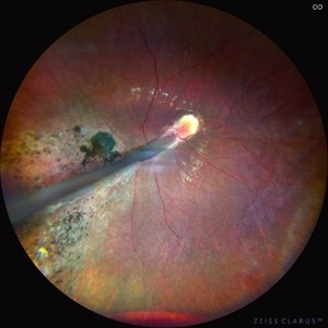
A widefield image of right eye of a 65 year old male showing large subretinal bleed at the posterior pole most likely in a case of PCV.
Photographer: Dr. Anjana Mirajkar- HV desai eye hospital ,Pune
Imaging device: optos
Condition/keywords: idiopathic polypoidal choroidal vasculopathy, subretinal blood
-
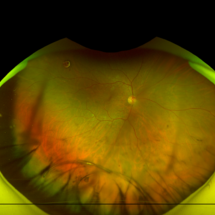 Dislocated IOL
Apr 23 2025 by Anjana Mirajkar, MS Ophthalmology
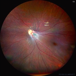
A widefield imaging of the right eye of a 55 year old male showing dislocated IOL inferiorly.
Photographer: Dr. Anjana Mirajkar- HV desai eye hospital ,Pune
Imaging device: Optos
Condition/keywords: dislocated intraocular lens (IOL)
-
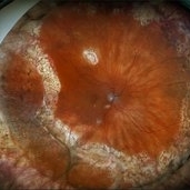
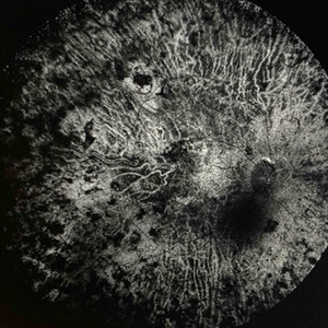 Extensive Chorioretinal Scarring in the Right Eye
Apr 22 2025 by Maxwell J Wingelaar, MD
Fundus autofluorescence of Extensive chorioretinal scarring in the right eye.
Photographer: Killian Roberts
Imaging device: Heidelberg Spectralis AF
Condition/keywords: chorioretinal atrophy, chorioretinal inflammations
-
 Extensive Chorioretinal Scarring in the Right Eye
Apr 22 2025 by Maxwell J Wingelaar, MD
A multicolor photo showing chorioretinal scarring with macular involvement in the right eye
Photographer: Killian Roberts
Imaging device: Heidelberg Spectralis Multicolor Photo
Condition/keywords: chorioretinal atrophy, chorioretinal inflammations
-
 Hourglass in an Eye
Apr 22 2025 by KRISHNENDU NANDI, MS
A twenty-five-year-young male presented with a decrease in vision in the right eye following a blunt trauma with a football. On examination the BCVA in the right eye was CFCF and the left eye was 6/6, N6. The anterior segment was within normal limits. AT was 12 and 10 mm of Hg in the right and left eyes, respectively. Fundus examination reveals subhyaloid haemorrhage in the right eye with an attached retina. The fundus of the left eye was within normal limits. YAG laser hyaloidotomy was done with an energy of 2 mJ in the right eye. After 3 weeks the BCVA in the right eye improved to 6/9, N6.
Photographer: Dr. Krishnendu Nandi
Imaging device: Topcon
Condition/keywords: Trauma, YAG HYALOIDOTOMY, Young Male
-
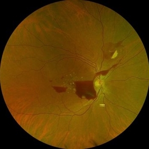
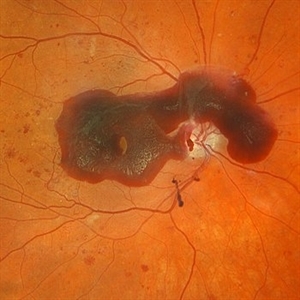 Severe NPDR with Subhyaloid Hemorrhage
Apr 9 2025 by Kimberly Wakester
Optomap RGB of an 47 year-old man with severe NPDR with subhyaloid hemorrhage in the right eye.
Photographer: Kimberly Wakester, COA, OCT-C
Imaging device: Optos California
Condition/keywords: severe NPDR, subhyaloid hemorrhage
-
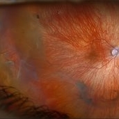
 Comets in the Eye (Retinopathy of Prematurity)
Apr 8 2025 by KANWALJEET HARJOT MADAN, M.S. (Ophthalmology), FAICO (Vitreous - Retina)
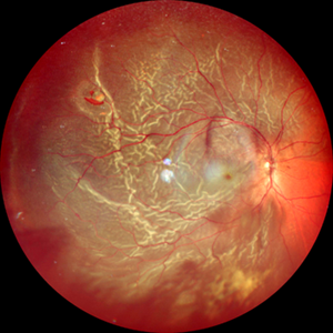
This is the fundus picture of right eye (RE) of a 4 years female child presented with outward deviation of right eye. Her parents also complained of diminution of vision in both eyes. On examination, her best corrected vision in RE was hand movements close to face and was 20/80 in LE. Posterior segment exam revealed presence of macular scar in RE and presence of dry retinal fold with dragging of retinal vessels. LE fundus revealed presence of nasal drag of optic disc. Parents gave history of untreated ROP as an infant. Retinopathy of Prematurity (ROP) is a Vaso proliferative disorder of Retina occurring in premature infants. Advances in neonatal care and ROP treatment has led these babies to live longer with this disease.
Photographer: Dr. Kanwaljeet Harjot Madan, Thind Eye Hospital, Jalandhar City (Punjab) INDIA.
Imaging device: Zeiss Fundus Camera
Condition/keywords: Retinopathy of Prematurity, Vaso proliferative disorder
-
 Comets in the Eye (Retinopathy of Prematurity)
Apr 8 2025 by KANWALJEET HARJOT MADAN, M.S. (Ophthalmology), FAICO (Vitreous - Retina)
This is the fundus picture of right eye (RE) of a 4 years female child presented with outward deviation of right eye. Her parents also complained of diminution of vision in both eyes. On examination, her best corrected vision in RE was hand movements close to face and was 20/80 in LE. Posterior segment exam revealed presence of macular scar in RE and presence of dry retinal fold with dragging of retinal vessels. LE fundus revealed presence of nasal drag of optic disc. Parents gave history of untreated ROP as an infant. Retinopathy of Prematurity (ROP) is a Vaso proliferative disorder of Retina occurring in premature infants. Advances in neonatal care and ROP treatment has led these babies to live longer with this disease.
Photographer: Dr. Kanwaljeet Harjot Madan, Thind Eye Hospital, Jalandhar City (Punjab) INDIA.
Imaging device: Zeiss Fundus Camera
Condition/keywords: Retinopathy of Prematurity
-
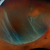
 Superior Rhegmatogenous Retinal Detachment (RRD) in the Right Eye, With a Retinal Tear Located Between the 1 and 2 O'clock Positions
Apr 4 2025 by Cesar Orlando Oviedo Vera
A 45-year-old male patient presented with a sudden onset of decreased visual acuity in the right eye, with a 24-hour progression. Upon examination, Image 1 revealed a superior rhegmatogenous retinal detachment in the right eye, with a retinal tear located between the 1 and 2 o'clock positions. Image 2: Pneumatic retinopexy by intravitreal injection of Sulfur Hexafluoride gas (SF6) at the time of diagnosis with subsequent application of 532 nm laser around the retinal tear.
Photographer: Cesar Orlando Oviedo Vera, Hospital Militar de Especialidades Oftalmológicas
Imaging device: Optos
Condition/keywords: Pneumatic Retinopexy, Retinal tear, Rhegmatogenous retinal detachment, SF6, Superior rhegmatogenous retinal detachment
-
 Pneumatic Retinopexy by Intravitreal Injection of Sulfur Hexafluoride Gas (SF6) at the Time of Diagnosis With Subsequent Application of 532 Nm Laser Around the Retinal Tear
Apr 4 2025 by Cesar Orlando Oviedo Vera
A 45-year-old male patient presented with a sudden onset of decreased visual acuity in the right eye, with a 24-hour progression. Upon examination, Image 1 revealed a superior rhegmatogenous retinal detachment in the right eye, with a retinal tear located between the 1 and 2 o'clock positions. Image 2: Pneumatic retinopexy by intravitreal injection of Sulfur Hexafluoride gas (SF6) at the time of diagnosis with subsequent application of 532 nm laser around the retinal tear.
Photographer: Cesar Orlando Oviedo Vera, Hospital Militar de Especialidades Oftalmológicas
Imaging device: Optos
Condition/keywords: Pneumatic Retinopexy, Retinal tear, Rhegmatogenous retinal detachment, SF6, Superior rhegmatogenous retinal detachment
-
 Chronic Central Serous Chorioretinopathy (CSCR)
Mar 31 2025 by Niloofar Piri, MD
Fundus Autofluorescence image of the right eye demonstrates classic guttering with hyper autofluorescence consistent with chronic CSCR. Guttering occurs where subretinal fluid migrates inferiorly due to gravity and stressed RPE cells accumulate lipofuscin material from high turnover of photoreceptor outer segments.
Photographer: Stefan Raev, COT; Saint Louis University
Condition/keywords: central serous chorioretinopathy (CSCR), Chronic CSR, Guttering
-
 Repaired Retinal Detachment with Scleral Buckle
Mar 25 2025 by Kimberly Wakester
Optomap RGB montage of an 64-year-old woman with a repaired retinal detachment with scleral buckle in the right eye. There is nasal and inferior pre-retinal membranes with traction. PPV was recommended but patient defers to proceed with sx at this time. Will continue to follow patient closely for worsening traction. Patient was educated on how to monitor their peripheral vision and was advised to report any changes immediately.
Photographer: Kimberly Wakester, COA, OCT-C
Imaging device: Optos California
Condition/keywords: pre-retinal membrane with traction, repaired RD, scleral buckle
-
 Repaired Retinal Detachment with PVR
Mar 25 2025 by Kimberly Wakester
Optomap RGB of a 79-year-old-woman with a repaired retinal detachment with PVR in the right eye. Patient is doing well over 7 months s/p vitrectomy with silicone oil and scleral buckle placement. Retina remains attached on the buckle under oil. Patient is to return in 6 months for follow up exam with repeat imaging.
Photographer: Kimberly Wakester, COA, OCT-C
Imaging device: Optos California
Condition/keywords: PVR, repaired RD, Retinal detachment under Silicon Oil, scleral buckle
-
 The Pouring RAM
Mar 25 2025 by Shrishti mishra
A 63 year old male with RAM lesion in the right eye associated with multilayered hemorrhage.
Imaging device: Optos nikon
Condition/keywords: FFA, retinal arterial macroaneurysm, subhyaloid hemorrhage
-
 Myopic Traction Maculopathy
Mar 17 2025 by Drew Mitchell
HD 1 line 100x 9 mm scan of a right eye with MTM at stage 3c. Macular Schisis Detachment.
Photographer: Drew Mitchell OCT-C
Imaging device: Zeiss Cirrus 5000
Condition/keywords: full thickness macular hole, Macular hole, myopic foveoschisis, myopic macular schisis, myopic traction maculopathy, PVD
-
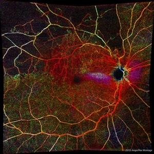 Branch Retinal Vein Occlusion with Macular Edema
Mar 14 2025 by Drew Mitchell
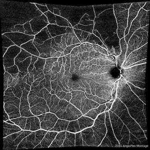
Zeiss Montage Angio 8x8 mm OCT Angiography Retina Depth Encoded Angioplex of a New BRVO in the right eye.
Photographer: Drew Mitchell, OCT-C
Imaging device: Zeiss Cirrus 6000
Condition/keywords: branch retinal vein occlusion (BRVO), macular edema, OCT Angiography
-
 Branch Retinal Vein Occlusion with Macular Edema
Mar 14 2025 by Drew Mitchell
Zeiss Montage Angio 8x8 mm OCT Angiography Superficial Angioplex of a New BRVO in the right eye.
Photographer: Drew Mitchell OCT-C
Imaging device: Zeiss Cirrus 6000
Condition/keywords: branch retinal vein occlusion (BRVO), macular edema, OCT Angiography
-
 Retinal Detachment Secondary to Anomalous PVD
Mar 13 2025 by Fabricio Dolores
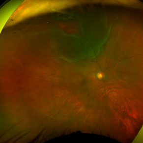
This color wide-field clinical image depicts the right eye of a female patient who experienced a sudden loss of vision one month earlier. She was initially diagnosed with a vitreous hemorrhage and managed with conservative treatment. Upon presentation to our institute one month later, a superior rhegmatogenous retinal detachment was identified, extending across the 12 o’clock meridian. This was accompanied by an inferior vitreous hemorrhage and a solitary superior retinal lesion located at M11 in the superior triangle of the ora serrata, in alignment with Lincoff's second law.
Photographer: Fabricio Dolores-Villanueva, MD
Imaging device: Nidek Mirante
Condition/keywords: Retinal Detachment
-
 Toxoplasma Macular Scar with CNVM
Mar 7 2025 by T. P . VIGNESH, MBBS,MS
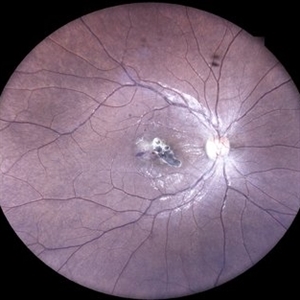
Fundus photograph of the right eye of a 28-year-old woman with a macular toxoplasma scar and CNVM.
Photographer: Sivanath
Imaging device: EIDON
Condition/keywords: CNVM, ocular toxoplasmosis
-
 Central Serous Chorioretinopathy
Mar 7 2025 by T. P . VIGNESH, MBBS,MS
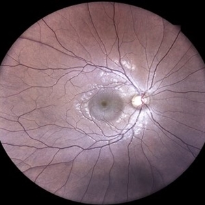
Fundus photograph of the right eye of an 32-year-old woman with CSCR.
Photographer: Sivanath
Imaging device: EIDON
Condition/keywords: central serous chorioretinopathy (CSCR)
-
 MACTEL
Mar 7 2025 by T. P . VIGNESH, MBBS,MS
Fundus photograph of the right eye of an 62-year-old woman with macular telangiectasia type 2.
Photographer: Sivanath
Imaging device: EIDON
Condition/keywords: IJT
-
 Multimodal Imaging in CHRPE
Mar 6 2025 by Gerardo - Montante Montelongo, MD
Fundus photograph of an 83-year-old male with a history of Diabetes, smoking, cataract surgery on the right eye in 2022, and open-angle glaucoma. Asymptomatic. Indirect ophthalmoscopy revealed 80% excavation, peripapillary atrophy, and a hyperpigmented perifoveal lesion with 35% atrophy, 10% drusen, and 5.1 mm diameter, corresponding to a CHRPE. At multimodal imaging, FFA shows hypoautofluorescence of the lesion, OCT shows preservation of internal retinal layers, atrophy of external retinal layer, with an RPE disruption, and posterior shadowing. USG shows a flat hyperechoic lesion 5.1 mm in diameter and 1.32 mm in thickness, solid and with high internal reflectance.
Photographer: Gerardo Montante-Montelongo, MD, Mexican Institute of Ophthalmology
Imaging device: Clarus 700
Condition/keywords: congenital hypertrophy of the retinal pigment epithelium (CHRPE), multimodal imaging
-
 Retinal Detachment with Multiple Breaks
Mar 5 2025 by Kimberly Wakester
Optomap RGB image of an 44-year-old man with a retinal detachment with a complex lattice break in the right eye. Surgery was recommended. Patient is to continue follow up care post operatively.
Photographer: Kimberly Wakester, COA
Imaging device: Optos California
Condition/keywords: Retinal Detachment, retinal tear
-
 Retinal Detachment
Mar 5 2025 by Kimberly Wakester
Optomap RGB image of an 9-year-old boy with a retinal detachment with retinal break at 9:00 in the right eye. Surgery was recommended. Patient is to continue follow up care post operatively.
Photographer: Kimberly Wakester, COA
Imaging device: Optos California
Condition/keywords: myopic eye, Retinal Detachment, retinal tear
-
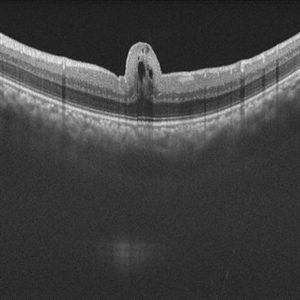 Retinal Fold in Posterior Microphthalmos
Mar 1 2025 by Hemanth Murthy, MBBS, MD, FASRS
Swept source OCT image of left eye of 34 year male patient with high hypermetropia(+14). BCVA 20/20 in right eye and 20/60 in left eye. Anterior segment was normal. There is loss of foveal pit with omega shaped elevation of inner retinal layers.
Photographer: Mr Veda Vyas
Condition/keywords: posterior microphthalmos
 Loading…