Search results (1327 results)
-
 Exudative Retinal Detachment
Aug 6 2025 by Aditya S Kelkar, MS, FRCS, FASRS,FRCOphth
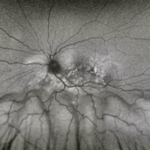
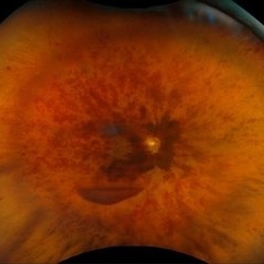
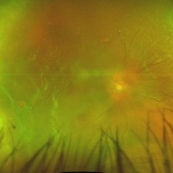
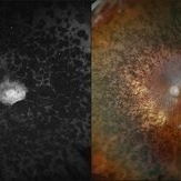
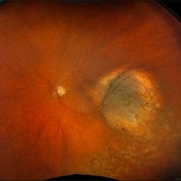
Fundus auto-fluorescence of a 41 year old female depicting retinal pigment epitheliopathy and exudative retinal detachment in case of ocular metastasis secondary to breast carcinoma.
Photographer: Dr.Rabia Naaz, National Institute of ophthalmology, Pune
Imaging device: OPTOS DAYTONA
Condition/keywords: Exudative retinal detachment, Retinal pigment epitheliopathy
-
 Total Retinal Detachment
Aug 6 2025 by Korey Starkey
59 year-old patient presents with total retinal detachment at first visit in OD. Recommending prompt surgical intervention.
Photographer: Korey Starkey
Imaging device: Optos
Condition/keywords: color fundus photograph, Optos, retinal detachment, total retinal detachment
-
 Horseshoe Retinal Tear
Aug 6 2025 by Korey Starkey
80 year-old patient presented with HSRT without detachment in the left eye and macula-off detachment in the right eye. Scheduled patient for prompt surgical repair OD and same day laser retinopexy OS to reduce risk of retinal detachment.
Photographer: Korey Starkey
Imaging device: Optos
Condition/keywords: color fundus photograph, fundus photography, horseshoe tear, Optos
-
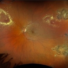 Retinal Aneurysms
Aug 6 2025 by Korey Starkey
54 year-old patient presents with scattered peripheral aneurysms with exudates. FA was performed showing peripheral nonperfusion and aneurysms. Treated patient with PRP and focal laser to aneurysms and continued observation.
Photographer: Kore Starkey
Imaging device: Optos
Condition/keywords: aneurysm, branch retinal vein occlusion (BRVO), chorioretinal scar, circinate ring, exudates, fundus photography, lesion, Optos, retinal aneurysms
-
 Central Retinal Vein Occlusion with Macular Edema
Aug 4 2025 by Virginia Gebhart
58 year old female with Central Retinal Vein Occlusion with Macular Edema. Diffuse retinal hemorrhages, central SRF and sub-hyaloid/vitreous hemorrhage, no obvious active NV. Will start patient on series of 4 monthly intravitreal anti-VEGF injections, followed by T&E protocol once stable.
Photographer: Virginia Gebhart, Retina Consultants of Carolina
Imaging device: Optos California
Condition/keywords: central retinal vein occlusion, CRVO, CRVO with macular edema, retinal hemorrhage, Sub hyaloid haemorrhage, vitreous hemorrhage
-
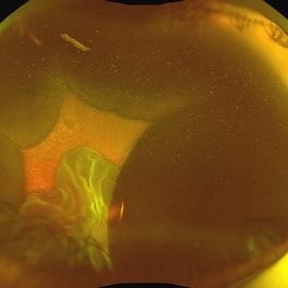 Suprachoroidal Hemorrhage
Aug 4 2025 by Anjana Mirajkar, MS Ophthalmology
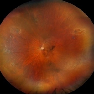
A fundus photograph of a 56 year old female with a 360 degree suprachoroidal hemorrhage with a 360 degree crumpled retina during cataract surgery.
Photographer: Dr. Anjana Mirajkar- HV Desai eye hospital ,Pune
Imaging device: optos
Condition/keywords: giant retinal tear, suprachoroidal hemorrhage
-
 Retinal Detachment Secondary to Large Temporal Tear
Aug 4 2025 by Anjana Mirajkar, MS Ophthalmology
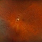
Fundus photograph of a 55 year old male with a retinal detachment with macula off with a large temporal tear.
Photographer: Dr. Anjana Mirajkar- HV desai eye hospital ,Pune
Imaging device: Optos
Condition/keywords: Retinal Detachment, retinal tear
-
 Unstable PDR s/p Laser
Aug 4 2025 by Anjana Mirajkar, MS Ophthalmology
Fundus photograph of a 60 year old male with an unstable PDR showing traction at the posterior pole with sub hyaloid hemorrhage. Peripheral PRP marks can be seen.
Photographer: Dr. Anjana Mirajkar- HV Desai eye hospital ,Pune
Imaging device: Optos
Condition/keywords: pan-retinal photocoagulation (PRP), proliferative diabetic retinopathy (PDR), subhyaloid hemorrhage, tractional retinal detachment
-
 Retinal Detachment
Aug 4 2025 by Anjana Mirajkar, MS Ophthalmology
Fundus photograph of a 40 year old male with a total retinal detachment with macula off with a HST at 1 o clock and a break at mid periphery at 8 o clock.
Photographer: Dr. Anjana Mirajkar- HV Desai eye hospital ,Pune
Imaging device: optos
Condition/keywords: retinal detachment, retinal holes
-
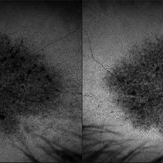
 Bear Tracks CHRPE - Red Channel
Jul 29 2025 by Drew Mitchell
Green Free UWF image of extensive bear track patterned CHRPE.
Photographer: Drew Mitchell, OCT-C
Imaging device: Optos California
Condition/keywords: bear tracks, CHRPE, congenital hypertrophy of the retinal pigment epithelium (CHRPE), Green Free, OPTOS CALIFORNIA
-
 Multiple Retinal Tears
Jul 25 2025 by Virginia Gebhart
60 year old male referred for horseshoe tear of the retina. Scleral depressed exam revealed 7 tears in the same eye. Prophylaxis laser performed to seal all tears.
Photographer: Virginia Gebhart, Retina Consultants of Carolina
Imaging device: Optos California
Condition/keywords: horseshoe tear, multiple retinal tears, operculated tear, Retinal tear
-
 Chronic RD with Retinal Dialysis
Jul 23 2025 by Virginia Gebhart
64 year old female with chronic retinal detachment from head trauma 41 years ago. Peripheral scarring from 6:00 to 11:00 with area of subretinal fluid inferotemporally, well demarcated with subretinal bands. Retinal dialysis inferotemporal from 7:00 to 9:00. No surgical repair needed or recommended at this time.
Photographer: Virginia Gebhart, Retina Consultants of Carolina
Imaging device: Optos California
Condition/keywords: chronic retinal detachment, demarcation, RD, Retinal Detachment, retinal dialysis, subretinal bands
-
 Retinal detachment with Single Break
Jul 18 2025 by Kimberly Wakester
Optomap RGB of a 62-year-old man with a retinal detachment with a single break in the left eye. Patient has a previously treated HSRT in the left eye. Surgery was recommended. Patient is to continue follow up care post operatively.
Photographer: Kimberly Wakester, COA, OCT-C
Imaging device: Optos California
Condition/keywords: RD, retinal tear
-
 Pigmentary Retinal Dystrophy
Jul 18 2025 by Kimberly Wakester
Optomap RGB and AF of the left eye of an 76-year-old woman with pigmentary retinal dystrophy. No progression of the bone spicules noted on exam and optos imaging. Will continue yearly follow care with dilated exam and optos imaging.
Photographer: Kimberly Wakester, COA, OCT-C
Imaging device: Optos California
Condition/keywords: pigmentary retinal dystrophy
-
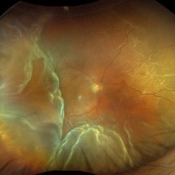
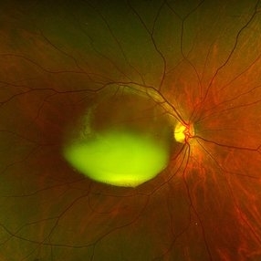
 Retinoschisis with Outer Layer Holes
Jul 18 2025 by Kimberly Wakester
Optomap RGB of an 56-year-old woman with retinoschisis with outer layer holes s/p laser in the left eye. Patient remains stable. Will continue follow up care with dilated exam and optos imaging.
Photographer: Kimberly Wakester, COA, OCT-C
Imaging device: Optos California
Condition/keywords: outer layer hole, retinoschisis
-
 Secondary Pigmentary Degeneration of Retina
Jul 18 2025 by Kimberly Wakester
Optomap RGB and AF of an 63-year-old man with secondary pigmentary degeneration of the retina. Patient's Spark genetic testing revealed heterozygous mutations of unknown significance in LRP5, COL18A1, CPLANE1, SLC24A1 and VCAN. Clinical findings most consistent with Wagner's Syndrome (VCAN mutation, autosomal dominant). Will continue follow up care every 6 months with dilated exam and repeat OCT and Optos imaging.
Photographer: Kimberly Wakester, COA, OCT-C
Imaging device: Optos California
Condition/keywords: secondary pigmentary degeneration, Wagner's Syndrome
-
 Secondary Pigmentary Degeneration of Retina
Jul 18 2025 by Kimberly Wakester
Optomap RGB and AF of an 63-year-old man with secondary pigmentary degeneration of the retina. Patient's Spark genetic testing revealed heterozygous mutations of unknown significance in LRP5, COL18A1, CPLANE1, SLC24A1 and VCAN. Clinical findings most consistent with Wagner's Syndrome (VCAN mutation, autosomal dominant). Will continue follow up care every 6 months with dilated exam and repeat OCT and Optos imaging .
Photographer: Kimberly Wakester, COA, OCT-C
Imaging device: Optos California
Condition/keywords: secondary pigmentary degeneration, Wagner disease
-
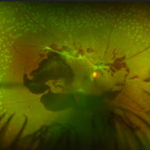
 New Choroidal Melanoma
Jul 16 2025 by Virginia Gebhart
78 year old male with a partially amelanotic dome-shaped lesion with RPE changes, hard exudates, overlying intraretinal fluid and minimal SRF temporally. Exam and ultrasound findings consistent with choroidal melanoma. Pt will be scheduled for brachytherapy pending CT scan results.
Photographer: Virginia Gebhart, Retina Consultants of Carolina
Imaging device: Optos California
Condition/keywords: amelanotic melanoma, choroidal melanoma
-
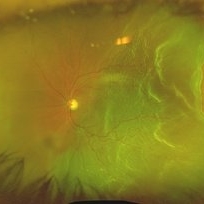
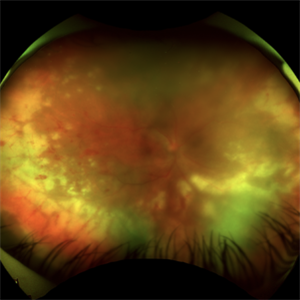
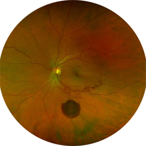 Large Subhyaloid Hemorrhage
Jul 11 2025 by Jessilla Phou
This is a fundus photograph depicting a large subhyaloid hemorrhage in the mid periphery of the left eye. The patient, a 53-year-old female, presented with a sudden onset of floaters, headache, and blurred vision. The image also demonstrates associated optic disc hemorrhage, vitreous hemorrhage, retinal hemorrhage, and venous tortuosity. Despite the extensive workup performed and the severity of the hemorrhage, no underlying cause was determined.
Photographer: Jessilla Phou
Imaging device: Optos California
Condition/keywords: fundus photograph, optic disc hemorrhage, retinal hemorrhage, venous tortuosity, vitreous hemorrhage
-
 Chronic Sub-Hyaloid Hemorrhage with Dehemoglobinized Blood
Jul 11 2025 by Aditya S Kelkar, MS, FRCS, FASRS,FRCOphth
Fundus photograph of an 38-year-old man with a long standing sub hyaloid hemorrhage with dehemoglobinized blood.
Photographer: Optom Salomi Sonawane, National Institute of Ophthalmology, Pune, India
Imaging device: Optos Daytona
Condition/keywords: chronic, dehemoglobinized hemorrhage, SUBHYALOID HEMORRHAGE
-
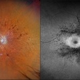
 Sialidosis
Jul 10 2025 by Jessilla Phou
These are fundus photographs capturing an 18 year old male with Type 1 Sialidosis, a rare inherited lysosomal storage disorder caused by a deficiency in the neuraminidase 1 (Neu1) enzyme. Currently, there are fewer than 1,000 people in the USA who have this disorder. It is characterized by a cherry red spot in the macula which occurs when lipids accumulate in the retinal ganglion cells. This causes the macula to appear red as seen in these fundus images. The patient presented at our office with ataxia, depth perception issues, and slow reaction time. His visual acuity was 20/40, suggestive of early stage Sialidosis.
Photographer: Jessilla Phou
Imaging device: Optos California
Condition/keywords: cherry red spot, fundus photograph, Sialidosis
-
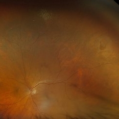
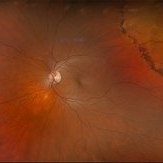
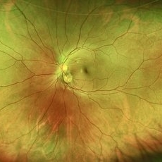 Pseudoduplication of the Optic Disc
Jul 9 2025 by Hrishikesh Naik, MS
A peripapillary colobomatous pseudo-duplication of the optic disc as seen in an asymptomatic 23 year old female with myopia referred for routine retinal periphery screening. Rest retinal exam was normal. Duplication of the optic disc can be classified as either true duplication or pseudoduplication, both of which are rare clinical conditions. Pseudodoubling of the optic disc is commonly caused by optic disc or peripapillary colobomas, characterized by a circumscribed, disc-like lesion with radiating vessels but only one normal optic nerve. A few cases have involved pathological myopia, moderate myopia, proliferative diabetic retinopathy and CHARGE syndrome. The lesion is often found inferior to the normal optic disc. The patient was advised regular follow ups.
Photographer: Hrishikesh Naik
Imaging device: Optos Daytona
Condition/keywords: Coloboma, Pseudoduplication of optic disc
-
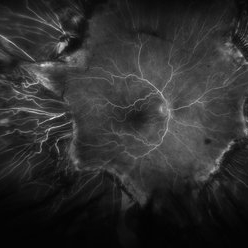 Fluorescein Angiogram of ROP With Cryo Scarring
Jul 7 2025 by Jenn Geelan
FA photo of a 34 year old male with prior stage 3 ROP with history of 360 degree cryotherapy.
Photographer: Jenn Geelan, Retina-Vitreous Surgeons of CNY
Imaging device: Optos California
Condition/keywords: cryotheraphy scar, fluorescein angiogram (FA), fundus photograph, retinopathy of prematurity (ROP), ROP, tilted disc
-
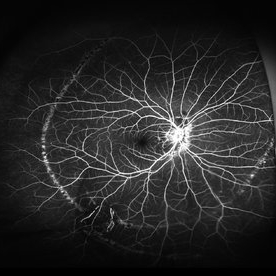 Schlaegel Line
Jul 7 2025 by César Adrián Gómez Valdivia, MD
Fluorescein Angiography showing a Schlaegel Line in a patient with suspected PHOS
Photographer: @eyemissu2
Imaging device: California ICG OPTOS
Condition/keywords: Schlaegel Line
-
 Acute Retinal Necrosis (ARN)
Jul 3 2025 by Heitor Nogueira
Fundus photograph of an 63-year-old woman who reported unilateral visual acuity loss for 10 days associated with ocular pain. He presented conjunctival hyperemia with temporal and nasal nodular scleritis, anterior chamber reaction 2+/4+, Koeppe nodules, granulomatous PKs, vitreitis 2+/4+, multiple areas of vasculitis in the arcades and periphery, associated with hemorrhages and necrotizing retinitis in the temporal, inferior and nasal periphery. Positive serology for Herpes Virus
Photographer: Heitor Nogueira, Penido Burnier Institute, Campinas, São Paulo, Brazil
Imaging device: Optos Daytona
Condition/keywords: ARN complications, Herpes, progressive outer retinal necrosis (PORN), Uveitis
 Loading…