Search results (84 results)
-
 Unexpected Sanctuary: Gas Bubble Entrapment in Morning Glory Disc
Sep 5 2025 by Danny Salgado Gómez
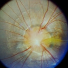
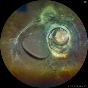
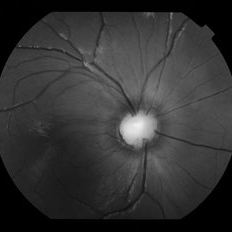
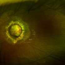
Fundus photograph of a 62-year-old male patient with Morning Glory syndrome in the right eye, who underwent vitrectomy, gas, and endolaser for posterior pole detachment. In the postoperative period, a gas bubble is observed within the optic disc, which persisted even after complete reabsorption of the intraocular gas.
Photographer: Dr. Danny Salgado, Retina and Vitreous Fellow, Clínica Oftalmológica del Caribe, Colombia.
Condition/keywords: gas bubble, intraocular gas, Morning Glory, Retinal Detachment, vitrectomy
-
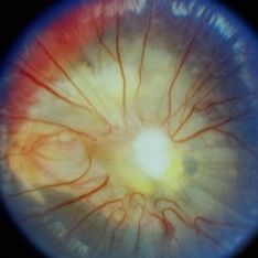
 Morning Glory Anomaly
Jan 5 2025 by César Adrián Gómez Valdivia, MD
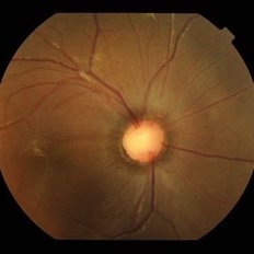
Morning Glory Anomaly found in a 10 year-old male patient with poor visual acuity and strabismus. Findings were unilateral.
Photographer: @eyemissu2
Imaging device: TOPCON TRC-50DX
Condition/keywords: Morning Glory Anomaly, Morning Glory Syndrome
-
 Coexistent Morning Glory Syndrome and Persistent Hyperplastic Primary Vitreous
Jul 15 2024 by Arthi Mohankumar , MS,MRCS ED, FICO,FAICO
Anterior segment and fundus photograph of a 9-year-old female revealing PHPC changes (Images A,B,C) and morning glory syndrome (image D).
Photographer: Arthi Mohankumar
Condition/keywords: Morning Glory Anomaly, Morning Glory Syndrome, persistent hyperplastic primary vitreous (PHPV)
-
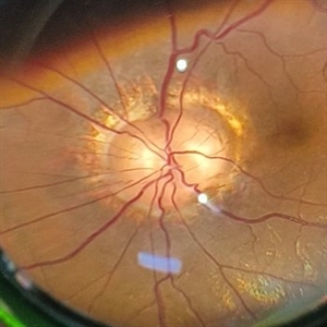
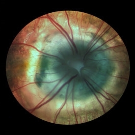
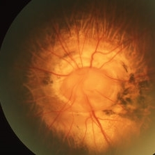
 Morning Glory Disc
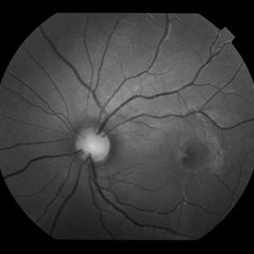
Sep 21 2023 by Ben Serar
Fundus photograph showing funnel shaped optic disc with radiating retinal vessels in a case of Morning glory syndrome.
Condition/keywords: Morning Glory Syndrome
-
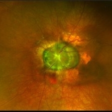
 Morning Glory disc
Sep 14 2023 by Ben Serar
Fundus photograph showing Funnel shaped optic disc in a case of Morning Glory Syndrome.
Condition/keywords: Morning Glory Syndrome
-
 Morning Glory Disc
Sep 12 2023 by Ben Serar
Fundus photograph showing Funnel shaped optic disc in a case of Morning Glory Syndrome.
Condition/keywords: Morning Glory Syndrome
-
 Morning Glory Syndrome
Jul 27 2023 by Karen Flores Guevara
Fundus photograph of a 30-year-old woman with Morning Glory Syndrome first diagnosed.
Photographer: Karen Flores-Guevara, MD, Asociación para Evitar la Ceguera en México I.A.P.
Condition/keywords: Morning Glory Syndrome
-
 Morning Glory Syndrome
May 12 2023 by Kalyan Singh
Fundus photograph of a 50 year old male presented with diminution of vision in right eye from last 2 years .
Photographer: Dr Kalyan Singh, GSVM medical college, Kanpur
Imaging device: Smartphone (1 plus 10R)
Condition/keywords: disc, Morning Glory Syndrome
-
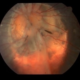
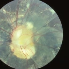
 Morning glory optic disc anomaly with retinal detachment
Sep 13 2022 by Min Kim, MD, PhD, MBA, FASRS
Fundus examination of this 5 year-old male shows large funneled optic nerve with conical excavation of the dysplastic optic disc. 360° macula-involving retinal detachment was observed. The best corrected visual acuity of the right eye was counting fingers 10cm.
Photographer: Min Kim, M.D.-Ph.D.-M.B.A. Gangnam Severance Hospital Yonsei University College of Medicine, Department of Ophthalmology
Imaging device: Optos Silverstone P200TxE
Condition/keywords: Morning Glory Anomaly, Morning Glory Syndrome
-
 Morning Glory
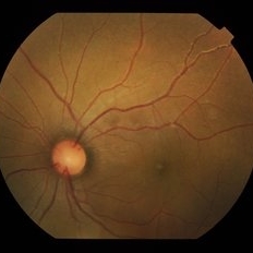
Jul 1 2022 by Geovanni Jassiel Rios, MD
Fundus Photograph 5 year old child with abnormal cup embryo-development. We can appreciate the radial vessel conformation and funnel shape nerve anomaly.
Photographer: Image Department Hospital de la Luz
Condition/keywords: Morning Glory Syndrome
-
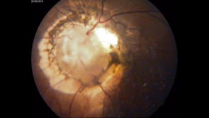
 RD operated with Morning Glory Optic Disc
Jul 22 2021 by Vishal Gupta, MBBS, MS
Fundus image of a 7-year-old patient status post Vitrectomy and Silicone oil insertion in a case of Retinal detachment with Morning glory Syndrome.
Photographer: Dr Shobhit Chawla, Prakash Netra Kendr, Lucknow, UP, INDIA
Imaging device: Zeiss Clarus
Condition/keywords: Morning Glory Syndrome, silicone oil
-
 Morning Glory Disc Anomaly
Nov 11 2020 by Yoshihiro Yonekawa, MD, FASRS
Color fundus photograph of a young boy with morning glory disc anomaly. Notice the concavity surrounding the enlarged disc, radial vasculature, and nasally dragged macula. MRI was negative for moyamoya disease, a known association.
Photographer: Alicia Thresher, Mid Atlantic Retina
Imaging device: Topcon
Condition/keywords: disc coloboma, Morning Glory Syndrome, pediatric retina
-
 Morning Glory Disc
Jan 20 2020 by Sarah Oelrich
Morning Glory Disc
Photographer: Sarah Oelrich CRA COT OCT-C Southeastern Retina Associates
Imaging device: Topcon
Condition/keywords: Morning Glory Syndrome
-
 Morning Glory Syndrome
Jan 6 2020 by Olivia Rainey
Ultra-wide field pseudocolor image of a 23-month-old male with morning glory syndrome affecting his left eye. Patient presented with esotropia affecting his left eye and strabismic amblyopia affecting both eyes. He could fix and follow on exam and his medical history was unremarkable.
Photographer: Olivia Rainey
Imaging device: Optos California
Condition/keywords: esotropia, left eye, macular, Morning Glory Syndrome, Optos, strabismic amblyopia, ultra-wide field imaging
-
 Morning Glory Disc Anomaly with Contractile Movements
Sep 30 2019 by Ashish Arjun Ahuja, DNB, FICO,(UK), FAICO (Retina)
A 16-year-old female patient presented at Orbit Eye Hospital, mumbai with complaints of diminished vision in the left eye since childhood. Vision in OS was finger counting 2 meters with myopic refraction (-8.00) and other eye was normal. Fundus examination revealed a morning glory disc anomaly with pulsatile movements of the tissue present in the optic disc.
Photographer: Zartash nasib ( B . Optometrist)
Imaging device: Zeiss
Condition/keywords: Morning Glory Syndrome
-
 Optic Disc Coloboma
Jul 24 2019 by Haider Ali
16-year-old boy with horizontal nystagmus and decreased vision in both eyes.
Photographer: Dr Haider Ali Chaudhry, Madinah Teaching Hospital, Faisalabad
Condition/keywords: coloboma, coloboma of optic disc, coloboma of the optic nerve, excavation, Morning Glory Syndrome
-
 Morning Glory Syndrome
Jun 19 2019 by Olivia Rainey
Ultra-wide field pseudocolor image of an 10-year-old girl with Morning Glory Syndrome affecting her left eye. Patient is able to count fingers at 4 feet.
Photographer: Olivia Rainey
Imaging device: Optos
Condition/keywords: left eye, Morning Glory Syndrome, Optos, pseudocolor, ultra-wide field imaging
-
 Morning Glory
Jun 17 2019 by Feyene Art
Oil on canvas painting inspired by a fundus photograph posted by Alex P. Hunyor, MD. https://imagebank.asrs.org/file/3048/optic-disc-dysplasia
Photographer: Feyene Art
Imaging device: Oil on Canvas Painting
Condition/keywords: Morning Glory Syndrome, optic disc dysplasia
-
 Morning Glory Syndrome
Apr 1 2019 by Gary R. Cook, MD, FACS
Young adult Vietnamese male with Morning Glory Syndrome OS.
Imaging device: Topcon VT-50
Condition/keywords: Morning Glory Syndrome
-
 Fellow Eye of Morning Glory Syndrome
Apr 1 2019 by Gary R. Cook, MD, FACS
Fellow eye (OD) of the young adult Vietnamese male with Morning Glory Syndrome of his disc OS.
Imaging device: Topcon VT-50
Condition/keywords: Morning Glory Syndrome
-
 Morning Glory Disc Anomaly
Apr 1 2019 by Gary R. Cook, MD, FACS
Morning glory disc anomaly with retinal detachment temporally.
Condition/keywords: Morning Glory Syndrome
-
 Slide 9-69
Feb 26 2019 by Lancaster Course in Ophthalmology
Noncystic peripheral retinal glial tuft.
Condition/keywords: Morning Glory Syndrome
 Loading…