Search results (112 results)
-
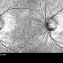
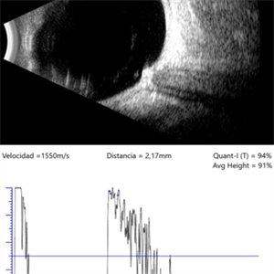 Melanocytoma
Mar 25 2025 by Gustavo Uriel Fonseca Aguirre
Longitudinal B-scan echogram shows mildly elevated lesion overlying surface of optic nerve. A-scan shows regular internal structure and high reflectivity of lesion.
Photographer: Gustavo U. Fonseca Aguirre, Hospital Conde de Valenciana, Ciudad de México
Condition/keywords: Melanocytoma
-
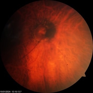
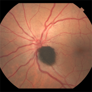
 Melanocytoma of Optic Disc
Jan 13 2025 by Virginia Gebhart
25 year-old female referred for melanocytoma of optic disc. Lesion is benign, no treatment necessary. Pt asymptomatic.
Photographer: Virginia Gebhart, Retina Consultants of Carolina
Imaging device: Topcon 50DX
Condition/keywords: benign melanocytoma, melanocytoma, optic disc melanocytoma
-
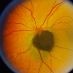
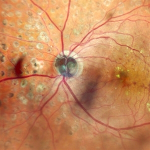 Melanocytoma of the Optic Nerve
Apr 6 2024 by Hector Gabriel Moreno Solano, MD, MHA
Optic Nerve laser scan image reconstruction of a 57-year-old male presented for an ophthalmological evaluation with a chief complaint of progressive visual loss. Indirect ophthalmoscopy revealed proliferative diabetic retinopathy, without macular edema, and a hyperpigmented lesion at the optic disc which corresponds to a melanocytoma.
Photographer: Héctor Gabriel Moreno-Solano, MD, MHA
Imaging device: Mirante
Condition/keywords: intraocular tumor, macular edema, melanocytoma, optic nerve
-
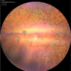 Melanocytoma of the Optic Nerve
Apr 6 2024 by Hector Gabriel Moreno Solano, MD, MHA
Fundus photograph of a 57-year-old male presented for an ophthalmological evaluation with a chief complaint of progressive visual loss. Indirect ophthalmoscopy revealed proliferative diabetic retinopathy, without macular edema, and a hyperpigmented lesion at the optic disc which corresponds to a melanocytoma.
Photographer: Héctor Gabriel Moreno-Solano
Imaging device: Clarus 700
Condition/keywords: diabetic retinopathy, intraocular tumor, melanocytoma, optic nerve
-
 Optic Nerve Melanocytoma
Mar 14 2024 by César Adrián Gómez Valdivia, MD
Benign neoplasm with seldom malignant transformation.
Photographer: Erika Paulina Ornelas Cazares
Imaging device: TOPCON TRC-50DX
Condition/keywords: disc, Melanocytoma, Nerve, Optic, optic disc melanocytoma
-
 Mirror Effect of OD-Melanocytoma
Mar 12 2024 by MEENAL SONI
SD-OCT of Optic disc melanocytoma with hyperintense homogenous flat lesion at disc and back shadowing.
Photographer: Dr. Meenal Soni, Fellow VR, ASG eye Hospital Jodhpur
Imaging device: OPTOPOL
Condition/keywords: optic disc melanocytoma
-
 Optic Disc Melanocytoma
Mar 12 2024 by MEENAL SONI
A 72 year old woman presented with DOV, on dilated examination was found to have PCO with optic disc melanocytoma. The vision improved with ND-YAG laser capsulotomy.
Photographer: Dr. Meenal Soni, Fellow VR, ASG eye Hospital Jodhpur
Imaging device: ZEISS Visucam 400
Condition/keywords: optic disc melanocytoma
-
 Melanocytoma of the disc
Dec 27 2023 by NIDHI PANWAR, MD FNB FICO
Fundus photograph of an otherwise healthy 41 year old female , with recently detected diabetes mellitus and came for fundoscopy was Detected to have left eye optic disc melanocytoma .
Photographer: Nidhi Panwar, NMC Royal hospital, Sharjah , UAE
Condition/keywords: melanocytoma, optic disc
-
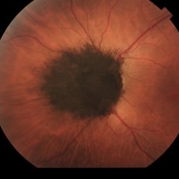 Melanocytoma of Optic Disc
Nov 3 2023 by Virginia Gebhart
69 year-old female with pigmented lesion that covers the optic nerve. Patient has been aware for over 30 years. Remains stable and unchanged
Photographer: Virginia Gebhart
Imaging device: Topcon
Condition/keywords: benign melanocytoma, Melanocytoma, optic disc melanocytoma
-
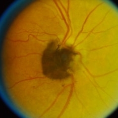
 Melanocytoma of the optic disc
Oct 10 2023 by Navneet Mehrotra, DNB
melanocytoma of the optic nerve head in a 48 year old female diagnosed on routine examination
Photographer: Dharti, Retina Care , Ahmedabad
Imaging device: Nidek RS 330
Condition/keywords: benign pigmented lesion, melanocytoma
-
 Optic Disc Melanocytoma
Sep 14 2023 by Ben Serar
Fundus photograph showing hyper pigmented lesion at the optic disc in a case of Optic disc melanocytoma.
Condition/keywords: optic disc melanocytoma
-
 Optic Disc Melanocytoma
Sep 14 2023 by Ben Serar
Fundus photograph showing hyper pigmented lesion at the optic disc in a case of Optic disc melanocytoma.
Condition/keywords: optic disc melanocytoma
-
 Optic Disc Melanocytoma
Sep 14 2023 by Ben Serar
Fundus photograph of the LE showing hyper pigmented lesion at the optic disc in a case of Optic disc melanocytoma.
Condition/keywords: optic disc melanocytoma
-
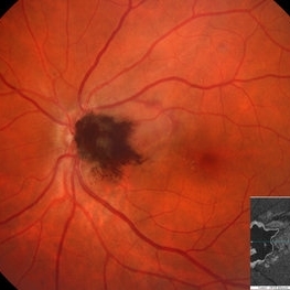
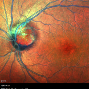
 Optic Nerve Melanocytoma
Apr 3 2023 by Gustavo Aguirre Suarez
Fundus photograph of a 36-year-old female with a lesion dependent on the optic nerve head with subretinal extension, elevated, about 1.5 disc diameters, dark brown to black in color, involving more than three quarters of the neuroretinal ring towards the inferonasal area.
Photographer: Dr. Gustavo Aguirre-Suarez
Imaging device: Zeiss Visucam 500
Condition/keywords: melanocytic lesion, Melanocytoma
-
 Melanocytoma
Nov 3 2022 by Stephanie Burke
Topcon fundus photograph of a 49-year old female with a melanocytoma of the left eye.
Photographer: Stephanie Burke, CRA, OCT-C
Condition/keywords: melanocytoma
-
 Optic Disc Melanocytoma
Aug 31 2022 by Vishal Agrawal, MD, FRCS,FACS,FASRS
Optic disc Melanocytoma detected on routine examination in the left eye of a 30 year old male .
Photographer: Vishal Agrawal
Imaging device: Clarus 700
Condition/keywords: Melanocytoma
-
 ONH Melanocytoma Multimodal Imaging
Mar 15 2021 by Deepak Bhojwani, MS
ONH melanocytoma multimodal imaging.
Condition/keywords: melanocytoma, optic nerve head
-
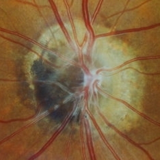
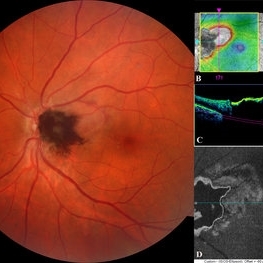
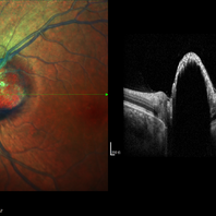
 Optic Disc Melanocytoma
Mar 15 2021 by Deepak Bhojwani, MS
Fundus photograph of a 49- year-old gentlemen with a characteristic dark brown elevated pigmented mass lesion centered on optic disc and extending into temporal peripapillary area classically suggestive of optic disc melanocytoma. Also note the pigment dispersion and retinal edema just superotemporal to the lesion secondary to tumor necrosis. Inset -Enface OCT image segmented at IS-OS ellipsoid zone level delineating exact horizontal & vertical extent of this tumor mass. Enface OCT imaging also helps in detailing the choroidal extension of such tumors.
Photographer: DEEPAK BHOJWANI; OCCURA EYE CARE & RESEARCH CENTER
Imaging device: OCT
Condition/keywords: enface imaging, melanocytoma, optic disc, optical coherence tomography (OCT)
-
 Choroidal Disc Melanoma
Dec 24 2020 by Aditya S Kelkar, MS, FRCS, FASRS,FRCOphth
Melanoma or Melanocytoma- A mystery
Condition/keywords: melanocytoma
-
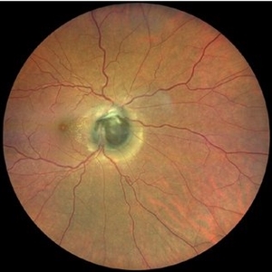
 Optic Disc Melanocytoma
-
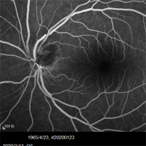
 Optic Disc Melanocytoma
-
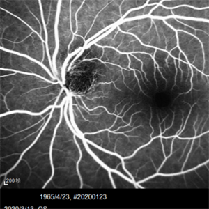
 Optic Disc Melanocytoma
-
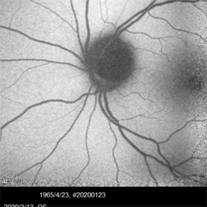
 Optic Disc Melanocytoma
-
 Optic Disc Melanocytoma
-
 Optic Disc Melanocytoma
May 25 2020 by Pengyao Lin
Optic disc melanocytoma.
Imaging device: multicolore image
Condition/keywords: optic disc melanocytoma
 Loading…