Search results (112 results)
-
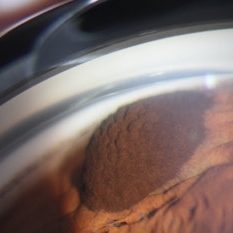 Iris Pigmented Lesion
Apr 27 2018 by Mark Lazcano
Gonio photograph of 20-year-old male with pigmented iris lesion consistent with melanocytoma
Photographer: mark Lazcano,University of Miami , Bascom Palmer Eye Institute
Imaging device: gonio Prism
Condition/keywords: pigmented lesion
-
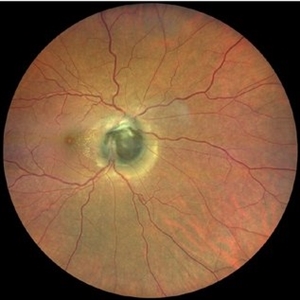
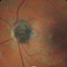
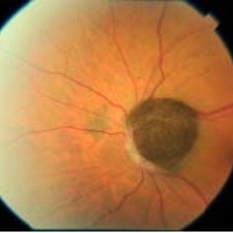
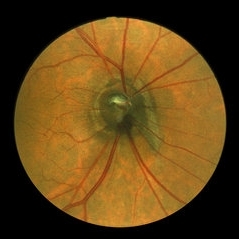 Optic Disc Melanocytoma
Jun 4 2014 by Henry J. Kaplan, MD
Optic disc melanocytoma with jet black pigmentation . #1
Condition/keywords: melanocytoma, optic disc melanocytoma
-
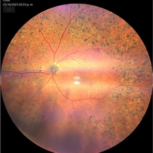
---thumb.jpg/image-square;max$300,300.ImageHandler) Melanocytoma
Melanocytoma
Feb 13 2013 by From the Collections of Thomas M. Aaberg, MD and Thomas M. Aaberg Jr., MD
Melanocytoma, color fundus photo, optic disc.
Condition/keywords: melanocytoma, optic disc
-
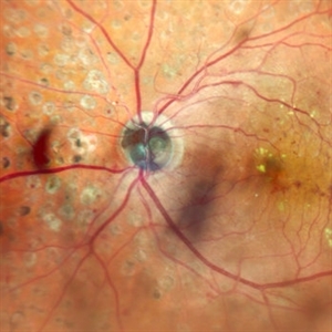
 Melanocytoma of the Optic Nerve
Apr 6 2024 by Hector Gabriel Moreno Solano, MD, MHA
Fundus photograph of a 57-year-old male presented for an ophthalmological evaluation with a chief complaint of progressive visual loss. Indirect ophthalmoscopy revealed proliferative diabetic retinopathy, without macular edema, and a hyperpigmented lesion at the optic disc which corresponds to a melanocytoma.
Photographer: Héctor Gabriel Moreno-Solano
Imaging device: Clarus 700
Condition/keywords: diabetic retinopathy, intraocular tumor, melanocytoma, optic nerve
-
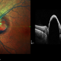
 Melanocytoma of the Optic Nerve
Apr 6 2024 by Hector Gabriel Moreno Solano, MD, MHA
Optic Nerve laser scan image reconstruction of a 57-year-old male presented for an ophthalmological evaluation with a chief complaint of progressive visual loss. Indirect ophthalmoscopy revealed proliferative diabetic retinopathy, without macular edema, and a hyperpigmented lesion at the optic disc which corresponds to a melanocytoma.
Photographer: Héctor Gabriel Moreno-Solano, MD, MHA
Imaging device: Mirante
Condition/keywords: intraocular tumor, macular edema, melanocytoma, optic nerve
-
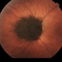
 Optic Nerve Melanocytoma
Apr 3 2023 by Gustavo Aguirre Suarez
Fundus photograph of a 36-year-old female with a lesion dependent on the optic nerve head with subretinal extension, elevated, about 1.5 disc diameters, dark brown to black in color, involving more than three quarters of the neuroretinal ring towards the inferonasal area.
Photographer: Dr. Gustavo Aguirre-Suarez
Imaging device: Zeiss Visucam 500
Condition/keywords: melanocytic lesion, Melanocytoma
-
 Choroidal Disc Melanoma
Dec 24 2020 by Aditya S Kelkar, MS, FRCS, FASRS,FRCOphth
Melanoma or Melanocytoma- A mystery
Condition/keywords: melanocytoma
-
 Melanocytoma
-
 Melanocytoma
May 2 2013 by Henry J. Kaplan, MD
Melanocytoma of the optic nerve head.
Condition/keywords: melanocytoma
-
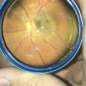 Melanocytoma of the Optic Nerve Head
Jan 13 2020 by Marlon García Roa, MD
Female 49-years-old, VA 20/100 OD and 20/20 OI. The patient does not associate low visual in right eye. Does not have heredofamiliar cancer background, and with a single pathographic unique personal background. Peripapilary injury of 3000 microns of diameter, elevated, with little defined edges, and orange pigment on the edge. in right eye.
Photographer: Marlon García Roa , INSTITUTO MEXICANO DE OFTALMOLOGIA, QUERETARO, MEXICO
Imaging device: SMARTPHONE
Condition/keywords: melanocytoma
-
 Optic Disc Melanocytoma
Sep 14 2023 by Ben Serar
Fundus photograph showing hyper pigmented lesion at the optic disc in a case of Optic disc melanocytoma.
Condition/keywords: optic disc melanocytoma
-
 Optic nerve melanocytoma 1 image 2
Jan 11 2013 by Alex P. Hunyor, MD
Optic nerve melanocytoma, left eye. Note: gradual enlargement over 12 years' followup. Color image 2, taken 1985.
Condition/keywords: optic disc melanocytoma
-
 Not All Vitreous Seeding Represents Malignancy: Case of Melanocytoma
Nov 18 2019 by Sophia El Hamichi, MD
Large optic disc melanocytoma with surrounding pigment dispersion. It is a benign lesion. The main differential in this case is melanoma with vitreous seeding.
Condition/keywords: melanocytoma, melanoma, vitreous seeding
-
 Melanocytoma
Sep 14 2012 by Michael P. Kelly, FOPS
Photographer: Michael P. Kelly, FOPS Director, Duke Eye Center Labs, Duke University Hospital
Imaging device: Zeiss FF450
Condition/keywords: melanocytoma
-
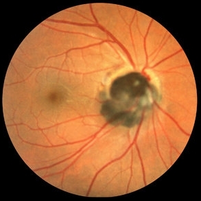
 Melanocytoma with Choroidal Melanoma
Oct 8 2012 by Susanna S. Park, MD, PhD
Fundus photograph of a 75-year-old woman with a slowly growing pigmented lesion.
Photographer: Ellen Redenbo, University of California Davis Eye Center
Condition/keywords: melanocytoma
-
 Melanocytoma
Oct 30 2012 by Lihteh Wu, MD
43-year-old hispanic female found to have on routine examination a melanocytoma of the optic nerve.
Condition/keywords: melanocytoma
-
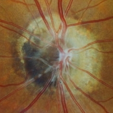
 Melanocytoma Case 2
Oct 5 2012 by Ronald C. Gentile, MD
Magnified photo of a melanocytoma of the optic disc. The elevated, black melanocytoma is seen impinging on the optic nerve with loss of the optic disc margins.
Photographer: The New York Eye & Ear Infirmary Department of Medical Imaging
Condition/keywords: melanocytoma
-
 Melanocytoma of Optic Disc
Nov 3 2023 by Virginia Gebhart
69 year-old female with pigmented lesion that covers the optic nerve. Patient has been aware for over 30 years. Remains stable and unchanged
Photographer: Virginia Gebhart
Imaging device: Topcon
Condition/keywords: benign melanocytoma, Melanocytoma, optic disc melanocytoma
-
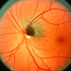
 Optic Disc Melanocytoma
Oct 16 2012 by Jeffrey G. Gross, MD, FASRS
Optic disc melanocytoma.
Condition/keywords: optic disc melanocytoma
-
 Melanocytoma and Vitreomacular Traction Syndrome Fundus Color Image
Mar 19 2018 by Nelson Antonio Segovia Rodríguez, MD
Color fundus image of an 68-year-old male with a optic dis melanocytoma and associated vitreomacular traction syndrome.
Photographer: Nelson Segovia, private practice
Imaging device: Zeiss
Condition/keywords: melanocytoma, optical coherence tomography (OCT), vitreomacular traction (VMT)
-
 Optic Disc Melanocytoma
-
---thumb.jpg/image-square;max$300,300.ImageHandler) Benign Melanocytoma of the Optic Disc
Benign Melanocytoma of the Optic Disc
Jan 11 2013 by Hyung-Woo Kwak, MD
Fundus photography of optic disc showing a dark pigmented lesion.
Photographer: Dongho Kang, Kyung Hee Univsersity Hospital, Seoul
Imaging device: Zeiss f 450 plus
Condition/keywords: benign melanocytoma
-
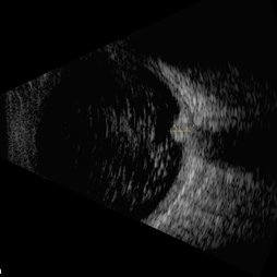
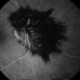 Giant Optic Disc Melanocytoma
Jun 12 2017 by Nabil Bouslous
Fundus photograph, angiography and B scan of an 50-year-old woman with a giant optic disc melanocytoma which has been involving over a past year.
Photographer: Nabil Bouslous, Mohamed VI university hospital, Marrakech, Morocco.
Condition/keywords: melanocytoma, optic disc
-
 Large Melanocytoma
Sep 2 2015 by Ali Tavallali, MD, FASRS
Fundus photograph of a 35-year-old female with large melanocytoma.
Photographer: Maryam Ravanshid
Condition/keywords: melanocytoma
 Loading…