Search results (189 results)
-
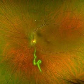
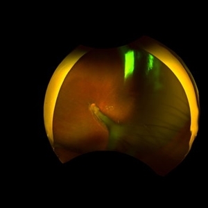
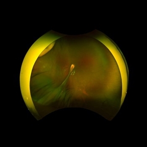
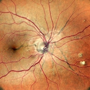
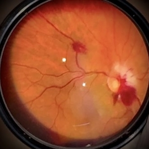 Bilateral Roth Spot in the Setting of Mitral Valve Endocarditis
Aug 18 2025 by Helder Vasconcelos
A 55-year-old man with chronic alcoholism presented with wasting and fever. The symptoms were preceded by a recent tooth extraction and gingivitis. Fundus examination in the ICU showed a retinal hemorrhage with a white spot (Roth spot) associated with peripapillary hemorrhage and cotton wool exudate. A similar Roth spot was observed in the contralateral eye.
Photographer: Helder Vasconcelos
Imaging device: Smartphone Fundoscopy
Condition/keywords: Infectious endocarditis, Roth Spots
-
 Oguchi Disease
Aug 12 2025 by Debarun Sharma
A 21 year old female presented with a history of night blindness for the past 16 years when she had difficulty in doing work and navigating places at night. BCVA OU was 6/6. Fundus examination showed the Mizuo-Nakamura phenomenon. ERG was done which showed extinguished rod responses with slightly diminished cone responses. A of Oguchi’s disease was made. The patient was advised for genetic testing and sibling screening. Oguchi’s disease is a rare cause of congenital stationary night blindness with characteristic fundus appearance.
Photographer: Debarun Sharma
Imaging device: Optos
Condition/keywords: Oguchi disease
-
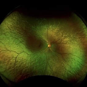
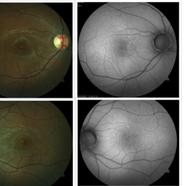
 Black Swan - Optic Disc Melanocytoma
Aug 5 2025 by SHRADDHA ASHOK CHANDORKAR, DNB DO FVRS
Just like the Black Swan which signifies an event that comes as a surprise, can have a major effect, and is often inappropriately rationalized after the fact with the benefit of hindsight, a 50 yr old presbyopic lady came to OPD with complains of diminution of vision - BCVA being 6/6 N6 in both eyes. Fundus examination revealed a pigmented nodule covering the optic disc .In most cases, fluorescein angiography of a melanocytoma of the optic disk demonstrates hypofluorescence throughout the angiogram. OCT disc showed elevated lesion, OCT macula normal and USG B scan with measurements were done to corroborate the posterior extension and to note increase in size if any on follow ups, perimetry was done to check for any field defects. All tests seemingly within normal limits - Patient was counselled and asked for 6 monthly follow up. Optic Disc Melanocytoma usually unilateral known to be a benign lesion that carries an excellent prognosis, the malignancy of this specific condition is rare 1-2%. The mean age at diagnosis of optic disk melanocytoma is 50 years with a median of 52 and range of 1–91 years. It is possible that melanocytoma is a congenital lesion but may not become clinically apparent until later in life, perhaps due to acquisition of pigment in a previously amelanotic lesion.
Photographer: Dr.Shraddha A. Chandorkar
Imaging device: topcon
Condition/keywords: optic disc melanocytoma
-
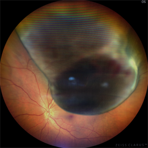
 Commotio Retinae
Jun 10 2025 by CUI YUELING
The patient presented 2 hours after sustaining a left eye injury caused by a stick. Visual acuity in the left eye was 0.2 without improvement upon correction, and intraocular pressure measured 15 mmHg. Examination of the anterior segment revealed ciliary conjunctival injection accompanied by patchy subconjunctival hemorrhage. The corneal surface remained smooth, and the anterior chamber was deep with hyphema characterized by blood-tinged aqueous humor predominantly settled inferiorly. The pupil was slightly irregular, approximately 3 mm in diameter, with a superotemporal notch; pupillary light reflex was intact. The lens appeared clear. Fundus examination showed well-defined optic disc margins with normal coloration and a cup-to-disc ratio of 0.2. Retinal arteries and veins were normally distributed with an artery-to-vein ratio of 2:3. At the posterior pole, the foveal reflex exhibited concentric ripple-like changes centered on the fovea, accompanied by localized pigment attenuation and reduced reflex intensity. Irregular reflectivity was noted in the superotemporal and inferotemporal nerve fiber layers.
Photographer: Yueling Cui
Imaging device: Zeiss Clarus 500
Condition/keywords: commotio retinae
-
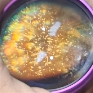
 Vitreous Cavity Inhabitant
Jun 2 2025 by Poornachandra B, MS, FVRS
A 36-year-old male presented with a 6-week history of intermittent ocular redness, now accompanied by the recent onset of floaters for the past 2 days. Fundus examination revealed the presence of a nematode in the vitreous cavity.
Photographer: Mr Dhikshith
Condition/keywords: parasite
-
 Bilateral Benign Yellow Dot Maculopathy
May 6 2025 by Amol yuvraj ganvir
A 37-year-old female patient presented for a routine eye examination. Her best-corrected visual acuity was 6/6 in both eyes. Fundus examination revealed multiple small yellow dots over the macula in both eyes. FAF imaging demonstrated characteristic hyperautofluorescence corresponding to these dots.
Photographer: Dr. Amol Ganvir, Vitreo-Retina Fellow, Ishwar Eye Centre, Rohtak, Haryana
Imaging device: Visucam-Zeiss
Condition/keywords: Autoflourescence, yellow dots
-
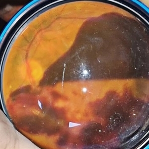
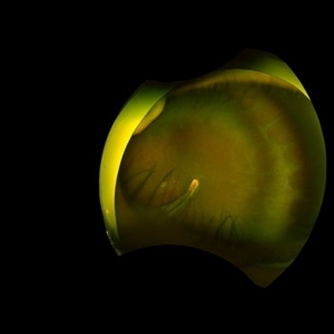
 Uveal Melanoma
Apr 26 2025 by Vishal Agrawal, MD, FRCS,FACS,FASRS
A 32 year-old male presented with complaints of perceiving a shadow in OS for 15-20 days. His BCVA was 20/20 OU. On Fundus examination, a large, elevated, well-defined, pigmented choroidal mass with few hemorrhages over the lesion was seen and a provisional diagnosis of uveal melanoma was made. urgent oncological consultation was recommended for further treatment.
Photographer: Dr Ayushi Gupta
Imaging device: Clarus 700
Condition/keywords: melanoma
-
 Hourglass in an Eye
Apr 22 2025 by KRISHNENDU NANDI, MS
A twenty-five-year-young male presented with a decrease in vision in the right eye following a blunt trauma with a football. On examination the BCVA in the right eye was CFCF and the left eye was 6/6, N6. The anterior segment was within normal limits. AT was 12 and 10 mm of Hg in the right and left eyes, respectively. Fundus examination reveals subhyaloid haemorrhage in the right eye with an attached retina. The fundus of the left eye was within normal limits. YAG laser hyaloidotomy was done with an energy of 2 mJ in the right eye. After 3 weeks the BCVA in the right eye improved to 6/9, N6.
Photographer: Dr. Krishnendu Nandi
Imaging device: Topcon
Condition/keywords: Trauma, YAG HYALOIDOTOMY, Young Male
-
 Proliferative Diabetic Retinopathy S/P Pan Retinal Photocoagulation
Mar 4 2025 by Prithvi Chandrakanth
A 52-year-old female patient presented with complaints of diminishing vision, compounded by uncontrolled diabetes mellitus. Her Fundus examination revealed proliferative diabetic retinopathy, characterized by neovascularization of the disc and elsewhere, and sclerosed vessels. To address this, Pan Retinal Photocoagulation was performed, and the condition stabilized, halting the progression of the disease.
Photographer: DR PRITHVI CHANDRAKANTH, DR CHANDRAKANTH NETHRALAYA, KOZHIKODE, KERALA, INDIA
Imaging device: EIDON
Condition/keywords: Diabetic Retinopathy, Neovascularisation at the Disc (NVD), neovascularization of the disc (NVD), NVD, pan-retinal photocoagulation (PRP), PDR, PDR with NVE (periphery), PRP
-
 Subhyaloid Hemorrhage
Mar 1 2025 by Vishal Agrawal, MD, FRCS,FACS,FASRS
A 37-year-old male presented with sudden diminution of vision in the right eye. On fundus examination boat shaped sub hyaloid hemorrhage was noted and a YAG hyaloidotomy was performed.
Photographer: Dr Ayushi Gupta
Imaging device: Clarus 700
Condition/keywords: Sub hyaloid haemorrhage, YAG HYALOIDOTOMY
-
 Vortex-pattern Exudative Retinal Detachment
Feb 22 2025 by CUI YUELING
Patient: Male, 40 years old. Chief Complaint: Blurred vision and metamorphopsia in the left eye for more than 10 days. Past Medical History Hypertension for 4 years, with a highest recorded blood pressure of 160/80 mmHg. Currently controlled with oral "Nifedipine Sustained-Release Tablets, 2 tablets daily." Long-term history of heavy alcohol consumption and smoking. Ophthalmic Examination: Visual Acuity: Right eye (OD): 0.4 (uncorrected, no improvement with correction). Left eye (OS): 0.5 (-1.5DS = 1.0). Intraocular Pressure (IOP): OD: 15 mmHg. OS: 17 mmHg. Anterior Segment:Unremarkable. Fundus Examination: Right eye: Optic disc margins are clear, with a slightly reddish hue. Cup-to-disc ratio (C/D) = 0.2. A scalloped, orange-red elevated lesion is observed superior to the optic disc, with anterior displacement of the focal point. This is accompanied by a secondary, turbine-like exudative retinal detachment centered around the optic disc, involving the macula. The macular region shows scattered punctate yellow-white exudates. Diagnosis: Choroidal hemangioma with secondary exudative retinal detachment(OD).
Photographer: Cui yueling The First People's Hospital of Zunyi, Guizhou, Zunyi, China
Imaging device: Zeiss Clarus 500
Condition/keywords: choroidal hemangioma, exudative retinal detachment
-
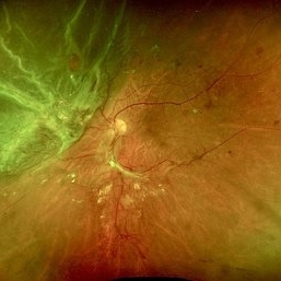
 Coat's Disease with Exudative RD
Feb 12 2025 by Tejaswita Verma
Fundus photo of a 7 year old boy with vision Counting fingers close to face in the right eye and intermittent outward deviation of the right eye observed by parents. Fundus examination shows subretinal exudates, telengiectatic vessels in superotemporal quadrant, intraretinal hemorrhages, macular scar, NVD.
Photographer: DR. TEJASWITA VERMA
Imaging device: MIRANTE
Condition/keywords: Coats' disease, exudative retinal detachment
-
 Sub Hyaloid Hemorrhage
Jan 29 2025 by Vishal Agrawal, MD, FRCS,FACS,FASRS
A 45-year-old male patient presented with decreased vision in Left Eye. On fundus examination, a boat-shaped sub hyaloid hemorrhage was noted. YAG hyaloidotomy was performed and the patient recovered with a vision of 20/20.
Photographer: Dr Ayushi Gupta
Imaging device: Clarus 700
Condition/keywords: Sub hyaloid haemorrhage, YAG HYALOIDOTOMY
-
 Subhyaloid Hemorrhage
Jan 22 2025 by DR Rohit Gupta
48 year old female presented with right eye diminution of vision, on fundus examination a large hemorrhage was seen in subhyaloid space with multiple retinal hemorrhages. Patients was known case of diabetes with uncontrolled blood sugar level.
Photographer: Dr Rohit gupta
Imaging device: Samsung S21
Condition/keywords: SUB ILM hemorrhage, subhyaloid blood, subhyaloid hemorrhage
-
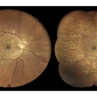
 A Classic Case of Retinal Ora Serrata Imaging
Jan 16 2025 by yuan duo
A 5-year-old girl, born full-term with no history of systemic disease, presented with poor vision since early childhood and underwent fundus examination. Anterior segments of both eyes showed no significant abnormalities. Fundus examination revealed retinal folds extending from the optic disc to the temporal peripheral retina, with blood vessels coursing through the folds (A, B). Avascular zones were observed in the peripheral retina, and the ora serrata’s boundaries were clearly visible, displaying dentate processes and bays (C, D). Retinal pigmentation was evident. Genetic testing confirmed the final diagnosis of bilateral Familial Exudative Vitreoretinopathy (FEVR).
Condition/keywords: Retinal Ora Serrata
-
 Familial Exudative Vitreoretinopathy
Jan 16 2025 by yuan duo
A 5-year-old girl, born full-term with no history of systemic disease, presented with poor vision since early childhood and underwent fundus examination. Anterior segments of both eyes showed no significant abnormalities. Fundus examination revealed retinal folds extending from the optic disc to the temporal peripheral retina, with blood vessels coursing through the folds (A, B). Avascular zones were observed in the peripheral retina, and the ora serrata’s boundaries were clearly visible, displaying dentate processes and bays (C, D). Retinal pigmentation was evident. Genetic testing confirmed the final diagnosis of bilateral Familial Exudative Vitreoretinopathy (FEVR).
Condition/keywords: Retinal Ora Serrata
-
 Synchysis Scintillans
Jan 6 2025 by DR Rohit Gupta
60 year old male presented with diminution of vision . On fundus examination multiple crystals deposits seen in vitreous cavity.
Photographer: Dr Rohit gupta
Imaging device: Samsung S21
Condition/keywords: asteroid hyalosis, synchysis scintillans, vitreous cavity
-
 Post Dengue Fever Retinitis
Dec 27 2024 by Tejaswita Verma
A 35 year old female presented with bilaterally decreased vision since a month post dengue fever. Vision was finger counting 6 mt. in the RE and 6/36 in the left eye .Fundus examination revealed severe vitritis in the RE with retinitis lesions and hemorrhages and macular star ,Fundus examination of the left eye revealed moderate vitritis with retinitis lesions with hemorrhages and macular star. She was started on tab doxycycline 100 mg BD for 2 weeks and oral steroids. She was also advised RE intravitreal implant Ozurdex with explained prognosis.
Photographer: DR. TEJASWITA VERMA
Imaging device: MIRANTE
Condition/keywords: bilateral dengue retinitis, Dengue Fever, POST FEVER RETINITIS
-
 Post Dengue Fever Retinitis
Dec 27 2024 by Tejaswita Verma
A 35 year old female presented with bilaterally decreased vision since a month post dengue fever. Vision was finger counting 6 mt. in the RE and 6/36 in the left eye .Fundus examination revealed severe vitritis in the RE with retinitis lesions and hemorrhages and macular star ,Fundus examination of the left eye revealed moderate vitritis with retinitis lesions with hemorrhages and macular star. She was started on tab doxycycline 100 mg BD for 2 weeks and oral steroids
Photographer: DR. TEJASWITA VERMA
Imaging device: MIRANTE
Condition/keywords: Dengue Fever, macular star, POST FEVER RETINITIS
-
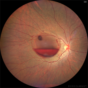
 Setting Sail: A Striking View of a Boat-Shaped Subhyaloid Hemorrhage
Nov 27 2024 by Jasmeet Kaur Chandi
A 22 year old female came with sudden drop in vision after exposure to laser show. Fundus examination revealed a large area of subhyaloid hemorrhage at macula .ND YAG Laser Hyaloidotomy was planned. Laser applied at an energy level of 1.2 mJ and employed just above the inferior apex of the bleeding. With increased energy, the blood bubble started to undulate. A rupture was finally achieved at 3.2 mJ. This fundus image was taken just after the laser therapy and it captured the outpouring.
Photographer: Dr. Jasmeet Kaur Chandi
Condition/keywords: subhyaloid haemorrhage, YAG HYALOIDOTOMY
-
 Combined Traction Rhegmatogenous Detachment
Oct 17 2024 by Hemanth Murthy, MBBS, MD, FASRS
A 68 year old male presented with a shadow in the left eye since 3 days. He was a known diabetic and hypertensive for 20 years. Vision was 20/40 in right eye and 20/60 in left eye. Fundus examination showed Proliferative diabetic retinopathy in right eye and Proliferative diabetic retinopathy with combined traction rhegmatogenous detachment in left eye.
Photographer: Mr Veda Vyas
Condition/keywords: combined retinal detachment, proliferative diabetic retinopathy (PDR)
-
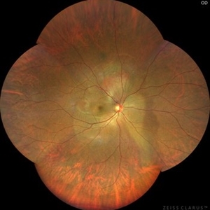
 Angioid Streaks
Sep 29 2024 by Tejaswita Verma
Fundus photograph of a 35 year-old female with 6/6 vision in RE , unremarkable anterior segment and family history of angioid streaks and pseudoxanthoma elasticum in sister. Fundus examination revealed angioid streaks radiating from disc , sparing the fovea .Her Sister had received multiple anti VEGF injections for angioid streaks with CNVM.
Photographer: DR. TEJASWITA VERMA
Imaging device: MIRANTE
Condition/keywords: angioid streaks
-
 Pathological Myopia
Sep 25 2024 by DR Rohit Gupta
Fundus photograph of a 28 year-old male having high myopia on fundus examination Degenerative changes are seen in retina suggestive of pathological myopia.
Photographer: Dr Rohit gupta
Imaging device: Samsung S21
Condition/keywords: choroidal degeneration, degeneration of optic disc, lacquer cracks, myopia, Myopia macular degeneration CNVM foster fuch spot, pathologic myopia, staphyloma
 Loading…