Search results (150 results)
-
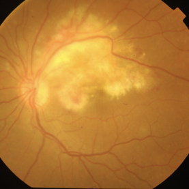
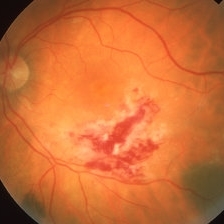
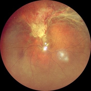 RD with PVR in CMV Retinitis in an HIV Positive Patient
Jul 31 2024 by Tejaswita Verma
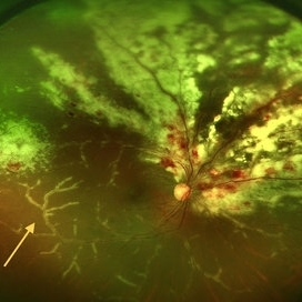
Fundus photograph of a 48 year old male with CF 1.5 mt vision having history of CMV retinitis, on HAART with CD4 count 81, showing retinal detachment with proliferative vitreoretinopathy changes. He was advised pars plana vitrectomy with silicon oil infusion.
Photographer: DR. TEJASWITA VERMA
Imaging device: MIRANTE
Condition/keywords: CMV retinitis with retinal detachment, HIV
-
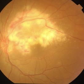
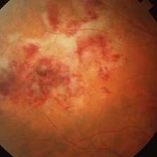
 Cheese Pizza Pie Appearance in CMV Retinitis
Mar 30 2024 by KANWALJEET HARJOT MADAN, M.S. (Ophthalmology), FAICO (Vitreous - Retina)
This is Fundus Photograph of left eye of 53 year male depicting an area of Retinal Necrosis with few Retinal Haemorrhages suggestive of CMV Retinitis. Areas of Perivascular Exudation also seen. On investigations, the patient was found to be HIV positive. He was started on Anti Retro Viral treatment after physician opinion.
Photographer: Dr. Kanwaljeet Harjot Madan, Thind Eye Hospital, Jalandhar City (Punjab) INDIA.
Imaging device: Zeiss Fundus Camera
Condition/keywords: AIDS, cytomegalovirus (CMV), retinitis
-
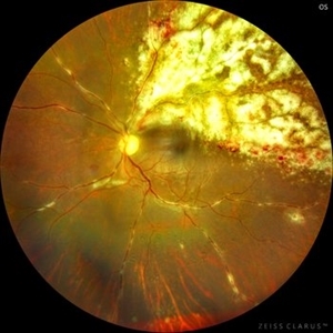
 CMV Retinitis
Feb 17 2024 by Eloy Mata-Cortes, MD
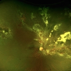
Fundus photograph of left eye showing Cytomegalovirus retinitis of a 40-year-old male with positive HIV history. He presented with CD4 cell count of 50 cells/mm3 and decreased vision of left eye. In the photograph we can see the three typical patterns in this retinitis: a hemorrhagic appearance in superior temporal arcade and between nasal arcades, granular pattern in superior temporal retina, and a “frosted branch” angiitis surrounding the retinal vessels in nasal and superior retina.
Photographer: Eloy Mata-Cortes, Instituto Mexicano de Oftalmologia, Queretaro, Mexico
Imaging device: Clarus 700
Condition/keywords: CMV retinitis, cytomegalovirus (CMV), frosted branch angiitis, Frosted Branch Angitis
-
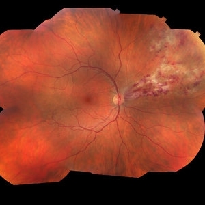
 Cytomegalovirus Retinitis
Jan 29 2024 by Isaac Agranoff
Widefield fundus photograph of a 73 year old male with Cytomegalovirus Retinitis. Patient presented with CMV retinitis after noticing visual changes the last 7 months. Vision was measured in office at HM.
Photographer: Isaac Agranoff
Imaging device: Optos California
Condition/keywords: branch retinal vein occlusion (BRVO), central retinal artery occlusion, CMV retinitis, cytomegalovirus (CMV)
-
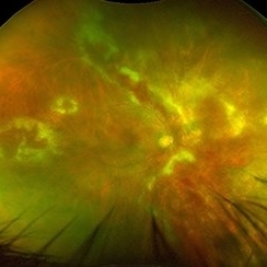
 CMV retintis
Sep 14 2023 by Ben Serar
Fundus photograph of LE showing superficial flame-shaped haemorrhages with surrounding retinal necrosis at the posterior pole along the superotemporal arcade in a case of fulminant type of CMV retinitis.
Condition/keywords: CMV retinitis, fulminant retinitis, pizza-pie appearance, viral retinitis
-
 CMV Retinitis with Shallow RD
Aug 21 2023 by rahul saradge
47 YEAR OLD MALE HAVING CMV RETINITIS WITH SHALLOW RD, VITRITIS
Photographer: Hitesh Rawlani , Isha Netralaya
Condition/keywords: cmv retinits with shallow RD
-
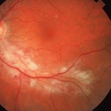
 Resolving CMV retinitis
Aug 10 2023 by Vaidehi Sathaye
Fundus photograph of LE of a 24 year old male patient showing resolving CMV retinitis lesion after initiation of Intravitreal Ganciclovir therapy
Photographer: Dr. Vaidehi Sathaye
Condition/keywords: CMV retinitis
-
 CMV retinitis
Aug 10 2023 by Vaidehi Sathaye
Fundus photograph of LE of a 24 year male patient with Fulminant type of CMV retinitis.
Photographer: Dr. Vaidehi Sathaye
Condition/keywords: CMV retinitis
-
 Progression of CMV Retinitis (click on image for GIF)
May 27 2023 by Jeremy Reitinger
Progression of treated CMV retinitis with intravitreal ganciclovir
Photographer: Jeremy Reitinger, Indiana University
Condition/keywords: CMV retinitis
-
 Progression of CMV Retinitis (click on image for GIF)
May 27 2023 by Jeremy Reitinger
Progression of treated CMV retinitis with intravitreal ganciclovir
Photographer: Jeremy Reitinger, Indiana University
Condition/keywords: CMV retinitis
-
 Cytomegalovirus Retinitis
May 8 2023 by Akansha Sharma
Colour fundus photograph of a 37 year old male with cytomegalovirus retinitis with macular edema
Photographer: Dr. Urmil Shah, Dr. Denish Patel, Dr. Akansha Sharma, Bharati Eye Clinic, Ahmedabad, Gujarat
Condition/keywords: CMV retinitis
-
 CMV Retinitis with Frosted Branch Angiitis
Sep 23 2020 by Nimesh A. Patel, MD, FASRS
Fundus photo showing peri-vascular inflammation of both arteries and veins with translucent exudation (yellow arrow). Superior nasally, there is classic retinal whitening with retinal hemorrhages superior. This patient was found to have a low CD4 count and a diagnosis of AIDS was made.
Condition/keywords: cytomegalovirus (CMV), HIV, uveitis
-
 CMV Retinitis
Sep 23 2020 by Nimesh A. Patel, MD, FASRS
Follow up funds photograph 2 weeks post initiation of treatment with intravenous ganciclovir and intravitreal ganciclovir demonstrating improvement of the vasculitis.
Condition/keywords: cytomegalovirus (CMV)
-
 CMV Retinitis in AIDS Patient
Dec 12 2019 by McGill University Health Centre
Histopathological examination of the retina showing no inflammation and a large multinucleated CMV infected cell.
Photographer: Miguel N. Burnier, McGill University Health Center-McGill University Ocular Pathology & Translational Research Laboratory
Imaging device: Zeiss
Condition/keywords: CMV retinitis, cytomegalovirus (CMV), histopathology, multinucleated giant cells, retina
-
 CMV Retinitis in AIDS Patient
Dec 12 2019 by McGill University Health Centre
Histopathological examination of the retina showing no inflammation and a large multinucleated CMV infected cell. Sections of the choroid shows no inflammation, the choroid capillaries and normal choroidal melanocytes.
Photographer: Miguel N. Burnier, McGill University Health Center-McGill University Ocular Pathology & Translational Research Laboratory
Imaging device: Zeiss
Condition/keywords: choroid, cytomegalovirus (CMV), histopathology, multinucleated giant cells, retina
-
 CMV Retinitis in AIDS Patient
Dec 12 2019 by McGill University Health Centre
Fundus photograph of a 32-year-old man with HIV infection and 100 CD4+ cells count. Several areas of retinal necrosis interspersed with areas of hemorrhage around blood vessels can be observed.
Photographer: Miguel N. Burnier, McGill University Health Center-McGill University Ocular Pathology & Translational Research Laboratory
Imaging device: Fundoscopy
Condition/keywords: AIDS, cytomegalovirus (CMV), HIV, retinitis
-
 Regressed CMV Retinitis
Mar 26 2019 by Gary R. Cook, MD, FACS
39-year-old white male with HIV/AIDS showing area of regressed CMV retinitis following therapy with IV ganciclovir.
Imaging device: Topcon VT-50
Condition/keywords: CMV retinitis
-
 CMV Retinitis
Mar 26 2019 by Gary R. Cook, MD, FACS
39-year-old white male with HIV/AIDS and active CMV retinitis along inferotemporal arcade.
Imaging device: Topcon VT-50
Condition/keywords: CMV retinitis
-
 CMV Retinitis
Mar 26 2019 by Gary R. Cook, MD, FACS
Left eye of a 49-year-old white male with HIV/AIDS and active CMV retinitis; V.A.= 20/30.
Imaging device: Topcon VT-50
Condition/keywords: CMV retinitis
-
 CMV Retinitis
Mar 26 2019 by Gary R. Cook, MD, FACS
49-year-old white male with HIV/AIDS and active CMV retinitis OD; V.A.= 20/25.
Imaging device: Topcon VT-50
Condition/keywords: CMV retinitis
-
 AIDS and CMV Retinitis
Mar 26 2019 by Gary R. Cook, MD, FACS
31-year-old white male with HIV/AIDS and CMV retinitis OS; VA= counting fingers.
Imaging device: Topcon VT-50
Condition/keywords: AIDS, cytomegalovirus (CMV)
-
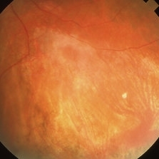
 Cytomegalovirus (CMV) Retinitis
Mar 26 2019 by Gary R. Cook, MD, FACS
31-year-old white male with active CMV retinitis along inferotemporal arcade OS; VA = 20/20.
Imaging device: Topcon VT-50
Condition/keywords: CMV retinitis
-
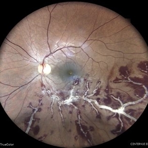
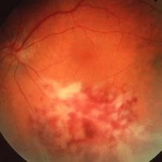
 CMV Retinitis
Jan 10 2019 by Rahul Komati, MD
63-year-old male with history of plasma cell leukemia, presenting with photopsias and 20/25 vision. Fundus photograph shows superior area of retinitis, intraretinal hemorrhage, and vessel sclerosis. Retinitis regressed with systemic valganciclovir and 5 intravitreal foscarnet injections over 3 weeks.
Photographer: Pamela Hulvey, University of Chicago
Imaging device: Optos
Condition/keywords: CMV retinitis
-
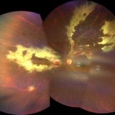
 Cytomegalovirus Retinitis
Jan 16 2018 by Olivia Rainey
Color fundus montage of an 37-year-old, HIV positive male with CMV retinitis affecting his right eye. Patient's vision was sc20/20-1. He received an intravitreal Ganciclovir injection as well. The referring physcian suspects his condition is secondary to his chemotherapy for large B cell lymphoma or stomach cancer. The patient had not started taking oral Valgancyclovir.
Photographer: Olivia Rainey
Imaging device: Topcon 50dx
Condition/keywords: CMV retinitis, color fundus photograph, cytomegalovirus (CMV), HIV, montage
 Loading…