-
 Sector Retinitis Pigmentosa
Mar 13 2014 by Hyung-Woo Kwak, MD
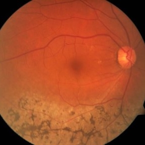
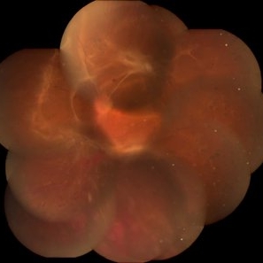
Fundus photograph of an 57-year-old woman with a sector retinitis pigmentosa. Regionalized areas of bone spicule pigmentation is in the inferior quadrants of the retina.
Photographer: Missok Lee, Kyung Hee University Hospital, Seoul, Korea
Imaging device: Zeiss F450 Plus
Condition/keywords: sector retinitis pigmentosa
-
 Sector Retinitis Pigmentosa
Mar 13 2014 by Hyung-Woo Kwak, MD
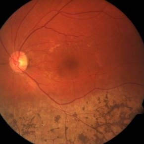
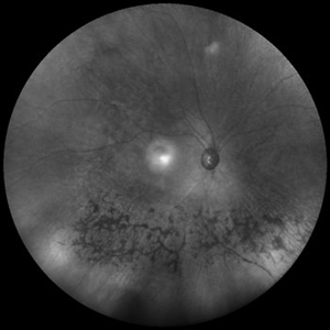
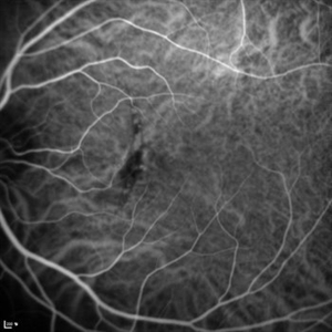
Wide field infrared image of an 57-year-old woman with a sector retinitis pigmentosa. Regionalized areas of bone spicule pigmentation is in the inferior quadrants of the retina.
Photographer: Missok Lee, Kyung Hee University Hospital, Seoul, Korea
Imaging device: Heidelberg Spectralis
Condition/keywords: sector retinitis pigmentosa
-
 Sector Retinitis Pigmentosa
Mar 13 2014 by Hyung-Woo Kwak, MD
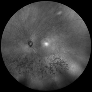
Wide field infrared image of an 57-year-old woman with a sector retinitis pigmentosa. Regionalized areas of bone spicule pigmentation is in the inferior quadrants of the retina.
Photographer: Missok Lee, Kyung Hee University Hospital, Seoul, Korea
Imaging device: Heidelberg Spectralis
Condition/keywords: sector retinitis pigmentosa
-
 Metallic Intraocular Foreign Body
Apr 16 2014 by Hyung-Woo Kwak, MD
Coronal CT scan of metallic intraocular foreign body (IOFB).
Photographer: Kyung Hee University Hospital
Imaging device: CT scan
Condition/keywords: intraocular foreign body
-
 Metallic Intraocular Foreign Body
Apr 16 2014 by Hyung-Woo Kwak, MD
Fundus photograph of intraocular foreign body (IOFB) from grass eliminator. IOFB was removed via vitrectomy (20G microforcep).
Photographer: Kyung Hee University Hospital
Imaging device: Surgical microscope (Zeiss OPMI LUMERA 700)
Condition/keywords: intraocular foreign body
-
 Cilioretinal Artery Occlusion
Sep 2 2012 by Hyung-Woo Kwak, MD
Cloudiness localized to the area of papillomacular bundle normally perfused by retinal vessel.
Imaging device: Zeiss F450 plus
Condition/keywords: cilioretinal artery occlusion
-
 Candida Endophthalmitis
Sep 2 2012 by Hyung-Woo Kwak, MD
A few localized epiretinal infiltrates of fluffy creamy white appearance in both fundus.
Imaging device: Zeiss F450 plus
Condition/keywords: candida endophthalmitis
-
 Vitelliform Macular Dystrophy
Sep 2 2012 by Hyung-Woo Kwak, MD
The typical appearance is of bilateral, round or oval, yellow, symmetrical, subretinal lesions, typically one-third to one-half optic disc diameter in size.
Imaging device: Zeiss F450 plus
-
 Vitelliform Macular Dystrophy
Sep 2 2012 by Hyung-Woo Kwak, MD
The typical appearance is of bilateral, round or oval, yellow, symmetrical, subretinal lesions, typically one-third to one-half optic disc diameter in size.
Imaging device: Zeiss F450 plus
Condition/keywords: Best disease
-
 Chronic Macular Hole
Sep 2 2012 by Hyung-Woo Kwak, MD
A large hole with rolled everted edges, adjacent cystoid intraretinal spaces, a shallow rim of subretinal fluids.
Imaging device: Zeiss F450 plus
Condition/keywords: macular hole
-
 Broad Subretinal Hemorrhage
Sep 2 2012 by Hyung-Woo Kwak, MD
A broad blotchy black area underlying the retinal blood vessels with the retina usually elevated the blood.
Imaging device: Zeiss F450 plus
Condition/keywords: subretinal hemorrhage
-
 Chorioretinal Fold
Sep 2 2012 by Hyung-Woo Kwak, MD
Chorioretinal folds are seen as coarse striations of the fovea surface after trauma.
Imaging device: Zeiss F450 plus
Condition/keywords: chorioretinal fold
-
 Retinitis Pigmentosa
Sep 2 2012 by Hyung-Woo Kwak, MD
A mild pigment epithelial atrophy in the mid-periphery with small white dots at the level of the RPE in fundus.
Imaging device: Zeiss F450 plus
Condition/keywords: retinitis pigmentosa
-
 Central Retinal Vein Occlusion
Sep 2 2012 by Hyung-Woo Kwak, MD
Multiple dense, dark, blotchy hemorrhages, cotton-wool spots, and pale optic disc are signs suggestive of retinal ischemia in CRVO.
Imaging device: Zeiss F450 plus
Condition/keywords: central retinal vein occlusion (CRVO)
-
 Choroidal Hemangioma
Oct 20 2012 by Hyung-Woo Kwak, MD
Fundus, ICG, and OCT examination showed a typical chorioretinal scar lying concentric to the optic disc. Typical choroidal rupture was performed after intravitreal gas injection under the diagnosis of submacular hemorrhage caused by trauma, after the absorption of submacular hemorrhage
Condition/keywords: chorioretinal scar, choroidal rupture, submacular hemorrhage
-
 Choroidal Hemangioma
Oct 20 2012 by Hyung-Woo Kwak, MD
Fundus, ICG and OCT examination showed a typical chorioretinal scar lying concentric to the optic disc. Typical choroidal rupture was performed after intravitreal gas injection under the diagnosis of submacular hemorrhage caused by trauma, after the absorption of submacular hemorrhage
-
 Hypertensive Retinopathy
Oct 20 2012 by Hyung-Woo Kwak, MD
The sudden appearance of cotton wool spots with hypertension retinopathy is known as accelerated hypertension. This patient has acute hypertension of rapid onset and measured systolic blood pressure was more than 200mmhg at this time.
Imaging device: Zeiss F450 plus
Condition/keywords: cotton wool spots, hypertension
-
 Circumscribed Choroidal Hemangioma
Oct 20 2012 by Hyung-Woo Kwak, MD
Fundus and OCT examination showed an oval mass at the posterior pole with indistinct margins that blend with surrounding choroid. FA early phase showed hyperfluorescence.
-
 Circumscribed Choroidal Hemangioma
Oct 20 2012 by Hyung-Woo Kwak, MD
Fundus and OCT examination showed an oval mass at the posterior pole with indistinct margins that blend with surrounding choroid. FA early phase showed hyperfluorescence.
-
 Circumscribed Choroidal Hemangioma
Oct 20 2012 by Hyung-Woo Kwak, MD
Fundus and OCT examination showed an oval mass at the posterior pole with indistinct margins that blend with surrounding choroid. FA early phase showed hyperfluorescence.
-
 Circumscribed Choroidal Hemangioma
Oct 20 2012 by Hyung-Woo Kwak, MD
Fundus and OCT examination showed an oval mass at the posterior pole with indistinct margins that blend with surrounding choroid. FA early phase showed hyperfluorescence.
-
 Multiple Evanescent White Dot Syndrome (MEWDS)
Oct 20 2012 by Hyung-Woo Kwak, MD
Numerous small deep ill-defined, grey-white dot were seen at the posterior pole and mid-periphery. Some lesions showed mild hyperfluorescence in autofluorescence (AF) but were of limited diagnostic value. ICG showed more numerous hypofluorescent spots than are apparent clinically or on AF/FA
Condition/keywords: hypofluorescent spots, multiple evanescent white dot syndrome (MEWDS)
-
 Multiple Evanescent White Dot Syndrome (MEWDS)
Oct 20 2012 by Hyung-Woo Kwak, MD
Numerous small deep ill-defined, grey-white dot were seen at the posterior pole and mid-periphery. Some lesions showed mild hyperfluorescence in autofluorescence (AF) but were of limited diagnostic value. ICG showed more numerous hypofluorescent spots than are apparent clinically or on AF/FA.
Condition/keywords: hypofluorescent spots, multiple evanescent white dot syndrome (MEWDS)
-
 Chorioretinal Fold
Oct 20 2012 by Hyung-Woo Kwak, MD
Fundus showed dense, white, well-demarcated, geographical areas of confluent opacification associated with vasculitis. This patients was constantly receiving immunosuppressants after pancreas transplant surgery.
Condition/keywords: chorioretinal fold
-
 Rhegmatous Retinal Detachment
Oct 20 2012 by Hyung-Woo Kwak, MD
Fundus showed a large tear in superior bullous retinal detachment.
Condition/keywords: retinal tear
-
 Toxoplasmosis
Oct 20 2012 by Hyung-Woo Kwak, MD
This is a typical quiescent toxoplasma scar in the posterior pole. It is sharply circumscribed with retinal hyperpigmentation and pigment epithelial atrophy.
Condition/keywords: pigment epithelial atrophy, retinal hyperpigmentation, toxoplasmosis
-
 Tractional Retinal Detachment
Oct 20 2012 by Hyung-Woo Kwak, MD
Observed following Fundus seems like a typical tractional retinal detachment in severe proliferative diabetic retinopathy. But during vitrectomy, another rhegmatous retinal detachment was observed.
-
 Choroidal Neovascularization
Oct 20 2012 by Hyung-Woo Kwak, MD
This 35-year-old young female patient has drusen-like lesions under the macular in both eyes. Such patients have a considerable risk of developing choroidal neovascular lesions.
Condition/keywords: choroidal neovascularization (CNV)
-
---thumb.jpg/image-square;max$300,300.ImageHandler) Choroidal Neovascularization
Choroidal Neovascularization
Oct 20 2012 by Hyung-Woo Kwak, MD
This 35-year-old young female patient has drusen-like lesions under the macular in both eyes. Such patients have a considerable risk of developing choroidal neovascular lesions.
Condition/keywords: choroidal neovascularization (CNV)
-
 Central Retinal Artery Occlusion
Oct 20 2012 by Hyung-Woo Kwak, MD
This is a typical recent central retinal artery occlusion with a ‘cherry red’ spot at the macula. The patient visited our hospital with sudden visual loss occurred after the filler injection around the eyes.
Condition/keywords: central retinal artery occlusion (CRAO), cherry red spot
-
 Vogt-Koyanagi-Harada Syndrome
Oct 20 2012 by Hyung-Woo Kwak, MD
Fundus and OCT examination showed multiple serous retinal detachment.
Photographer: Hyung Woo Kwak, Department of Ophthalmology, Kyung Hee University Hospital, Seoul, Korea
Imaging device: Zeiss f 450 plus, Heidelberg Spectralis
Condition/keywords: serous retinal detachment
-
---thumb.jpg/image-square;max$300,300.ImageHandler) Choroidal rupture
Choroidal rupture
Jan 11 2013 by Hyung-Woo Kwak, MD
There is a yellow or white crescent-shaped subretinal streak concentric with the optic nerve.
Photographer: Misook Lee, Kyung Hee Univsersity Hospital, Seoul
Imaging device: Zeiss f 450 plus
Condition/keywords: choroidal rupture
-
 Stargardt’s disease
Jan 11 2013 by Hyung-Woo Kwak, MD
Retinal pigment epithelium atrophy in the posterior pole bilaterally.
Photographer: Misook Lee, Kyung Hee Univsersity Hospital, Seoul
Imaging device: Zeiss f 450 plus
-
 Stargardt’s disease
Jan 11 2013 by Hyung-Woo Kwak, MD
Retinal pigment epithelium atrophy in the posterior pole bilaterally.
Photographer: Misook Lee, Kyung Hee Univsersity Hospital, Seoul
Imaging device: Zeiss f 450 plus
Condition/keywords: Stargardt disease
-
---thumb.jpg/image-square;max$300,300.ImageHandler) Myopic fundus
Myopic fundus
Jan 11 2013 by Hyung-Woo Kwak, MD
Myopic fundus reveals yellow-colored lacquer cracks and peripapillary atrophy. There was visible choroidal vessel due to thin retina.
Photographer: Misook Lee, Kyung Hee Univsersity Hospital, Seoul
Imaging device: Zeiss f 450 plus
Condition/keywords: myopic fundus
-
---thumb.jpg/image-square;max$300,300.ImageHandler) Coats disease
Coats disease
Jan 11 2013 by Hyung-Woo Kwak, MD
Fundus imaging shows hemorrhage and hard exudates from leaking blood vessel.
Photographer: Taegi Kim, Kyung Hee Univsersity Hospital, Seoul
Imaging device: Zeiss f 450 plus
Condition/keywords: argon photocoagulation
-
---thumb.jpg/image-square;max$300,300.ImageHandler) Benign Melanocytoma of the Optic Disc
Benign Melanocytoma of the Optic Disc
Jan 11 2013 by Hyung-Woo Kwak, MD
Fundus photography of optic disc showing a dark pigmented lesion.
Photographer: Dongho Kang, Kyung Hee Univsersity Hospital, Seoul
Imaging device: Zeiss f 450 plus
Condition/keywords: benign melanocytoma
-
 White Dot Syndrome
Jan 11 2013 by Hyung-Woo Kwak, MD
The fundus showed multiple scattered white spots
Photographer: Misook Lee, Kyung Hee Univsersity Hospital, Seoul
Imaging device: Zeiss f 450 plus
Condition/keywords: white dot syndrome
-
 Large retinal dialysis with retinal detachment
Jan 11 2013 by Hyung-Woo Kwak, MD
A bullous retinal detachment result from from giant retinal dialysis (2 o’clock position) and retinal tear (7 o’clock).
Photographer: Misook Lee, Kyung Hee Univsersity Hospital, Seoul
Imaging device: Zeiss f 450 plus
Condition/keywords: retinal dialysis
-
 Bruch’s membrane rupture
Jan 11 2013 by Hyung-Woo Kwak, MD
An area of Bruch’s membrane rupture involving the fovea is seen on color photograph (left).
Photographer: Misook Lee, Kyung Hee Univsersity Hospital, Seoul
Imaging device: Zeiss f 450 plus
Condition/keywords: myopic choroidal neovascularization (CNV)
-
 Bruch’s membrane rupture
Jan 11 2013 by Hyung-Woo Kwak, MD
An area of Bruch’s membrane rupture involving the fovea is seen on indocyanine green angiography: late phase (right).
Photographer: Misook Lee, Kyung Hee Univsersity Hospital, Seoul
Imaging device: Zeiss f 450 plus
Condition/keywords: Bruch's membrane, myopic choroidal neovascularization (CNV)
A project from the American Society of Retina Specialists