-
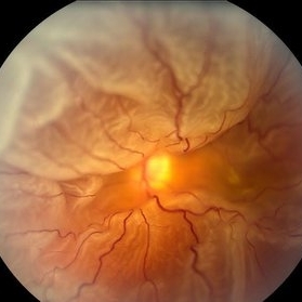
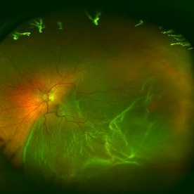
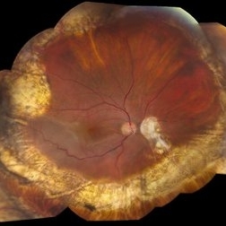
 Giant Retinal Tear With RD
Jun 29 2013 by Jason S. Calhoun
Middle aged patient comes in with sudden loss of vision inferior, nasally. Patient presents plus 2 APD in the right eye. Fundus exam reveals a giant retinal tear at 10-11 o'clock with RD. Vitrectomy with laser and gas exchange of C3F8 scheduled.
Photographer: Jason S. Calhoun, Mayo Clinic Jacksonville, Florida
Imaging device: TOPCON TRC 50-EX
Condition/keywords: retinal tear
-
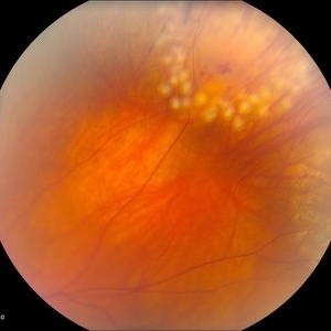
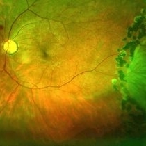
 360 Degree Retinal Detachment
Jun 29 2013 by Jason S. Calhoun
Total retinal detachment in the left eye.
Photographer: Jason S. Calhoun, Mayo Clinic Jacksonville, Florida
Imaging device: TOPCON TRC 50-EX
-
---thumb.JPG/image-square;max$300,300.ImageHandler) Stereo Image of Retinal Tear
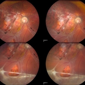
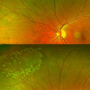
Stereo Image of Retinal Tear
Jun 30 2013 by Jason S. Calhoun
Four up image of a stereo 3-D pair of retinal tears.
Photographer: Jason S. Calhoun, Mayo Clinic Jacksonville, Florida
Condition/keywords: retinal tear, stereo pair
-
---thumb.JPG/image-square;max$300,300.ImageHandler) Stereo 3-D Image of Retinal Tear
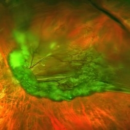
Stereo 3-D Image of Retinal Tear
Jun 30 2013 by Jason S. Calhoun
Stereo imaging of a retinal tear superior, temporal at 2-o'clock
Photographer: Jason S. Calhoun, Mayo Clinic Jacksonville, Florida
Condition/keywords: retinal tear
-
 Retinal Detachment With Dislocated IOL Lens
Jun 30 2013 by Jason S. Calhoun
47-year-old male who had trauma to the right eye. Patient had retinal detachment surgery in the past (scleral buckle), to the right eye. Patient came in with another retinal detachment with dislocated PC IOL lens. Notice the haptics tearing the retina. Patient underwent vitrectomy with gas exchange. VA was hand motion 1 day post-op.
Photographer: Jason S. Calhoun, Mayo Clinic Jacksonville, Florida
Condition/keywords: dislocated posterior chamber intraocular lens (PCIOL), retinal tear
-
---thumb.JPG/image-square;max$300,300.ImageHandler) Retinal Detachment With Dislocated IOL Lens
Retinal Detachment With Dislocated IOL Lens
Jun 30 2013 by Jason S. Calhoun
47-year-old male who had trauma to the right eye. Patient had retinal detachment surgery in the past (scleral buckle), to the right eye. Patient came in with another retinal detachment with dislocated PC IOL lens. Notice the haptics tearing the retina. Patient underwent vitrectomy with gas exchange. VA was hand motion 1 day post-op.
Photographer: Jason S. Calhoun, Mayo Clinic Jacksonville, Florida
Condition/keywords: dislocated posterior chamber intraocular lens (PCIOL), retinal tear
-
---thumb.JPG/image-square;max$300,300.ImageHandler) Giant Retinal Tear Treated With Laser
Giant Retinal Tear Treated With Laser
Jul 8 2013 by Jason S. Calhoun
Patient in with a shower of floaters. VA was 20/30 and fundus exam shows giant retinal tear temporally. Patient was treated with laser retinopexy to prevent retinal detachment.
Photographer: Jason S. Calhoun, Department of Ophthalmology, Mayo Clinic Jacksonville, Florida
Condition/keywords: laser retinopexy, retinal tear
-
---thumb.JPG/image-square;max$300,300.ImageHandler) Superior Retinal Detachment.
Superior Retinal Detachment.
Jul 10 2013 by Jason S. Calhoun
Fundus photograph shows small retinal detachment at 12-o'clock superiorly in the right eye.
Photographer: Jason S. Calhoun, Department of Ophthalmology, Mayo Clinic Jacksonville, Florida
-
---thumb.JPG/image-square;max$300,300.ImageHandler) Retinal Detachment with Horseshoe Tear
Retinal Detachment with Horseshoe Tear
Jul 11 2013 by Jason S. Calhoun
Fundus photo shows horseshoe tear superior-temporal in the left eye with retinal detachment.
Photographer: Jason S. Calhoun, Department of Ophthalmology, Mayo Clinic Jacksonville, Florida
-
---thumb.JPG/image-square;max$300,300.ImageHandler) Horseshoe Tear With Detachment
Horseshoe Tear With Detachment
Jul 13 2013 by Jason S. Calhoun
Retinal tear with retinal detachment.
Photographer: Jason S. Calhoun, Department of Ophthalmology, Mayo Clinic Jacksonville, Florida
-
---thumb.JPG/image-square;max$300,300.ImageHandler) Giant Retinal Tear
Giant Retinal Tear
Jul 13 2013 by Jason S. Calhoun
Giant retinal tear with retinal detachment.
Photographer: Jason S. Calhoun, Department of Ophthalmology, Mayo Clinic Jacksonville, Florida
Condition/keywords: retinal tear
-
---thumb.JPG/image-square;max$300,300.ImageHandler) Multiple Retinal Tear
Multiple Retinal Tear
Jul 13 2013 by Jason S. Calhoun
Retinal tear with retinal detachment.
Photographer: Jason S. Calhoun, Department of Ophthalmology, Mayo Clinic Jacksonville, Florida
Condition/keywords: retinal tear
-
---thumb.JPG/image-square;max$300,300.ImageHandler) Giant Retinal Tear
Giant Retinal Tear
Jul 13 2013 by Jason S. Calhoun
Giant Retinal Tear with Retinal Detachment inferiorly.
Photographer: Jason S. Calhoun, Department of Ophthalmology, Mayo Clinic Jacksonville, Florida
Condition/keywords: retinal tear
-
---thumb.JPG/image-square;max$300,300.ImageHandler) Small Tear in the Retina
Small Tear in the Retina
Jul 13 2013 by Jason S. Calhoun
Retinal tear with retinal detachment.
Photographer: Jason S. Calhoun, Department of Ophthalmology, Mayo Clinic Jacksonville, Florida
Condition/keywords: retinal tear
-
---thumb.JPG/image-square;max$300,300.ImageHandler) Retinal Detachment
Retinal Detachment
Jul 13 2013 by Jason S. Calhoun
Fundus photo shows retinal detachment.
Photographer: Jason S. Calhoun, Department of Ophthalmology, Mayo Clinic Jacksonville, Florida
-
---thumb.JPG/image-square;max$300,300.ImageHandler) Retinal Detachment
Retinal Detachment
Jul 13 2013 by Jason S. Calhoun
Fundus photo shows retinal detachment.
Photographer: Jason S. Calhoun, Department of Ophthalmology, Mayo Clinic Jacksonville, Florida
-
---thumb.JPG/image-square;max$300,300.ImageHandler) Retinal Detachment
Retinal Detachment
Jul 13 2013 by Jason S. Calhoun
Fundus photo shows retinal detachment.
Photographer: Jason S. Calhoun, Department of Ophthalmology, Mayo Clinic Jacksonville, Florida
-
---thumb.JPG/image-square;max$300,300.ImageHandler) Retinal Detachment
Retinal Detachment
Jul 13 2013 by Jason S. Calhoun
Fundus photo shows retinal detachment.
Photographer: Jason S. Calhoun, Department of Ophthalmology, Mayo Clinic Jacksonville, Florida
Condition/keywords: retinal detachment with retinal defect
-
---thumb.JPG/image-square;max$300,300.ImageHandler) Horseshoe Tear Before Laser Treatment
Horseshoe Tear Before Laser Treatment
Jul 13 2013 by Jason S. Calhoun
Retinal tear temporally, proceeded with laser retinopexy.
Photographer: Jason S. Calhoun, Department of Ophthalmology, Mayo Clinic Jacksonville, Florida
Condition/keywords: retinal tear
-
---thumb.JPG/image-square;max$300,300.ImageHandler) Horseshoe Tear With Laser Treatment
Horseshoe Tear With Laser Treatment
Jul 13 2013 by Jason S. Calhoun
Retinal tear which was treated with a laser retinopexy.
Photographer: Jason S. Calhoun, Department of Ophthalmology, Mayo Clinic Jacksonville, Florida
Condition/keywords: laser retinopexy, retinal tear
-
---thumb.JPG/image-square;max$300,300.ImageHandler) Retinal Tear with Retinal Detachment
Retinal Tear with Retinal Detachment
Jul 13 2013 by Jason S. Calhoun
Retinal tear with retinal detachment.
Photographer: Jason S. Calhoun, Department of Ophthalmology, Mayo Clinic Jacksonville, Florida
Condition/keywords: retinal tear
-
---thumb.JPG/image-square;max$300,300.ImageHandler) SF6 Gas Bubbles
SF6 Gas Bubbles
Jul 13 2013 by Jason S. Calhoun
Patient with retinal detachment superiorly at 11-o'clock. SF6 gas was injected then laser was performed the nest day.
Photographer: Jason S. Calhoun, Department of Ophthalmology, Mayo Clinic Jacksonville, Florida
Imaging device: TOPCON TRC 50-EX
Condition/keywords: gas pneumatic displacement
-
 Lasered Retinal Tear
Jul 14 2013 by Jason S. Calhoun
Patient with increased floaters. Fundus photos show retinal tear at 2-o'clock. Laser retinopexy was performed to prevent retinal detachment
Photographer: Jason S. Calhoun, Department of Ophthalmology, Mayo Clinic Jacksonville, Florida
Imaging device: TOPCON TRC 50-EX
Condition/keywords: laser retinopexy, laser treatment, retinal tear
-
 Retinal Detachment After Vitrectomy
Jul 14 2013 by Jason S. Calhoun
Post-op patient that had vitrectomy 2-days earlier has reoccurring retinal detachment inferiorly.
Photographer: Jason S. Calhoun, Department of Ophthalmology, Mayo Clinic Jacksonville, Florida
Condition/keywords: post-op
-
 Retinal Detachment With Macula Detached
Jul 15 2013 by Jason S. Calhoun
Patient went to the ER over the weekend with loss of vision in the right eye. VA was count fingers in the right eye. Noticed a curtain in the right eye. Horseshoe tear was seen at 10-o'clock temporally, right eye. Proceeded with surgery the next day.
Photographer: Jason S. Calhoun, Department of Ophthalmology, Mayo Clinic Jacksonville, Florida
Imaging device: TOPCON TRC 50-EX
-
 Retinal Detachment with Retinal Hole
Sep 30 2013 by Jason S. Calhoun
Patient in with complaints of floaters in the right eye. VA was 20/40 with no improvement. Fundus exam shows retinal detachment from 9-12 o'clock with hole at 10:30 posteriorly. Pneumatic retinopexy was performed with C3F8 Gas bubble and laser around the retinal tear in the right eye.
Photographer: Jason S. Calhoun, Department of Ophthalmology, Mayo Clinic Jacksonville, Florida
Imaging device: TOPCON TRC 50-EX
Condition/keywords: retinal hole
-
 Retinal Detachment with Retinal Hole (3-D)
Sep 30 2013 by Jason S. Calhoun
Patient with complaints of floaters in the right eye. VA was 20/40 with no improvement. Fundus exam shows retinal detachment from 9-12 o'clock with hole at 10:30 posteriorly. Pneumatic retinopexy was performed with C3F8 Gas bubble and laser around the retinal tear in the right eye.
Photographer: Jason S. Calhoun, Department of Ophthalmology, Mayo Clinic Jacksonville, Florida
Imaging device: TOPCON TRC 50-EX
Condition/keywords: retinal hole
-
 Rhegmatogenous Retinal Detachment
Oct 11 2013 by Jason S. Calhoun
Patient in for a second opinion on RD, right eye. VA is NLP in the right eye. Fundus photography shows inferior retinal detachment with holes and subretinal fibrosis. No further surgery is suggested at this time.
Photographer: Jason S. Calhoun, Ophthalmic Photographer, Department of Ophthalmology, Mayo Clinic Jacksonville
Imaging device: TOPCON TRC 50-EX
-
 Rhegmatogenous Retinal Detachment
Oct 11 2013 by Jason S. Calhoun
Patient in for second opinion on RD, right eye. VA is NLP in the right eye. Fundus photography shows inferior retinal detachment with holes and subretinal fibrosis. No further surgery is suggested at this time.
Photographer: Jason S. Calhoun, Ophthalmic Photographer, Department of Ophthalmology, Mayo Clinic Jacksonville
Imaging device: TOPCON TRC 50-EX
-
 Horseshoe Tear With Scleral Buckle
Oct 31 2013 by Jason S. Calhoun
Patient had a retinal detachment with retinal tear superior temporally. Underwent surgery and had a scleral buckle placed with good support of the tear. VA is count fingers and will return in 2-months for follow up.
Photographer: Jason S. Calhoun, Ophthalmic Photographer, Department of Ophthalmology, Mayo Clinic Jacksonville
Imaging device: TOPCON TRC 50-EX
Condition/keywords: retinal tear, scleral buckle
-
 Large Retinal Detachment
Sep 17 2015 by Jason S. Calhoun
Large retinal detachment in the right eye with the macula detached.
Photographer: Jason Calhoun, Mayo Clinic Jacksonville, Department of Opthalmolgy
Imaging device: OPTOS 200TX
Condition/keywords: chronic retinal detachment
-
 Retinal Detachment
Sep 17 2015 by Jason S. Calhoun
Retinal detachment with retinal tear temporal in the left eye.
Photographer: Jason Calhoun, Mayo Clinic Jacksonville, Department of Opthalmology
Imaging device: OPTOS 200TX
-
 Horseshoe Tear
Sep 17 2015 by Jason S. Calhoun
Horseshoe tear with sub retinal fluid present superior temporal in the left eye.
Photographer: Jason Calhoun, Mayo Clinic, Department of Ophthalmology
Imaging device: OPTOS 200TX
-
 Retinal Tear
Sep 17 2015 by Jason S. Calhoun
Retinal tear temporal about 4 o'clock in the left eye. Post laser treatment is present.
Photographer: Jason Calhoun, Mayo Clinic Jacksonville, Department of Ophthalmology
Imaging device: OPTOS 200TX
Condition/keywords: retinal tear
-
 Retinal Tear with Laser Retinopexy
Sep 17 2015 by Jason S. Calhoun
Retinal tear superior nasal in the left eye. Laser retinopexy was performed.
Photographer: Jason Calhoun, Mayo Clinic, Department of Ophthalmology
Imaging device: OPTOS 200TX
Condition/keywords: laser retinopexy, retinal tear
-
 Full Retinal Detachment
Sep 17 2015 by Jason S. Calhoun
Large retinal detachment in the left eye.
Photographer: Jason Calhoun, Mayo Clinic, Department of Ophthalmology
Imaging device: OPTOS 200 TX
-
 Full Retinal Detachment
Sep 17 2015 by Jason S. Calhoun
Large retinal detachment in the left eye.
Photographer: Jason Calhoun, Mayo Clinic, Department of Ophthalmology
Imaging device: TOPCON-TRC50EX
-
 Reoccurring Retinal Detachment
Jun 27 2013 by Jason S. Calhoun
Patient comes in for second opinion for RD in the right eye. Patient's VA was count fingers at 2-ft in the right eye and 20/20 in the left eye. Patient is aphakic and has had 5- retinal surgeries in the past in the right eye including a scleral buckle. RD present with the macula off. Will consider surgery with silicone oil.
Photographer: Jason S. Calhoun, Mayo Clinic Jacksonville, Florida
Imaging device: TOPCON TRC 50-EX
-
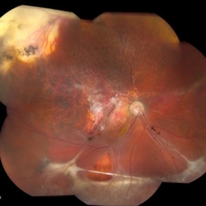
 Large Retinal Tear
Jun 27 2013 by Jason S. Calhoun
Patient with lattice degeneration came in with retinal detachment with a large retinal tear at 12 o'clock in the right eye. VA was 20/400. Macula was detached as well. Patient underwent surgery to re-attach the retina.
Photographer: Jason S. Calhoun, Mayo Clinic Jacksonville, Florida
Imaging device: TOPCON TRC 50-EX
Condition/keywords: retinal tear
-
 C3F8 Gas Bubble for Retinal Tear/ Pneumatic Retinopexy
Jun 27 2013 by Jason S. Calhoun
Patient comes in with retinal detachment and a pneumatic retinopexy was performed. Gas bubble is visible with optic nerve reflected.
Photographer: Jason S. Calhoun, Mayo Clinic Jacksonville, Florida
Imaging device: TOPCON TRC 50-EX
Condition/keywords: pneumatic retinopexy
-
 Horseshoe Retinal Tear With Bridging Blood Vessel
Jun 27 2013 by Jason S. Calhoun
Patient came in with curtain appearing in the superior part of vision. Retinal tear superior visible with detachment. Surgery is scheduled.
Photographer: Jason S. Calhoun, Mayo Clinic Jacksonville, Florida
Imaging device: TOPCON TRC 50-EX
Condition/keywords: retinal tear
-
 Horseshoe Retinal Tear
Jun 27 2013 by Jason S. Calhoun
Patient came in with retinal detachment. Surgery is scheduled.
Photographer: Jason S. Calhoun, Mayo Clinic Jacksonville, Florida
Imaging device: TOPCON TRC 50-EX
Condition/keywords: retinal tear
A project from the American Society of Retina Specialists