-
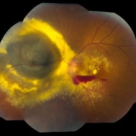 Choroidal Melanoma
Jun 30 2013 by Jason S. Calhoun
Post-op radioactive implant with hard exudates surrounding the melanoma with hemorrhage present.
Photographer: Jason S. Calhoun, Mayo Clinic Jacksonville, Florida
-
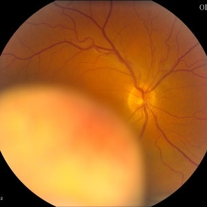
---thumb.JPG/image-square;max$300,300.ImageHandler) Choroidal Melanoma
Choroidal Melanoma
Jun 30 2013 by Jason S. Calhoun
Preoperative patient before radioactive implant surgery.
Photographer: Jason S. Calhoun, Mayo Clinic Jacksonville, Florida
-
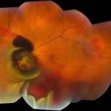 Choroidal Melanoma
Jun 30 2013 by Jason S. Calhoun
Choroidal melanoma with retinal detachment present inferior.
Photographer: Jason S. Calhoun, Mayo Clinic Jacksonville, Florida
-
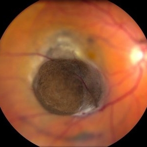 Choroidal Melanoma Anterior
Jun 30 2013 by Jason S. Calhoun
Choroidal melanoma anterior photo
Photographer: Jason S. Calhoun, Mayo Clinic Jacksonville, Florida
-
 Choroidal Melanoma With Radiation Retinopathy
Jul 8 2013 by Jason S. Calhoun
Patient came with follow up on choroidal melanoma. Right eye that was treated back in June of 2009 with a radioactive implant. Vein occlusion is also present with VA - hand motion. Hemorrhages visible with hard exudates from the radiation retinopathy.
Photographer: Jason S. Calhoun, Department of Ophthalmology, Mayo Clinic Jacksonville, Florida
Condition/keywords: radiation retinopathy
-
---thumb.JPG/image-square;max$300,300.ImageHandler) Choroidal Melanoma, Post-op Radioactive Implant
Choroidal Melanoma, Post-op Radioactive Implant
Jul 8 2013 by Jason S. Calhoun
Male patient who had a radioactive implant for choroidal melanoma in the left eye. VA is 20/150. VA has remained the same for the last 3-months.
Photographer: Jason S. Calhoun, Department of Ophthalmology, Mayo Clinic Jacksonville, Florida
-
---thumb.JPG/image-square;max$300,300.ImageHandler) Choroidal Melanoma With Retinal Detachment
Choroidal Melanoma With Retinal Detachment
Jul 8 2013 by Jason S. Calhoun
57-year-old female who underwent proton beam therapy for choroidal melanoma. VA was 20/400 at post-op visit.
Photographer: Jason S. Calhoun, Department of Ophthalmology, Mayo Clinic Jacksonville, Florida
-
 Metastasis Choroidal Melanoma
Jul 8 2013 by Jason S. Calhoun
71-year-old female who has undergone chemotherapy treatment for breast cancer. Choroidal melanoma was from metastasis from her breast cancer. VA is 20/20 as the tumor is in the superior aspect above the optic nerve.
Photographer: Jason S. Calhoun, Department of Ophthalmology, Mayo Clinic Jacksonville, Florida
Condition/keywords: choroidal metastasis
-
---thumb.JPG/image-square;max$300,300.ImageHandler) Choroidal Melanoma
Choroidal Melanoma
Jul 11 2013 by Jason S. Calhoun
Choroidal melanoma covering the optic nerve. Pre and post-op photos after radioactive implant surgery.
Photographer: Jason S. Calhoun, Department of Ophthalmology, Mayo Clinic Jacksonville, Florida
-
---thumb.JPG/image-square;max$300,300.ImageHandler) Choroidal Melanoma
Choroidal Melanoma
Jul 11 2013 by Jason S. Calhoun
Choroidal melanoma temporal of macula with retinal vein indenting through a pit of the melanoma.
Photographer: Jason S. Calhoun, Department of Ophthalmology, Mayo Clinic Jacksonville, Florida
-
---thumb.JPG/image-square;max$300,300.ImageHandler) Treated Choroidal Melanoma
Treated Choroidal Melanoma
Jul 11 2013 by Jason S. Calhoun
Radioactive retinopathy present with hard exudates at 11 o'clock in the right eye. Patient had radioactive implant surgery.
Photographer: Jason S. Calhoun, Department of Ophthalmology, Mayo Clinic Jacksonville, Florida
-
---thumb.JPG/image-square;max$300,300.ImageHandler) Treated Choroidal Melanoma
Treated Choroidal Melanoma
Jul 13 2013 by Jason S. Calhoun
Fundus photo shows radiation retinopathy after radioactive treatment to melanoma in the right eye. Cotton wool spots and hemorrhages visible around the optic nerve.
Photographer: Jason S. Calhoun, Department of Ophthalmology, Mayo Clinic Jacksonville, Florida
-
---thumb.JPG/image-square;max$300,300.ImageHandler) Choroidal Melanoma With Retinal Detachment
Choroidal Melanoma With Retinal Detachment
Jul 14 2013 by Jason S. Calhoun
Choroidal melanoma with retinal detachment present inferior.
Photographer: Jason S. Calhoun, Department of Ophthalmology, Mayo Clinic Jacksonville, Florida
Imaging device: TOPCON TRC 50-EX
-
 Choroidal Melanoma With Retinal Detachment
Jul 14 2013 by Jason S. Calhoun
Choroidal melanoma with retinal detachment present inferior.
Photographer: Jason S. Calhoun, Department of Ophthalmology, Mayo Clinic Jacksonville, Florida
Imaging device: TOPCON TRC 50-EX
-
 Choroidal Mass
Jul 14 2013 by Jason S. Calhoun
Fundus photo shows yellowish choroidal mass in the left eye.
Photographer: Jason S. Calhoun, Department of Ophthalmology, Mayo Clinic Jacksonville, Florida
Imaging device: TOPCON TRC 50-EX
Condition/keywords: choroidal nevus, indolent choroidal mass
-
---thumb.JPG/image-square;max$300,300.ImageHandler) Choroidal Melanoma
Choroidal Melanoma
Jul 14 2013 by Jason S. Calhoun
Choroidal melanoma located superior-temporally in the left eye.
Photographer: Jason S. Calhoun, Department of Ophthalmology, Mayo Clinic Jacksonville, Florida
Imaging device: TOPCON TRC 50-EX
-
 Treated Melanoma
Jul 14 2013 by Jason S. Calhoun
Choroidal melanoma treated with radiation. Hard exudates formed due to radiation retinopathy.
Photographer: Jason S. Calhoun, Department of Ophthalmology, Mayo Clinic Jacksonville, Florida
Imaging device: TOPCON TRC 50-EX
Condition/keywords: radiation retinopathy
-
---thumb.JPG/image-square;max$300,300.ImageHandler) Treated Melanoma
Treated Melanoma
Jul 14 2013 by Jason S. Calhoun
Choroidal melanoma treated with radiation. Hard exudates formed due to radiation retinopathy.
Photographer: Jason S. Calhoun, Department of Ophthalmology, Mayo Clinic Jacksonville, Florida
Imaging device: TOPCON TRC 50-EX
Condition/keywords: radiation retinopathy
-
 Melanoma with Subretinal Fluid
Jul 14 2013 by Jason S. Calhoun
52-year-old female who comes in for eye exam. Fundus exam shows yellowish elevated spot superior at 12-o'clock in the right eye. FA shows sub retinal fluid with melanoma in the right eye.
Photographer: Jason S. Calhoun, Department of Ophthalmology, Mayo Clinic Jacksonville, Florida
Imaging device: TOPCON TRC 50-EX
Condition/keywords: melanoma, subretinal fluid
-
---thumb.JPG/image-square;max$300,300.ImageHandler) Choroidal Mass 1
Choroidal Mass 1
Jul 14 2013 by Jason S. Calhoun
Fundus photo shows large elevated choroidal melanoma centrally in the left eye.
Photographer: Jason S. Calhoun, Department of Ophthalmology, Mayo Clinic Jacksonville, Florida
Imaging device: TOPCON TRC 50-EX
-
 Melanoma With RD
Jul 14 2013 by Jason S. Calhoun
Fundus photo shows choroidal melanoma with retinal detachment inferior in the left eye. Hemorrhage visible, nasally to the optic nerve.
Photographer: Jason S. Calhoun, Department of Ophthalmology, Mayo Clinic Jacksonville, Florida
Imaging device: TOPCON TRC 50-EX
-
 Melanoma With Radiation Retinopathy
Jul 14 2013 by Jason S. Calhoun
Fundus photo shows treated choroidal melanoma superior, nasal in the left eye. Patient was treated with radioactive implant. Radiation retinopathy with hard exudates present.
Photographer: Jason S. Calhoun, Department of Ophthalmology, Mayo Clinic Jacksonville, Florida
Imaging device: TOPCON TRC 50-EX
Condition/keywords: radiation retinopathy
-
---thumb.JPG/image-square;max$300,300.ImageHandler) Melanoma
Melanoma
Jul 14 2013 by Jason S. Calhoun
Fundus photo shows elevated choroidal melanoma superior, temporal in the left eye.
Photographer: Jason S. Calhoun, Department of Ophthalmology, Mayo Clinic Jacksonville, Florida
Imaging device: TOPCON TRC 50-EX
-
 Choroidal Melanoma With Pigment
Jul 14 2013 by Jason S. Calhoun
Fundus photo shows elevated choroidal melanoma inferior, temporal in the left eye. Notice the fine orange pigment in the melanoma.
Photographer: Jason S. Calhoun, Department of Ophthalmology, Mayo Clinic Jacksonville, Florida
Imaging device: TOPCON TRC 50-EX
-
 Melanoma
Jul 14 2013 by Jason S. Calhoun
Fundus photo shows elevated choroidal melanoma inferior, temporal in the left eye.
Photographer: Jason S. Calhoun, Department of Ophthalmology, Mayo Clinic Jacksonville, Florida
Imaging device: TOPCON TRC 50-EX
-
 Choroidal Melanoma (Treated)
Sep 11 2013 by Jason S. Calhoun
Fundus photography shows choroidal melanoma at 2-o'clock superiorily. Patient had a radioactive implant back in August 2011.
Photographer: Jason S. Calhoun, Department of Ophthalmology, Mayo Clinic Jacksonville, Florida
Imaging device: TOPCON TRC 50-EX
Condition/keywords: melanoma
-
 Choroidal Melanoma
Sep 11 2013 by Jason S. Calhoun
Fundus photography shows choroidal melanoma superiorly at 12-o'clock. Retinal hemorrhages scattered around the periphery.
Photographer: Jason S. Calhoun, Department of Ophthalmology, Mayo Clinic Jacksonville, Florida
Imaging device: TOPCON TRC 50-EX
-
 Choroidal Melanoma
Sep 23 2013 by Jason S. Calhoun
Patient with spot in vision in the right eye. Fundus photography shows medium size melanoma adjacent to the optic nerve and measures 13mm across horizontally and 6mm diameter in thickness. Patient will undergo proton beam therapy.
Photographer: Jason S. Calhoun, Department of Ophthalmology, Mayo Clinic Jacksonville, Florida
Imaging device: TOPCON TRC 50-EX
-
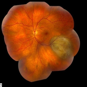
 Choroidal Melanoma
Jun 27 2013 by Jason S. Calhoun
Young male patient noticed a visual field defect in the right eye. Patient's VA was 20/60 with no improvement with pinhole. No history of cancer. Patient will be followed up in one week to discuss surgery.
Photographer: Jason S. Calhoun, Mayo Clinic Jacksonville, Florida
Imaging device: TOPCON TRC 50-EX
-
 Choroidal Tumor Consistent With Uveal Melanoma
Jun 27 2013 by Jason S. Calhoun
Patient comes in for second opinion for 2-melanomas in the right eye. Patient's VA is 20/20, both eyes. Fundus exam reveals melanoma superior to the macula which measured at 0.7-mm thick. Second melanoma is nasally which measured at 4.1-mm thick and has low internal reflectivity. Patient will be followed up with MRI and ocular protocol.
Photographer: Jason S. Calhoun, Mayo Clinic Jacksonville, Florida
Imaging device: TOPCON TRC 50-EX
-
 Uveal Choroidal Melanoma
Jun 27 2013 by Jason S. Calhoun
Patient came in for evaluation on a choroidal melanoma in the right eye. VA was 20/25 in both eyes. The melanoma is in the temporal aspect of the right eye. It measured at 0.7mm elevated after doing a BSCAN Ultrasound.
Photographer: Jason S. Calhoun, Mayo Clinic Jacksonville, Florida
Imaging device: TOPCON TRC 50-EX
A project from the American Society of Retina Specialists