 Initializing download.
-
 By Jason S. Calhoun
Mayo Clinic Jacksonville, Florida - Uploaded on Sep 23, 2013.
- Last modified by Caroline Bozell on Sep 23, 2013.
- Rating
- Appears in
- Choroidal Melanomas
- Photographer
- Jason S. Calhoun, Department of Ophthalmology, Mayo Clinic Jacksonville, Florida
- Imaging device
-
Fundus camera
TOPCON TRC 50-EX - Description
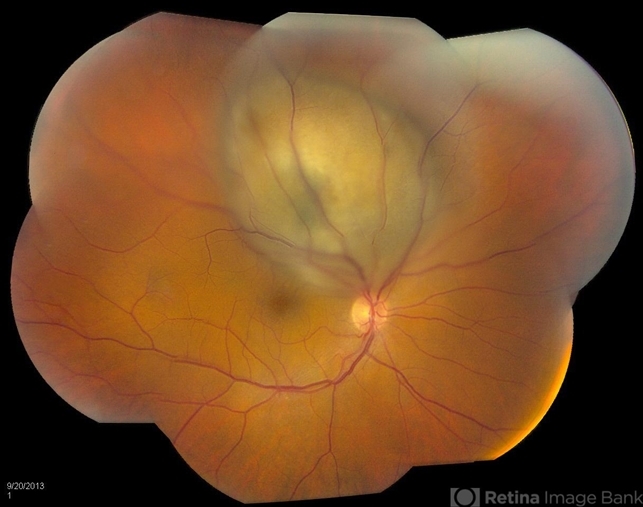
- Patient with spot in vision in the right eye. Fundus photography shows medium size melanoma adjacent to the optic nerve and measures 13mm across horizontally and 6mm diameter in thickness. Patient will undergo proton beam therapy.


---thumb.JPG/image-square;max$79,0.ImageHandler "Treated Melanoma")


")

---thumb.JPG/image-square;max$79,0.ImageHandler "Toxoplasmosis Vitreoretinal Traction")
---thumb.JPG/image-square;max$79,0.ImageHandler "Toxoplasmosis Vitreoretinal Traction")
