 Initializing download.
-
 By Jason S. Calhoun
Mayo Clinic Jacksonville, Florida - Uploaded on Sep 11, 2013.
- Last modified by Caroline Bozell on Sep 11, 2013.
- Rating
- Appears in
- Choroidal Melanomas
- Photographer
- Jason S. Calhoun, Department of Ophthalmology, Mayo Clinic Jacksonville, Florida
- Imaging device
-
Fundus camera
TOPCON TRC 50-EX - Description
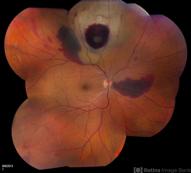
- Fundus photography shows choroidal melanoma superiorly at 12-o'clock. Retinal hemorrhages scattered around the periphery.


---thumb.JPG/image-square;max$79,0.ImageHandler "Treated Melanoma")


")

---thumb.JPG/image-square;max$79,0.ImageHandler "Toxoplasmosis Vitreoretinal Traction")
---thumb.JPG/image-square;max$79,0.ImageHandler "Toxoplasmosis Vitreoretinal Traction")
