-
 Multi-modal Imaging of Type - 1 CNVM
May 30 2025 by Shivankar Sen, MS, FVRS
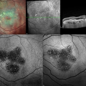
Multimodal Imaging of a case of Polypoidal Choroidal Vasculopathy Multicolor Reflectance showing multiple green-hyper-fringent lesions in the macular region (Up Left) Infra-red Autofluorescence and Blue Autofluorescence showing hypo-autofluorescent areas correspondingly revealing the exact extent of the sub-RPE Lesion (Down left and right respectively) Optical Coherence Tomography - Enhanced Depth Imaging showing Thumb-shaped Pigment Epithelial Detachment with presence of Sub-retinal fluid and Hyper-reflective foci (Top Right)
Photographer: Dr. Shivankar Sen
Imaging device: Heidelberg Spectralis HRA+OCT
Condition/keywords: Blue autofluroscence, CNVM, multicolor, near infrared autofluorescence (NIRAF), PCV, reflectance
-
 Berlins
Jun 25 2025 by Shivankar Sen, MS, FVRS
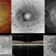
A 22 year old female came with history of injury to her left eye with a badminton racquet butt cap an hour before presentation On examination, she was found to have right eye 6/6;N6 vision and within normal limits, left eye 6/9;N6 vision, cells1+ in the anterior chamber, brisk pupillary response, no vitreous reaction and sub-clinical berlin's edema at the posterior pole. Multimodal imaging revealed frank boundaries of Berlin's edema more pronounced in the nasal parafoveal region. Figure details Top (Left to Right) Multicolor Reflectance showing bright yellow ring surrounding the perifovea; Blue Reflectance (Black on white contrast) showing corresponding black ring; Green Reflectance showing a characteristic white ring (all pronounced nasally); Bottom (Left-Right) Transverse structural OCT enface image showing white ring consistent with edema OCTA inner layer segmentation from ILM to GCL
Photographer: Gayathri M S
Imaging device: Heidelberg Spectralis HRA+OCT
Condition/keywords: blue reflectance, En Face OCTA, multicolor
-
 Berlins Edema - Multimodal Imaging
Jun 25 2025 by Shivankar Sen, MS, FVRS
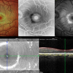
A 22 year old female came with history of injury to her left eye with a badminton racquet butt cap an hour before presentation On examination, she was found to have right eye 6/6;N6 vision and within normal limits, left eye 6/9;N6 vision, cells1+ in the anterior chamber, brisk pupillary response, no vitreous reaction and sub-clinical berlin's edema at the posterior pole. Multimodal imaging revealed frank boundaries of Berlin's edema more pronounced in the nasal parafoveal region. Figure details Top (Left to Right) Multicolor Reflectance showing bright yellow ring surrounding the perifovea; Blue Reflectance (Black on white contrast) showing corresponding black ring; Green Reflectance showing a characteristic white ring (all pronounced nasally); Bottom (Left-Right) Transverse structural OCT enface image showing white ring consistent with edema OCTA inner layer segmentation from ILM to GCL Transverse corresponding OCTA revealing faint hypo ring within perifoveal capillary bed
Photographer: Gayathri M S
Imaging device: Heidelberg Spectralis HRA+OCT
Condition/keywords: blue reflectance, En Face OCTA, enface imaging, multicolor, oct, reflectance
-
 CRAO With Cilio-retinal Sparing-MMI
Jun 25 2025 by Shivankar Sen, MS, FVRS
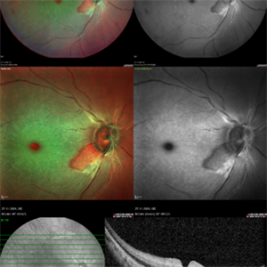
A 41 year old male came with complaints of Right eye blurring of vision since a day associated with watering and redness. He had no systemic illness, though gave a history of fall from bike 1 month back at the time of which he had blunt force trauma to the right side of the face. BCVA was 3/60, less than N36 in the right eye and 6/6, N6 in the left eye. Right eye had Marcus Gunn Pupil with clear lens, Left eye was within normal limits. IOP was normal; 16 in OD and 18 in OS. Retina evaluation revealed CRAO in the right eye with cilio-retinal artery sparing. Left eye was unremarkable Image Details Left to Right (Top 2 rows) Multicolor Reflectance Image (Blue-green enhanced 55 degree) revealing cilioretinal spared retinal stroma and a characteristic Cherry Red Spot; Green Reflectance showing corresopnding dark gray area with spared perfusion and black spot consistent with Cherry Red Spot on multicolor 2nd Row - 35 degree image (Multicolor Standard Reflectance and Green Reflectance) 3rd Row - SD-OCT revealing acute moderate CRAO findings with Middle retinal layer opacification and prominent middle limiting membrane (p-MLM) sign; Inner retinal layer opacification and prominent retinal pigment epithelium at the fovea with Diminished inner retinal layer stratification
Photographer: Gayathri M S
Imaging device: Heidelberg Spectralis HRA+OCT
Condition/keywords: CRAO with cilioretinal sparing, multicolor, multimodal imaging, OCT biomarkers, reflectance
A project from the American Society of Retina Specialists