-
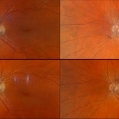
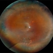
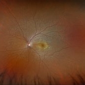
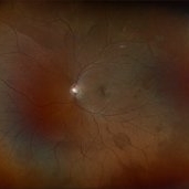
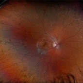
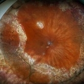
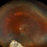
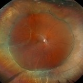
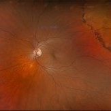
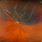
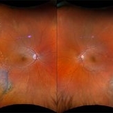
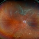 Retinal Detachment with Multiple Breaks
Nov 4 2024 by Kimberly Wakester
Ultra-widefield Fundus photograph of an 18-year-old woman with a Retinal detachment with multiple breaks in the right eye. Patient has high Myopia in both eyes. Patient was treated with scleral buckle placement with cryo laser in the right eye and is doing we post operatively.
Photographer: Kimberly Wakester, COA
Imaging device: Optos
-
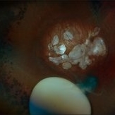
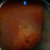
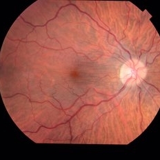
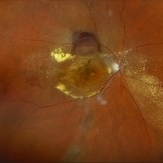
 Multifocal PC IOL
Nov 5 2024 by Kimberly Wakester
Anterior segment photograph of a 74-year-old woman with a Multifocal PC IOL in place in the left eye.
Photographer: Kimberly Wakester, COA
Imaging device: Topcon TRC-50DX
Condition/keywords: IOL, Multifocal
-
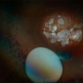
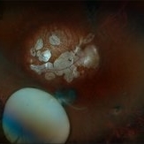
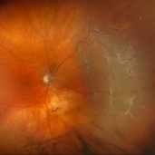
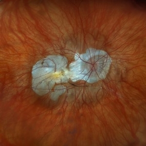
 ICC-8 Pinhole IOL
Nov 5 2024 by Kimberly Wakester
Anterior segment photograph of a 74-year-old woman with an ICC-8 pinhole IOL with stellate areas on the pinhole in the right eye.
Photographer: Kimberly Wakester, COA
Imaging device: Topcon TRC-50DX
Condition/keywords: Pinhole IOL
-
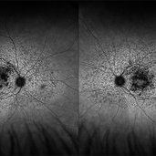
 Subluxation of the Lens
Dec 12 2024 by Kimberly Wakester
Ultra-wide field fundus photos of an 53-year-old man with a Subluxation of the Lens in the posterior vitreous cavity of the right eye after a trauma that happened many years ago. Patient remains stable with no adverse reaction to the lens at this time. No surgical intervention is recommended at this time. Patient also has myopic degeneration and lattice degeneration that will require patient to have follow up care.
Photographer: Kimberly Wakester, COA
Imaging device: Optos California
Condition/keywords: lattice degeneration, myopic degeneration, peripapillary atrophy, posterior staphyloma, Subluxation of the Lens
-
 Subretinal Fibrosis
Jan 14 2025 by Kimberly Wakester
Fundus photograph of an 86-year-old woman with the end stage of Age-related Macular Degeneration in the left eye. Patient went unseen for 3-4 years prior to establishing care at our practice. Due to the significant amount of subretinal fibrosis, treatment was not recommended due to limited visual recovery. Patient was advised of monocular vision and the importance of follow up care.
Photographer: Kimberly Wakester, COA
Imaging device: Optos California
Condition/keywords: AMD, subretinal fibrosis
-
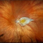
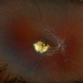
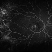
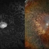
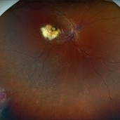 Coat's Disease
Jan 14 2025 by Kimberly Wakester
Fundus photographs of an 7-year-old boy with Coat's Disease in the right eye. There is subfoveal lipid end scarring in the macula and "light bulb" type telangiectasias temporally noted on exam and shown in Optos color photos. FA findings show anastomoses, capillary dropout, and "light bulb" type telangiectasias temporally with mild late leakage. Patient will be monitored at this time and have repeat imaging in 4 months.
Photographer: Kimberly Wakester, COA
Imaging device: Optos California
Condition/keywords: Coat's disease
-
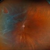
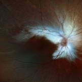
 Weiss Ring
Jan 21 2025 by Kimberly Wakester
Fundus photographs of a 70-year-old woman with a PVD with Weiss ring present in the left eye. Doing sweeps of the left eye shows how changing the patient's gaze can reposition the Weiss ring in the patient's eye.
Photographer: Kimberly Wakester, COA
Imaging device: Optos California
Condition/keywords: PVD, Weiss ring
-
 Systemic Lupus Erythematosus (SLE) Vasculitis
Jan 29 2025 by Kimberly Wakester
Fundus photographs of an 13-year-old boy with Systemic Lupus Erythematosus (SLE) Vasculitis in both eyes s/p PRP laser. Patient is doing well s/p PRP Laser OU and with continued use of oral medications. Patient will be monitored with follow up exams to check for recurring vasculitis or recurring/worsening NVE/NVD. Patient is to continue ongoing management with Rheumatologist.
Photographer: Kimberly Wakester, COA
Imaging device: Optos California
Condition/keywords: NVD, NVE, occlusive vasculitis, pan-retinal photocoagulation (PRP), Systemic Lupus Erythematosus (SLE) Vasculitis
-
 Central Retinal Vein Occlusion with Macular Edema
Jan 29 2025 by Kimberly Wakester
Fundus photograph of a 62-year-old man with central retinal vein occlusion with macular edema and a new PVD with an operculated retinal tear in the left eye. Laser to retinal tear was completed. Patient will return in 2-3 weeks for follow up exam with possible intravitreal injection for the CRVO with edema and to follow up on the operculated retinal tear s/p retinal tear laser.
Photographer: Kimberly Wakester, COA
Imaging device: Optos California
Condition/keywords: central retinal vein occlusion (CRVO), operculated tear, PVD
-
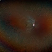
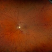
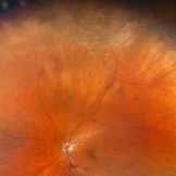
 Lattice Degeneration With Atrophic Retinal Holes
Jan 30 2025 by Kimberly Wakester
Ultra-wide field montage fundus photograph of a 56-year-old woman with lattice degeneration with atrophic holes statues post laser. Patient also has a small CHRPE temporal to macula and trace ERM that is not visually significant. Will continue follow up care to monitor and treat as needed.
Photographer: Kimberly Wakester, COA
Imaging device: Optos California
Condition/keywords: atrophic retinal hole, CHRPE, epiretinal membrane (ERM), lattice degeneration, montage photo
-
 Macular Hole
Jan 30 2025 by Kimberly Wakester
Fundus photograph of a 37-year-old man with an anteriorly dislocated lens in the left eye. The natural lens has displaced anteriorly in the AC secondary to trauma to the eye. There is also a Macular hole present with vitreous hemorrhage. Patient was recommended to proceed with lensectomy, iris repair and MH repair in the left eye.
Photographer: Kimberly Wakester, COA
Imaging device: Optos California
Condition/keywords: dislocated lens, macular hole, vitreous hemorrhage
-
 Dislocated Lens
Jan 30 2025 by Kimberly Wakester
Fundus photograph of a 37-year-old man with an anteriorly dislocated lens in the left eye. The natural lens has displaced anteriorly in the AC secondary to trauma to the eye. There is also a Macular hole present with vitreous hemorrhage. Patient was recommended to proceed with lensectomy, iris repair and MH repair in the left eye.
Photographer: Kimberly Wakester, COA
Imaging device: Topcon TRC-50DX
Condition/keywords: dislocated lens, iridodialysis
-
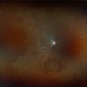
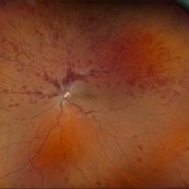
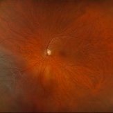
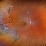
 Retinal Detachment with Multiple Breaks
Feb 3 2025 by Kimberly Wakester
Fundus photograph of a 67-year-old man with a retinal detachment with multiple breaks in the right eye. Patient is doing well s/p PPV and will continued to be observed during PO period.
Photographer: Kimberly Wakester, COA
Imaging device: Optos California
Condition/keywords: horseshoe tear, multiple retinal tears, retinal detachment
-
 Multifocal Pattern Dystrophy
Feb 5 2025 by Kimberly Wakester
Optomap RGB and AF photograph of an 37-year-old woman with multifocal pattern dystrophy in both eyes. Previously believed to be Stargardts, but genetic testing returned positive for PRPH2 mutation. Likely Multifocal Pattern Dystrophy given phenotypical appearance of SGD. There is stable NVE in the left eye. Will continue to monitor both eyes and consider treatment with PRP laser if needed for NVE in the left eye.
Photographer: Kimberly Wakester, COA
Imaging device: Optos California
Condition/keywords: multifocal pattern dystrophy, NVE, PRPH2 Positive
-
 Central Retinal Vein Occlusion with Macular Edema
Feb 11 2025 by Kimberly Wakester
Optomap RGB image of an 61-year-old man with a central retinal vein occlusion with macular edema in the left eye. Will continue monthly follow up care with repeat OCT and treatment with intravitreal anti-VEGF injections as needed.
Photographer: Kimberly Wakester, COA
Imaging device: Optos California
Condition/keywords: CRVO with macular edema, DBH
-
 Diabetic Macular Edema
Feb 12 2025 by Kimberly Wakester
Horizontal OCT scan of a 63-year-old woman with diabetic macular edema in the right eye. When reviewing the scan, one of the intraretinal cyst (IRC) appears heart shaped. A fun scan to see just a few day's before Valentine's day.
Photographer: Kimberly Wakester, COA
Imaging device: Heidelberg
Condition/keywords: diabetic macular edema, intraretinal cyst
-
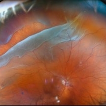
 Retinal Detachment with Horseshoe Retinal Tear
Feb 17 2025 by Kimberly Wakester
Optomap RGB image of a 62-year-old woman with a retinal detachment with a horseshoe retinal tear in the left eye. Patient had emergent surgery same day. She is doing well post operatively. Will continue follow up care as directed.
Photographer: Kimberly Wakester, COA
Imaging device: Optos California
Condition/keywords: horseshoe tear, retinal detachment
-
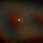
 Sickle Cell Retinopathy
Feb 24 2025 by Kimberly Wakester
Optomap RGB image of an 24-year-old woman with sickle cell retinopathy in both eyes. There is overall progression of the ischemic vessels and vascular drops out compared to previous images completed in 2021. Oral FA was completed and shows possible progression of peripheral non-perfusion but difficult to determine due to drinking FA dye and images not being as bright. On Clinical exam there is no evidence of NV, RD, or RT in either eye. Patient understands the need for continued follow up care and the likely need for PRP laser in both eyes.
Photographer: Kimberly Wakester, COA
Imaging device: Optos California
Condition/keywords: sickle cell retinopathy
-
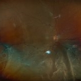
 Retinal Detachment with PVR
Feb 24 2025 by Kimberly Wakester
Optomap RGB of an 48-year-old man with a retinal detachment with PVR. Patient is 6 weeks s/p RD repair with giant HSRT. Patient has new PVR noted on post op exam causing the retina to re-detach. Patient is having to have a 2nd surgery to remove the scar tissue and have silicone oil placement. Will continue close follow up care.
Photographer: Kimberly Wakester, COA
Imaging device: Optos California
Condition/keywords: gas bubble, PVR, retinal detachment
-
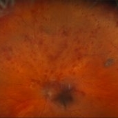
 Retinoschisis
Feb 26 2025 by Kimberly Wakester
Optomap RGB of a 56-year-old woman with bullous retinoschisis in the right eye. The patient remains stable with very mild progression. Patient is to continue follow up care at 6 month intervals to monitor for worsening progression.
Photographer: Kimberly Wakester, COA
Imaging device: Optos California
Condition/keywords: bullous retinoschisis
-
 Hereditary Retinal Dystrophy
Feb 27 2025 by Kimberly Wakester
Optomap RGB image of a 7-year-old girl with Hereditary retinal dystrophy. Biological mother is a CHM gene carrier and biological father is diagnosed with RP. Patient had genetic testing and was also confirmed to be a CHM gene carrier and also has the TTC21B gene. There is linear pigmentary changes on clinical exam and fundus photos. Atypical appearance of Retinitis Pigmentosa. Patient will continue follow up care with repeat imaging.
Photographer: Kimberly Wakester, COA
Imaging device: Optos California
Condition/keywords: CHM gene, hereditary retinal dystrophy, linear pigmentary changes
-
 Central Retinal Vein Occlusion With Macular Edema
Feb 27 2025 by Kimberly Wakester
Optomap RGB image of a 34-year-old woman with central retinal vein occlusion with macular edema in the left eye. Patient has had a fairly acute onset central retinal vein occlusion in her left eye with dense superior IRH and macular edema. Modest ischemic changes are seen on exam and fundus photos. Patient was educated on the etiology of CRVOs and the relationship to systemic risk factors. Recommended hypercoagulable work-up with her PCP and bloodwork was ordered. Treatment with intravitreal injections was recommended to reduce the macular edema. Patient is to continue monthly follow ups with repeat OCT.
Photographer: Kimberly Wakester, COA
Imaging device: Optos California
Condition/keywords: CRVO with macular edema
-
 Retinal Detachment
Mar 5 2025 by Kimberly Wakester
Optomap RGB image of an 9-year-old boy with a retinal detachment with retinal break at 9:00 in the right eye. Surgery was recommended. Patient is to continue follow up care post operatively.
Photographer: Kimberly Wakester, COA
Imaging device: Optos California
Condition/keywords: myopic eye, Retinal Detachment, retinal tear
-
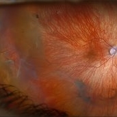
 Retinal Detachment with Multiple Breaks
Mar 5 2025 by Kimberly Wakester
Optomap RGB image of an 44-year-old man with a retinal detachment with a complex lattice break in the right eye. Surgery was recommended. Patient is to continue follow up care post operatively.
Photographer: Kimberly Wakester, COA
Imaging device: Optos California
Condition/keywords: Retinal Detachment, retinal tear
-
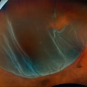
 Repaired Retinal Detachment with PVR
Mar 25 2025 by Kimberly Wakester
Optomap RGB of a 79-year-old-woman with a repaired retinal detachment with PVR in the right eye. Patient is doing well over 7 months s/p vitrectomy with silicone oil and scleral buckle placement. Retina remains attached on the buckle under oil. Patient is to return in 6 months for follow up exam with repeat imaging.
Photographer: Kimberly Wakester, COA, OCT-C
Imaging device: Optos California
Condition/keywords: PVR, repaired RD, Retinal detachment under Silicon Oil, scleral buckle
-
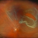
 Repaired Retinal Detachment with Scleral Buckle
Mar 25 2025 by Kimberly Wakester
Optomap RGB montage of an 64-year-old woman with a repaired retinal detachment with scleral buckle in the right eye. There is nasal and inferior pre-retinal membranes with traction. PPV was recommended but patient defers to proceed with sx at this time. Will continue to follow patient closely for worsening traction. Patient was educated on how to monitor their peripheral vision and was advised to report any changes immediately.
Photographer: Kimberly Wakester, COA, OCT-C
Imaging device: Optos California
Condition/keywords: pre-retinal membrane with traction, repaired RD, scleral buckle
-
 Retinal Detachment with Retinal Tear
Mar 31 2025 by Kimberly Wakester
Optomap RGB of an 48-year-old woman with a retinal detachment with retinal tear in the left eye. Surgery was recommended. Patient is to continue follow up care post operatively.
Photographer: Kimberly Wakester, COA, OCT-C
Imaging device: Optos California
Condition/keywords: Retinal Detachment, retinal tear
-
 Severe NPDR with Subhyaloid Hemorrhage
Apr 9 2025 by Kimberly Wakester
Optomap RGB of an 47 year-old man with severe NPDR with subhyaloid hemorrhage in the right eye.
Photographer: Kimberly Wakester, COA, OCT-C
Imaging device: Optos California
Condition/keywords: severe NPDR, subhyaloid hemorrhage
-
 Repaired Retinal Detachment
May 7 2025 by Kimberly Wakester
Optomap RGB montage of an 56-year-old woman with a repaired retinal detachment with scleral buckle and cryotherapy in the left eye. Patient remains stable s/p Vitreo-retinal surgery in 2007. Patient is to return in 1 year for follow up exam with repeat imaging.
Photographer: Kimberly Wakester, COA, OCT-C
Imaging device: Optos California
Condition/keywords: cryotherapy, repaired RD, scleral buckle
-
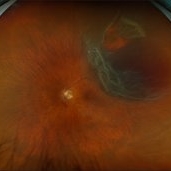
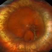
 Retinal Detachment with Giant Retinal Tear
May 14 2025 by Kimberly Wakester
Optomap RGB of an 66-year-old man with a retinal detachment with a giant retinal tear in the right eye. Surgery was recommended. Patient is to continue follow up care post operatively. Also noted in the image is a vitreous opacity that was caught at the right moment and appears to look like a smiley face.
Photographer: Kimberly Wakester, COA, OCT-C
Imaging device: Optos California
Condition/keywords: giant retinal tear, RD
-
 Neovascular AMD with Active CNV
May 22 2025 by Kimberly Wakester
Optomap RGB of an 82-year-old man with Neovascular AMD with Active CNV and Dry AMD in the right eye. There is advanced atrophic changes without subfoveal involvement located temporally to the fovea. Patient is to continue follow up care with dilated exam, repeat OCT, and treatment of intravitreal injection of Vabysmo every 5 weeks at this time.
Photographer: Kimberly Wakester, COA, OCT-C
Imaging device: Optos California
Condition/keywords: advanced geographic atrophy, dry age-related macular degeneration (dry AMD), neovascular age-related macular degeneration (AMD)
-
 Retinal Detachment with PVR
Jun 24 2025 by Kimberly Wakester
Optomap RGB of a 61-year-old man with Retinal Detachment with PVR in the right eye. There are multiple small holes present. Surgery was recommended. Patient is to continue follow up care post operatively.
Photographer: Kimberly Wakester, COA, OCT-C
Imaging device: Optos California
Condition/keywords: PVR, RD
-
 Repaired Retinal Detachment
Jun 24 2025 by Kimberly Wakester
Optomap RGB of an 45-year-old woman with a repaired retinal detachment in the right eye. The operative eye is doing well three-month s/p surgery. Retina is attached 360 on SB. There is resolving residual SRF at 6:00. Discussed the possible need for added laser. Will continue to observe and will return in 3 months for follow up exam.
Photographer: Kimberly Wakester, COA, OCT-C
Imaging device: Optos California
Condition/keywords: repaired RD, scleral buckle
-
 Epiretinal Membrane
Jun 25 2025 by Kimberly Wakester
Fundus photograph of a 32-year-old woman with a stable epiretinal membrane in the right eye. Patients vision remains stable. No intervention is required at this time.
Photographer: Kimberly Wakester, COA, OCT-C
Imaging device: Topcon TRC 50DX
Condition/keywords: ERM
-
 Secondary Pigmentary Degeneration of Retina
Jul 18 2025 by Kimberly Wakester
Optomap RGB and AF of an 63-year-old man with secondary pigmentary degeneration of the retina. Patient's Spark genetic testing revealed heterozygous mutations of unknown significance in LRP5, COL18A1, CPLANE1, SLC24A1 and VCAN. Clinical findings most consistent with Wagner's Syndrome (VCAN mutation, autosomal dominant). Will continue follow up care every 6 months with dilated exam and repeat OCT and Optos imaging .
Photographer: Kimberly Wakester, COA, OCT-C
Imaging device: Optos California
Condition/keywords: secondary pigmentary degeneration, Wagner disease
-
 Secondary Pigmentary Degeneration of Retina
Jul 18 2025 by Kimberly Wakester
Optomap RGB and AF of an 63-year-old man with secondary pigmentary degeneration of the retina. Patient's Spark genetic testing revealed heterozygous mutations of unknown significance in LRP5, COL18A1, CPLANE1, SLC24A1 and VCAN. Clinical findings most consistent with Wagner's Syndrome (VCAN mutation, autosomal dominant). Will continue follow up care every 6 months with dilated exam and repeat OCT and Optos imaging.
Photographer: Kimberly Wakester, COA, OCT-C
Imaging device: Optos California
Condition/keywords: secondary pigmentary degeneration, Wagner's Syndrome
-
 Retinoschisis with Outer Layer Holes
Jul 18 2025 by Kimberly Wakester
Optomap RGB of an 56-year-old woman with retinoschisis with outer layer holes s/p laser in the left eye. Patient remains stable. Will continue follow up care with dilated exam and optos imaging.
Photographer: Kimberly Wakester, COA, OCT-C
Imaging device: Optos California
Condition/keywords: outer layer hole, retinoschisis
-
 Pigmentary Retinal Dystrophy
Jul 18 2025 by Kimberly Wakester
Optomap RGB and AF of the left eye of an 76-year-old woman with pigmentary retinal dystrophy. No progression of the bone spicules noted on exam and optos imaging. Will continue yearly follow care with dilated exam and optos imaging.
Photographer: Kimberly Wakester, COA, OCT-C
Imaging device: Optos California
Condition/keywords: pigmentary retinal dystrophy
-
 Retinal detachment with Single Break
Jul 18 2025 by Kimberly Wakester
Optomap RGB of a 62-year-old man with a retinal detachment with a single break in the left eye. Patient has a previously treated HSRT in the left eye. Surgery was recommended. Patient is to continue follow up care post operatively.
Photographer: Kimberly Wakester, COA, OCT-C
Imaging device: Optos California
Condition/keywords: RD, retinal tear
-
 Retinal Detachment with Multiple Breaks
Aug 12 2025 by Kimberly Wakester
Optomap RGB of a 59-year-old man with a retinal detachment with multiple breaks in the left eye. Surgery was recommended. Patient is to continue follow up care post operatively.
Photographer: Kimberly Wakester, COA, OCT-C, Retina Consultants of Carolina
Imaging device: Optos California
Condition/keywords: lattice degeneration, left eye, Retinal Detachment with Multiple Breaks
-
 Retinal Detachment
Aug 12 2025 by Kimberly Wakester
Optomap RGB of a 63-year-old man with a bullous overhanging Retinal Detachment with superonasal tuft-associated tear, macula detached in the left eye. Surgery was recommended. Patient is to continue follow up care post operatively.
Photographer: Kimberly Wakester, COA, OCT-C, Retina Consultants of Carolina
Imaging device: Optos California
Condition/keywords: bullous retinal detachment, left eye, Mac off
-
 Myelinated Nerve Fiber Layer
Aug 13 2025 by Kimberly Wakester
Optomap RGB of a 3-year-old girl that presents with extensive myelinated never fiber in the right eye sparing the fovea. Patient is to return in 6 months for follow up visit with repeat Optos imaging.
Photographer: Kimberly Wakester, COA, OCT-C, Retina Consultants of Carolina
Imaging device: Optos California
Condition/keywords: myelinated nerve fiber layer
-
 Retinal Tear
Sep 4 2025 by Kimberly Wakester
Optomap RBG of a 55-year-old woman with a retinal tear at 12 with bridging vessel and some fluid. Treatment with prophylaxis laser was recommended. Patient is to continue follow up care post operatively.
Photographer: Kimberly Wakester, COA, OCT-C
Imaging device: Optos California
Condition/keywords: left eye, PVD, Retinal tear
-
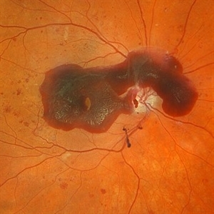
 Total Retinal Detachment
Sep 30 2025 by Kimberly Wakester
Optomap RGB of a 70-year-old woman with a total retinal detachment in the right eye. Exam confirms a chronic appearing retinal detachment with bare LP vision. Thorough scleral depressed exam was performed, revealing IT retinoschisis with large outer and inner holes as the likely causative break. Additionally, there is a full thickness macular hole. Surgery was recommended. Patient is to continue follow up care post operatively.
Photographer: Kimberly Wakester, COA, OCT-C
Imaging device: Optos California
Condition/keywords: macular hole, Retinoschisis, total retinal detachment
-
 Neovascular AMD
Oct 3 2025 by Kimberly Wakester
Optomap RGB of an 80-year-old-woman with Neovascular AMD in the right eye. There is recurrent migration of fluid and exudate inferiorly on exam. There is also a superior hemorrhagic PED. Last Avastin treatment was in 2015. Recommended treatment in her right eye with Avastin. Patient will return for follow up care and repeat OCT.
Photographer: Kimberly Wakester, COA, OCT-C
Imaging device: Optos California
Condition/keywords: Hemorrhagic PED, migration of fluid, Neovascular AMD
-
 Bullous Retinoschisis
Oct 3 2025 by Kimberly Wakester
Optomap RGB of an 29-year-old-woman with Bullous Retinoschisis in both eyes. All areas of retinoschisis remain completely stable on fundus photos and clinical exam. Recommended observation. Will continue yearly follow care with repeat imaging.
Photographer: Kimberly Wakester, COA, OCT-C
Imaging device: Optos California
Condition/keywords: bullous retinoschisis
-
 Bullous Retinoschisis with Pigmentary Changes
Oct 3 2025 by Kimberly Wakester
Optomap RGB of an 29-year-old-woman with Bullous Retinoschisis with pigmentary changes in the right eye. The retinoschisis remains completely stable on fundus photos and clinical exam. Recommended observation. Will continue yearly follow care with repeat imaging.
Photographer: Kimberly Wakester, COA, OCT-C
Imaging device: Optos California
Condition/keywords: bullous retinoschisis
-
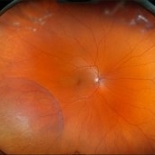
 Dry AMD, Advanced Atrophic with Subfoveal Involvement
Oct 3 2025 by Kimberly Wakester
Optomap RGB of an 76-year-old woman with Dry AMD, Advanced Atrophic with Subfoveal Involvement in the right eye. Longstanding, more likely due to myopic/choroidal atrophy. End-stage disease, would not recommend aggressive intervention. Patient is to continue follow up care and repeat OCT/imaging as directed per doctor. Just a fun mention about the image, if you look at the optic nerve and the vessels it appears to look like an eye.
Photographer: Kimberly Wakester, COA, OCT-C
Imaging device: Optos California
Condition/keywords: Advanced Atrophic with Subfoveal Involvement, dry age-related macular degeneration (dry AMD), Myopic Degeneration
-
 Retinal Detachment with Multiple Breaks
Oct 3 2025 by Kimberly Wakester
Optomap RGB of an 60-year-old man with a retinal detachment with multiple breaks in the left eye. Surgery was recommended. Patient is to continue follow up care post operatively.
Photographer: Kimberly Wakester, COA, OCT-C
Imaging device: Optos California
Condition/keywords: Mac off, Retinal Detachment with multiple breaks
-
 Elmiron Toxicity
Oct 30 2025 by Kimberly Wakester
Optomap RGB and Optomap AF of an 52-year-old woman with Elmiron toxicity from the use of Elmiron for 19 years. Patient is to continue follow up care every 3-4 months with repeat exam and testing to continue monitoring the progression of the degeneration.
Photographer: Kimberly Wakester, COA, OCT-C
Imaging device: Optos California
Condition/keywords: Elmiron Toxicity
-
 Pigmentary Retinal Dystrophy
Oct 30 2025 by Kimberly Wakester
Optomap RGB of an 77-year-old-woman with Pigmentary Retinal Dystrophy in the left eye. Patient is to continue follow up care yearly with dilated exam and diagnostic testing.
Photographer: Kimberly Wakester, COA, OCT-C
Imaging device: Optos California
Condition/keywords: bone spicules, Pigmentary Retinal Dystrophy
-
 Chorioretinitis
Oct 30 2025 by Kimberly Wakester
Optomap RGB of an 37-year-old-woman with stable Chorioretinitis in the right eye. Patient is to return in 6 months for dilated exam and repeat diagnostic testing.
Photographer: Kimberly Wakester, COA, OCT-C
Imaging device: Optos California
Condition/keywords: chorioretinitis
-
 Best Disease
Dec 9 2025 by Kimberly Wakester
Optomap RBG and AF photograph of an 65-year-old man with Best disease in the left eye. The hypopigmented lesions appear stable on clinical exam and fundus photos compared to previous images. Patient is to continue yearly follow up care with dilated exam and repeat imaging.
Photographer: Kimberly Wakester, COA, OCT-C
Imaging device: Optos California
Condition/keywords: Best Disease, Dystrophies of the Retinal Pigment Epithelium
-
 Chronic RD with Retinoschisis
Dec 9 2025 by Kimberly Wakester
Optomap RGB of an 67-year-old woman with a chronic retinal detachment with retinoschisis in the left eye. Surgical intervention vs observation was discussed with patient. Recommended close observation at this time to monitor the progression of the SRF prior to surgical intervention. Patient will return in 6-8 weeks to repeat dilated exam and imaging.
Photographer: Kimberly Wakester, COA, OCT-C
Imaging device: Optos California
Condition/keywords: Chronic RD, retinoschisis
-
 Chronic RD with Retinoschisis
Dec 9 2025 by Kimberly Wakester
B-scan Ultrasound of an 67-year-old woman with a chronic retinal detachment with retinoschisis in the left eye. Surgical intervention vs observation was discussed with patient. Recommended close observation at this time to monitor the progression of the SRF prior to surgical intervention. Patient will return in 6-8 weeks to repeat dilated exam and imaging.
Photographer: Kimberly Wakester, COA, OCT-C
Imaging device: Accutome
Condition/keywords: Chronic RD, retinoschisis
-
 Retinal Detachment with PVR
Dec 9 2025 by Kimberly Wakester
Optomap RGB of an 69-year-old man with a retinal detachment with PVR formation inferotemporal in the right eye s/p RD repair. Surgery was recommended. Patient is to continue follow up care post operatively.
Photographer: Kimberly Wakester, COA, OCT-C
Imaging device: Optos California
Condition/keywords: gas bubble, retinal detachment with PVR
-
 Retinal Detachment with HSRT
Dec 9 2025 by Kimberly Wakester
Optomap RGB of an 43-year-old woman with a retinal detachment with HSRT in the right eye. Surgery was recommended. Patient is to continue follow up care post operatively.
Photographer: Kimberly Wakester, COA, OCT-C
Imaging device: Optos California
Condition/keywords: retinal detachment with HSRT
-
 Retinitis Pigmentosa
Dec 9 2025 by Kimberly Wakester
Optomap RGB and AF of an 78-year-old woman with Retinitis Pigmentosa. Patient is to continue follow up care every 6 months to monitor progression.
Photographer: Kimberly Wakester, COA, OCT-C
Imaging device: Optos California
Condition/keywords: retinitis pigmentosa
-
 Chorioretinitis
Dec 16 2025 by Kimberly Wakester
Optomap RGB of a 37 year-old woman with Chorioretinitis. The chorioretinitis remains largely stable in both eyes on exam and compared to prior photos. Clinical and diagnostic findings in both eyes continue to be most consistent with punctate inner choroidopathy (PIC) /Multifocal choroiditis and panuveitis (MCP). Will continue follow up care every 6 months with dilated exam and repeat testing.
Photographer: Kimberly Wakester, COA, OCT-C
Imaging device: Optos California
Condition/keywords: chorioretinitis, multifocal chorioretinitis (MCP), punctate inner choroidopathy (PIC)
-
 Retinal Detachment with Giant Retinal Tear
Dec 16 2025 by Kimberly Wakester
Optomap RGB of a 66-year-old man with a Retinal Detachment with Giant Retinal Tear OS. Surgery was recommended. Patient is to continue follow up care post operatively.
Photographer: Kimberly Wakester, COA, OCT-C
Imaging device: Optos California
Condition/keywords: giant retinal tear, Retinal detachment
-
 Retinal Detachment with Single Break
Dec 16 2025 by Kimberly Wakester
Optomap RGB of a 45-year-old man with a retinal detachment with a large horseshoe retinal tear in the right eye. Surgery was recommended. Patient is to continue follow up care post operatively.
Photographer: Kimberly Wakester, COA, OCT-C
Imaging device: Optos California
Condition/keywords: HSRT, Retina detachment
-
 Retinal Detachment with Single Break
Dec 16 2025 by Kimberly Wakester
Optomap RGB of a 45-year-old man with a retinal detachment with a large horseshoe retinal tear in the right eye. Surgery was recommended. Patient is to continue follow up care post operatively.
Photographer: Kimberly Wakester, COA, OCT-C
Imaging device: Optos California
Condition/keywords: horseshoe tear, Retinal Detachment
-
 Best Disease
Dec 16 2025 by Kimberly Wakester
Optomap RGB and AF of a 49-year-old man with Dystrophies of the Retinal Pigment Epithelium that is consistent with Best's Disease in both eyes. Will continue yearly follow up care with dilated exam and testing to monitor progression.
Photographer: Kimberly Wakester, COA, OCT-C
Imaging device: Optos California
Condition/keywords: Best Disease, Dystrophies of the Retinal Pigment Epithelium
-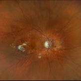
 Toxoplasmosis Chorioretinitis
Dec 16 2025 by Kimberly Wakester
Optomap RGB image of a 22-year-old man with Toxoplasmosis Chorioretinitis in both eyes. Recommended yearly examinations.
Photographer: Kimberly Wakester, COA, OCT-C
Imaging device: Optos California
Condition/keywords: toxoplasmosis chorioretinitis
-
 CHRPE with Lacunae
Dec 22 2025 by Kimberly Wakester
Optomap RGB image of an 48-year-old man with a CHRPE with lacunae in the right eye. Recommended yearly observation.
Photographer: Kimberly Wakester, COA, OCT-C
Imaging device: Optos California
Condition/keywords: congenital hypertrophy of the retinal pigment epithelium (CHRPE)
-
 Retinal Detachment with PVR
Dec 22 2025 by Kimberly Wakester
Optomap RGB montage image of a 54-year-old man with retinal detachment with PVR in the left eye. Surgery was recommended. Patient is to continue follow up care post operatively.
Photographer: Kimberly Wakester, COA, OCT-C
Imaging device: Optos California
Condition/keywords: retinal detachment with PVR
-
 Retinal Detachment with Single Break OD
Dec 22 2025 by Kimberly Wakester
Optomap RGB image of a 65-year-old man with a retinal detachment with a horseshoe retinal tear in the right eye. Surgery was recommended. Patient is to continue follow up care post operatively.
Photographer: Kimberly Wakester, COA, OCT-C
Imaging device: Optos California
Condition/keywords: horseshoe tear, RD
-
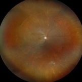
 Retinal Detachment
Jan 8 2026 by Kimberly Wakester
Optomap RGB image of a 64-year-old man with a retinal detachment with a single break in the right eye. Surgery was recommended. Patient is to continue follow up care post operatively.
Photographer: Kimberly Wakester, COA, OCT-C
Imaging device: Optos California
Condition/keywords: Mac off, retinal detachment, Right Eye
-
 Retinitis Pigmentosa
Feb 5 2026 by Kimberly Wakester
Optomap RGB and AF of a 72-year-old woman with Retinitis Pigmentosa. Patient is to continue follow up care every 12 months to monitor progression.
Photographer: Kimberly Wakester, COA, OCT-C
Imaging device: Optos California
Condition/keywords: autofluorescence imaging, Diffuse RPE atrophy, retinitis pigmentosa
-
 Retinal Detachment With a Single Break
Feb 5 2026 by Kimberly Wakester
Optomap RGB image of a 64-year-old man with a retinal detachment with a single break in the right eye. Surgery was recommended. Patient is to continue follow up care post operatively.
Photographer: Kimberly Wakester, COA,
Imaging device: Optos California
Condition/keywords: Retinal Detachment, right eye
-
 Chorioretinal Scar OD
Feb 5 2026 by Kimberly Wakester
Optomap RGB image of a 12-year-old boy with a Chorioretinal Scar in the right eye. Observation was recommended. Will continue follow up care yearly.
Photographer: Kimberly Wakester, COA, OCT-C
Imaging device: Optos California
Condition/keywords: chorioretinal scar, right eye
-
 Retinal Detachment with Multiple Breaks
Feb 5 2026 by Kimberly Wakester
Optomap RGB image of a 58-year-old man with a retinal detachment with multiple breaks in the left eye. Patient presents with macula-involved RD extending from 3:00-8:00, tear from 4:00-5:00 within area of lattice degeneration. Surgery was recommended. Patient is to continue follow up care post operatively.
Photographer: Kimberly Wakester, COA, OCT-C
Imaging device: Optos California
Condition/keywords: left eye, macula-involved RD, Retinal Detachment with Multiple Breaks
-
 Treated Horseshoe Tear of Retina Without Detachment
Feb 5 2026 by Kimberly Wakester
Optomap RGB image of an 55-year-old man with a treated horseshoe retinal tear in the right eye. The horseshoe tear remains well barricaded with laser; no extension of the tear past the laser on clinical exam and fundus photos. Observation recommended. Patient is to continue follow up care yearly.
Photographer: Kimberly Wakester, COA, OCT-C
Imaging device: Optos California
Condition/keywords: right eye, Treated Horseshoe Tear of Retina Without Detachment
-
 Branch Retinal Vein Occlusion and Hypertensive Retinopathy
Feb 5 2026 by Kimberly Wakester
Optomap RGB and FA image of a 75-year-old woman with Branch retinal vein occlusion and Hypertensive retinopathy in the left eye. There is an area of significant partial venous occlusion with fine collateral vessels versus neovascularization along the STA in the left eye. On fluorescein angiogram there was no leakage seen from the telangiectatic vessels, however there is some vascular leakage super temporally. Since there is no edema or neovascularization present, observation was recommended. She is to return in 3-4 months for recheck, prn sooner if changes.
Photographer: Kimberly Wakester, COA, OCT-C
Imaging device: Optos California
Condition/keywords: BRVO, fluorescein angiogram (FA), hypertensive retinopathy, left eye, Optomap RGB
A project from the American Society of Retina Specialists