 Initializing download.
-
 By Kimberly Wakester
Retina Consultants of Carolina, P.A.
Co-author(s): Samuel Feldman, MD - Uploaded on Sep 30, 2025.
- Last modified by Joshua Friedman on Oct 1, 2025.
- Rating
- Appears in
- Miscellaneous
- Condition/keywords
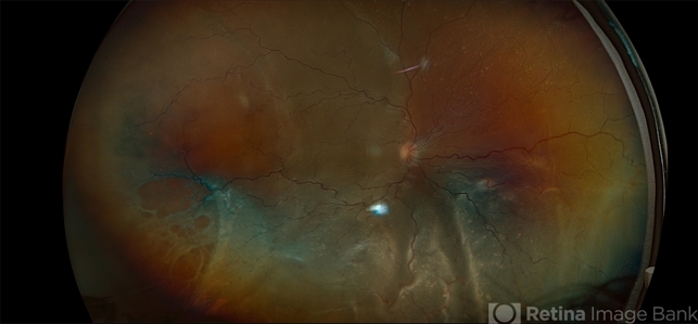
- total retinal detachment, macular hole, Retinoschisis
- Photographer
- Kimberly Wakester, COA, OCT-C
- Imaging device
-
Fundus camera
Optos California - Description
- Optomap RGB of a 70-year-old woman with a total retinal detachment in the right eye. Exam confirms a chronic appearing retinal detachment with bare LP vision. Thorough scleral depressed exam was performed, revealing IT retinoschisis with large outer and inner holes as the likely causative break. Additionally, there is a full thickness macular hole. Surgery was recommended. Patient is to continue follow up care post operatively.


