-
 Representative Full Field Electroretinography Responses
May 13 2024 by Gabrielle Hallai
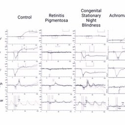
The left most column are control full field ERG responses from an individual with no known retinal pathology. In the second column is an example from a patient with autosomal recessive retinitis pigmentosa. This is an example of an intermediate case where rod function is extinguished but some cone function remains. In more advanced cases, full field ERG responses are typically extinguished to both scotopic and photopic stimuli. The third column is an example of congenital stationary night blindness (CSNB). While full field ERG responses can vary greatly depending on the specific subtype, this example of “complete CSNB” demonstrates extinguished rod pathway responses with the classic electronegative response for the scotopic 3.0 and 10.0 responses, consistent with bipolar cell dysfunction. Photopic cone responses are largely normal in this instance, but ”incomplete CSNB” can cause reduced photopic responses. In the final column, an example of full field ERG responses from a patient with achromatopsia. In achromatopsia, cone function is extinguished early in life, while rod pathway function is largely normal. ERG testing was completed using the Diagnosys ColorDome.
Photographer: Gabrielle Hallai, PhD, Cleveland Clinic Cole Eye Institute
Imaging device: Diagnosys ColorDome
Condition/keywords: achromatopsia, congenital stationary night blindness (CSNB), electroretinography, full field ERG, retinitis pigmentosa
-
 Representative Multifocal Electroretinography Responses
May 13 2024 by Gabrielle Hallai
Multifocal ERG responses from a control individual with no known retinal pathology is shown on the left. The topographical maps (left of each panel) demonstrate the patient’s pattern of responses. The trace arrays (right of each panel) demonstrate the patient’s multifocal ERG responses. The middle set of images demonstrates responses from a patient with Stargardt disease. The topographical map shows decreased patterns throughout the macula. The traces show decreased central response with preserved, but diminished responses in the periphery. The final set of images is from a patient with retinitis pigmentosa. In this case, the topographical map shows a small, green peak in the center. In the trace array, there are extinguished responses in the periphery with a diminished response in the center. Multifocal ERG testing was completed using the Diagnosys LCD Pattern Stimulator.
Photographer: Gabrielle Hallai, PhD, Cleveland Clinic Cole Eye Institute
Imaging device: Diagnosys LCD Pattern Stimulator
Condition/keywords: electroretinography, multifocal ERG (MFERG), retinitis pigmentosa, Stargardt disease
-
 Representative Pattern Electroretinography Responses
May 13 2024 by Gabrielle Hallai
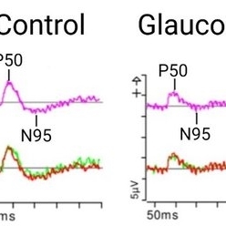
The pattern ERG response on the left is from a control individual with no known retinal pathology. There is a clearly discernable P50 and N95 peak. On the right, there is a representative image from a patient with end-stage glaucoma. In advanced glaucoma, there is often a lack of a clearly discernable N95 peak along with a diminished N95:P50 amplitude ratio, due to retinal ganglion cell dysfunction or degeneration. The top traces are averages from two independent trials shown on the bottom. Pattern ERG testing was completed using the Diagnosys pattern ERG protocol on a CRT monitor.
Photographer: Gabrielle Hallai, PhD, Cleveland Clinic Cole Eye Institute
Condition/keywords: electroretinography, glaucoma, pattern ERG (PERG)
-
 Representative Traces From Different Types of Visual Evoked Potentials (VEP)
May 13 2024 by Gabrielle Hallai
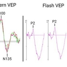
The left most figure demonstrates a normal pattern VEP response. The waveform is composed of a negative N75 peak, a positive P100 peak, and another negative N135 peak. The timing and amplitude of this response can be compared to determine if optic nerve/post-retinal visual pathways are functioning normally or abnormally. The second figure contains examples of a normal flash VEP response. In this case, a three channel VEP was completed which can help to determine chiasmal abnormalities. The major consistent component to assess in the flash VEP is the positive P2 peak that occurs around 120 ms. Pattern VEP responses were recorded using the Diagnosys pattern VEP protocol on a CRT monitor. Flash VEP responses were recorded using the Diagnosys ColorDome stimulator.
Photographer: Gabrielle Hallai, PhD, Cleveland Clinic Cole Eye Institute
Condition/keywords: flash VEP, pattern VEP, VEP, visual evoked potential
-
 Representative Electrooculogram Responses
May 13 2024 by Gabrielle Hallai
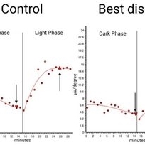
Electrooculogram responses on the left from a control individual with no known retinal pathology. There is a clear dark trough around 13 minutes (arrow down) and a light peak around 25 minutes (arrow up). The Arden ratio, or the light peak to dark trough ratio, is 2.54, indicative of normal retinal pigment epithelium function (normal > 1.80, abnormal < 1.65). On the right-hand side, there is a representative image from an individual with Best macular dystrophy. Note the reduced responses for both the dark and light phase. There is a reduced Arden ratio of 1.23, suggestive of abnormal retinal pigment epithelium function. An abnormal Arden ratio is universal in Best vitelliform macular dystrophy and is the most common electroretinographic change in this disease. Other bestrophinopathies such as autosomal recessive bestrophinopathy may have normal EOG. EOG testing was completed on the Diagnosys ColorDome.
Photographer: Gabrielle Hallai, PhD, Cleveland Clinic Cole Eye Institute
Imaging device: Diagnosys ColorDome
Condition/keywords: Best disease, electrooculogram, electroretinography, EOG
-
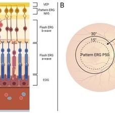 Summary of Retinal Origins of Various Electroretinographic Components
May 13 2024 by Gabrielle Hallai
Each layer of the retina can be assessed using various electroretinographic techniques. A) A summary of the retinal origins of each response is shown here. B) Macular function can be assessed by pattern and multifocal ERG recording and provide a specific assessment of central vision, while flash tests are dominated by the more peripheral retina.
Photographer: Gabrielle Hallai, PhD, Cleveland Clinic Cole Eye Institute
Condition/keywords: electroretinography, ERG
A project from the American Society of Retina Specialists