 Initializing download.
-
 By Gabrielle Hallai
Cleveland Clinic - Uploaded on May 13, 2024.
- Last modified by Joshua Friedman on May 14, 2024.
- Rating
- Appears in
- Miscellaneous
- Condition/keywords
- electroretinography, multifocal ERG (MFERG), Stargardt disease, retinitis pigmentosa
- Photographer
- Gabrielle Hallai, PhD, Cleveland Clinic Cole Eye Institute
- Imaging device
- Diagnosys LCD Pattern Stimulator
- Description
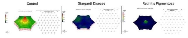
- Multifocal ERG responses from a control individual with no known retinal pathology is shown on the left. The topographical maps (left of each panel) demonstrate the patient’s pattern of responses. The trace arrays (right of each panel) demonstrate the patient’s multifocal ERG responses. The middle set of images demonstrates responses from a patient with Stargardt disease. The topographical map shows decreased patterns throughout the macula. The traces show decreased central response with preserved, but diminished responses in the periphery. The final set of images is from a patient with retinitis pigmentosa. In this case, the topographical map shows a small, green peak in the center. In the trace array, there are extinguished responses in the periphery with a diminished response in the center. Multifocal ERG testing was completed using the Diagnosys LCD Pattern Stimulator.


---thumb.jpg/image-square;max$79,0.ImageHandler "Retinitis Pigmentosa")


